BIOACTIVE POROUS PEG-PEPTIDE COMPOSITE HYDROGELS WITH TUNABLE MECHANICAL PROPERTIES
A THESIS
SUBMITTED TO THE MATERIALS SCIENCE AND NANOTECHNOLOGY PROGRAM OF GRADUATE SCHOOL OF ENGINEERING AND SCIENCE
OF BILKENT UNIVERSITY
IN PARTIAL FULFILLMENT OF THE REQUIREMENTS FOR THE DEGREE OF
MASTER OF SCIENCE
By Melis Göktaş August, 2014
I certify that I have read this thesis and that in my opinion it is fully adequate, in scope and in quality, as a thesis of the degree of Master of Science.
……….
Assoc. Prof. Dr. Mustafa Özgür Güler (Advisor)
I certify that I have read this thesis and that in my opinion it is fully adequate, in scope and in quality, as a thesis of the degree of Master of Science.
………. Assist. Prof. Dr. Ayşe Begüm Tekinay
I certify that I have read this thesis and that in my opinion it is fully adequate, in scope and in quality, as a thesis of the degree of Master of Science.
………. Assoc. Prof. Dr. Çağdaş Devrim Son
Approved for the Graduate School of Engineering and Science: ……….
Prof. Dr. Levent Onural Director of the Graduate School
i ABSTRACT
BIOACTIVE POROUS PEG-PEPTIDE COMPOSITE HYDROGELS WITH TUNABLE MECHANICAL PROPERTIES
Melis Göktaş
M.S. in Materials Science and Nanotechnology Supervisor: Assoc. Prof. Mustafa Özgür Güler
August, 2014
Mimicking the instructive cues of native extracellular matrix (ECM) is fundamental to understand and control the processes regulating cell function and cell fate. Extensive research on the structure and biological complexity of ECM has shown that three types of critical information from the ECM have influence on cellular behaviour: (1) biophysical properties (elasticity, stiffness), (2) biochemical properties (bioactive peptide epitopes of ECM molecules), and (3) nanoarchitecture (nanofibrillar structure, porosity) of ECM. Recent efforts have therefore focused on the construction of ECM mimetic materials to modulate tissue specific cell functions. Advances in biomaterial platforms include artificial ECM mimics of peptide conjugated synthetic polymer hydrogels presenting bioactive ligands produced with covalent chemistry. These materials have already found application in tissue engineering, however, these biomaterial platforms represent oversimplified mimics of cellular microenvironment and lack the complexity and multifunctional aspects of native ECM.
ii
In this work, we developed a novel polyethylene glycol (PEG)-peptide nanofiber composite hydrogel system with independently tunable biochemical, mechanical and physical cues that does not require any chemical modification of polymer backbone to create synthetic ECM analogues. This approach allows non-interacting modification of multifactorial niche properties (i.e. bioactive ligands, stiffness, porosity), since no covalent conjugation method was used to modify PEG monomers for the incorporation of bioactivity and porosity. Combining the self-assembled peptide nanofibers with crosslinked polymer network simply by facile mixing followed by photo-polymerization resulted in the formation of porous hydrogel systems. Resulting porous network can be functionalized with desired bioactive signalling epitopes by simply altering the amino acid sequence of peptide amphiphile molecules. In addition, the mechanical properties of the composite system can be precisely controlled by changing the PEG concentration. Ultimately, multifunctional PEG-peptide composite scaffolds reported in this work, can fill a critical gap in the available biomaterials as versatile synthetic mimics of ECM with independently tunable properties. Such a system could provide a useful tool allowing the investigation of how complex niche cues interplay to influence cellular behaviour and tissue formation both in 2D and 3D platforms.
Keywords: Extracellular Matrix (ECM), Peptide Nanofibers, Self Assembly, Polyethylene Glycol (PEG), Hydrogel
iii ÖZET
MEKANİK ÖZELLİKLERİ AYARLANABİLİR BİYOAKTİF POROZ PEG-PEPTİT KOMPOZİT HİDROJELLERİN ÜRETİMİ
Melis Göktaş
Malzeme Bilimi ve Nanoteknoloji Programı, Yüksek Lisans Tez danışmanı: Doç. Dr. Mustafa Özgür Güler
Ağustos, 2014
Hücre davranışını ve hücre fonksiyonlarını düzenleyen mekanizmaların anlaşılması ve kontrol edilmesi amacıyla, doğal hücrelerarası matris ortamının yönlendirici özelliklerinin taklit edilmesi önem taşımaktadır. Doğal hücrelerarası matrisin yapısı ve biyolojik kompleksitesi üzerine yapılan çalışmalar hücrelerarası matrise ait üç tip kritik bilginin hücre davranışı üzerinde etkili olduğunu göstermiştir: (1) biyofiziksel özellikler (elastisite, sertlik), (2) biyokimyasal özellikler (biyoaktif peptit sinyalleri), ve (3) nanoyapı (nanofibriler yapı, porozite). Bu sebeple, günümüzde doku spesifik hücre davranışlarının yönlendirilmesi amacıyla gerçekleştirilen çalışmalar hücrelerarası matris ortamını taklit eden biyomalzemelerin geliştirilmesi üzerine odaklanmıştır. Biyomalzeme alanında en önemli yeniliklerden biri, kovalent kimya metotları kullanılarak biyoaktif peptit epitopları ile modifiye edilmiş sentetik polimer hidrojellerin geliştirilmesidir. Sentetik polimerler günümüzde doku mühendisliği alanında uygulama bulmalarına rağmen, bu malzemeler hücre mikro-ortamının oldukça basitleştirilmiş modelleri olarak kalmakta ve çok fonksiyonlu doğal hücrelerarası matrisin kompleks yapısını taklit edememektedirler.
iv
Bu çalışmada, bağımsız olarak ayarlanabilir biyokimyasal, mekanik ve fiziksel özelliklere sahip özgün bir polietilen glikol (PEG)-peptit nanofiber kompozit hidrojel sistemi geliştirilmiştir. Geliştirilen kompozit hidrojel sistemi polimer yapısında herhangi bir kimyasal modifikasyona gerek duyulmaksızın sentetik ESM analoglarının üretimine olanak sağlamaktadır. Biyoaktivite ve porozitenin sağlanması için herhangi bir kovalent konjugasyon metodu kullanılmaması sayesinde üretilen hidrojellerin özellikleri birbirinden etkilenmeksizin çok yönlü olarak modifiye edilebilmektedir. Kendiliğinden biraraya gelen peptit nanofiberlerin, foto-polimerizasyon yöntemi ile çapraz bağlanan polimer ağı ile karıştırılması, porlu hidrojel sistemlerinin oluşturulmasını sağlamıştır. Elde edilen porlu yapılar basit bir şekilde peptit amfifil moleküllerinin amino asit dizilimleri değiştirilerek biyoaktif sinyallerle fonksiyonalize edilebilmektedir. Ayrıca oluşan kompozit sistemin mekanik özellikleri polimer konsantrasyonu değiştirilerek kolayca ayarlanabilmektedir. Sonuç olarak, üretilen çok fonksiyonlu PEG-peptit kompozit iskeleler doğal hücrelerarası matrisi taklit eden, özellikleri ayarlanabilir biyomalzeme platformları alanında önemli bir eksikliği giderebilecektir. Elde edilen bu sistem, iki boyutlu (2D) ve üç boyutlu (3D) ortamlarda hücrelerarası matris benzeri kompleks faktörlerin hücre davranışını ve doku oluşumunu nasıl etkilediğinin araştırılması için kullanışlı bir araç olarak işlev görebilir.
Anahtar kelimeler: Hücrelerarası Matris, Peptit Nanofiberler, Kendiliğinden Biraraya Gelme, Polietilen Glikol (PEG), Hidrojel.
v
ACKNOWLEDGEMENTS
I have spent two years in a great research environment with many valuable people. First of all, I would like to thank my supervisor Prof. Mustafa Özgür Güler for his support and guidance. He improved my scientific perspective and taught me to ask the right questions during my research. I also would like to thank Prof. Ayşe Begüm Tekinay for her guidance and support throughout my research. This work could not be accomplished without her precious contribution.
I would like to thank Hakan Ceylan, whose support and motivation contributed a lot to this work. I also want to thank Göksu Çınar for her companionship during two years.
I would like to thank Gülcihan Gülseren and Gülistan Tansık for always cheering me up and for their support. It was great to know you and work with you all: Melis Şardan, Reşad Mammadov, Aref Khalily, Ceren Garip, Elif Arslan, Didem Mumcuoğlu, Yasin Tümtaş, Öncay Yaşa, Gözde Uzunallı, Berna Şentürk.
I would like to thank UNAM (National Nanotechnology Research Center) for giving me this opportunity and TUBITAK (The Scientific and Technological Research Council of Turkey) for financial support, BIDEB 2228-A MSc fellowship, and grant 213M406.
vi
TABLE OF CONTENTS
TABLE OF CONTENTS ... VI LIST OF ABBREVIATIONS ... IX LIST OF FIGURES ... XI LIST OF TABLES ... XIII
1. CHAPTER 1: INTRODUCTION ... 1
1.1. MICROENVIRONMENT OF CELLS: EXTRACELLULER MATRIX ... 2
1.2. ECM STRUCTURE AND FUNCTION ... 3
1.2.1. Macromolecular components of ECM. ... 4
1.2.1.1.Collagen ... 4
1.2.1.2. Adhesive glycoproteins ... 5
1.2.1.2.1. Fibronectin ... 6
1.2.1.2.2. Vitronectin ... 6
1.2.1.2.3. Laminin ... 7
1.2.1.3. Matricellular proteins and glycoproteins ... 8
1.3. CELL-ECM INTERACTIONS ... 9
1.3.1. Adhesive properties of ECM: Integrin-binding epitopes ... 11
1.3.2. Mechanical properties of ECM ... 12
1.3.2.1. Cell probing of ECM stiffness: Mechanotransduction ... 13
1.3.3. Nanostructure and porosity of ECM ... 15
1.4. HYDROGELS AS ECM MIMICS ... 16
1.4.1. Bioengineering approaches to create synthetic ECM analogues ... 18
vii
1.4.1.2. Controlling the mechanical properties ... 21
1.4.1.3. Tuning the porosity and permeability ... 23
1.4.1.4. Self-assembly as a strategy for structural and bioactive ECM mimics ... 24
2. CHAPTER 2: BIOACTIVE POROUS PEG-PEPTIDE COMPOSITE HYDROGELS WITH TUNABLE MECHANICAL PROPERTIES. ... 27
2.1. INTRODUCTION ... 28
2.2. MATERIALS & METHODS ... 33
2.2.1. Materials ... 33
2.2.2. Synthesis and Characterization of Peptide Amphiphiles ... 33
2.2.3. Transmission Electron Microscopy (TEM) Imaging of PA Nanofibers ... 34
2.2.4. Preparation of 2D Hydrogels ... 35
2.2.5. Preparation of 3D Hydrogels ... 35
2.2.6. Scanning Electron Microscopy (SEM) ... 36
2.2.7. Oscilatory Rheology ... 36
2.2.8. Brunauer-Emmett-Teller (BET) Analysis ... 37
2.2.9. Cell Culture and Maintanance ... 37
2.2.10. Viability of Saos-2 Cells on PEG and PEG-Peptide Substrates ... 38
2.2.11. Adhesion of Saos-2 Cells on PEG and PEG-Peptide Substrates ... 38
2.2.12. Spreading and Cytoskeletal Organization Analysis of Saos-2 Cells on PEG and PEG-Peptide Substrates ... 39
2.2.13. Immunocytochemisty (ICC) ... 39
2.2.14. Quantitative Reverse Transcription Polymerase Chain Reaction ... 40
viii
2.3. RESULTS & DISCUSSIONS ... 41
2.3.1. Peptide Amphiphiles ... 41
2.3.2. Self-Assembly of PA Nanofibers ... 43
2.3.3. Synthesis of 2D Hydrogels ... 43
2.3.4. Material Characterizations ... 47
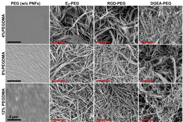
2.3.4.1. SEM Imaging of Resulting Networks ... 47
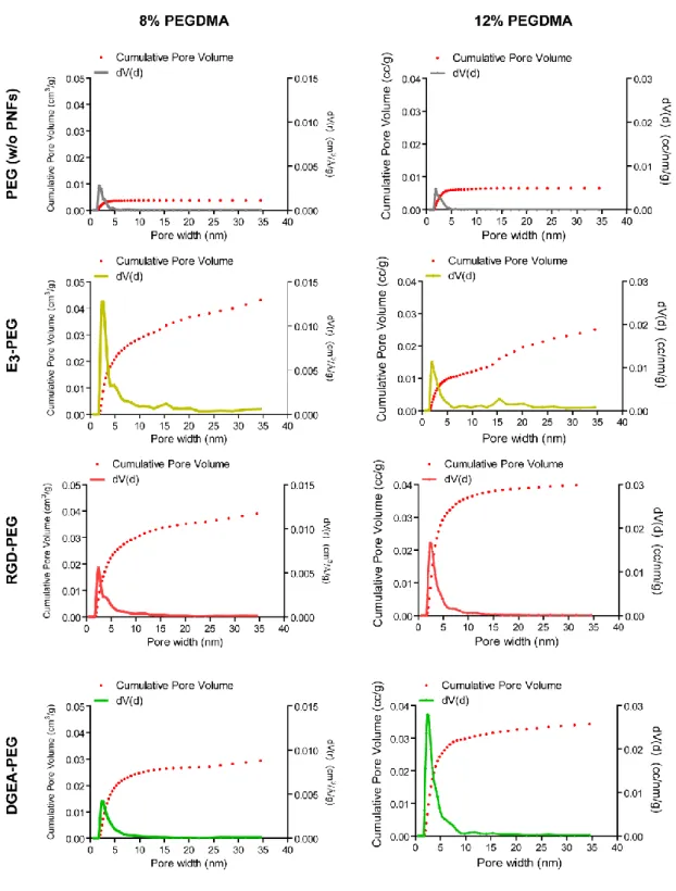
2.3.4.2. Porosity and Surfaces Area Analysis with BET Method ... 49
2.3.4.3. Mechanical Characterizations – Oscillatory Rheology ... 51
2.3.4.3.1. Time Sweep Test ... 51
2.3.4.3.2. Amplitude Sweep Test ... 53
2.3.5. Investigation of Cellular Behavior ... 55
2.3.5.1. Live/Dead Assay ... 55
2.3.5.2. Adhesion Assay ... 56
2.3.5.3. Spreading and Cytoskeletal Organization Analysis ... 59
2.3.5.4. Gene Expression Analysis ... 60
2.3.5.4.1. ICC Staining ... 64
2.3.5.4.2. qRT-PCR Analysis ... 64
2.3.6. Preperation of 3D Hydrogels ... 69
2.3.6.1. Viability Analysis within the 3D Hydrogels ... 69
2.4. CONCLUSION & FUTURE PERSPECTIVES ... 72
ix
LIST OF ABBREVIATIONS
1D: 2D: 3D: BET: BIS: COL1: DDR: DIEA: ECM: ESI: FACIT: FAK: FBS: GAG: GAPDH: HBTU: HPLC: hMSCs: ICC: LC-MS: LSA: LVR: MuSCs: One-Dimensional Two-Dimensional Three-Dimensional Brunauer-Emmett-Teller N,N′-methylenebis(acrylamide) Collagen-1Discoidin Domain Tyrosine Kinase Receptor Diisopropylethylamine
Extracellular Matrix Electrospray Ionization
Fibril Associated Collagens with Interrupted Triple Helices Focal Adhesion Kinase
Fetal Bovine Serum Glycosaminoglycan
Glyceraldehyde 3-Phosphate Dehydrogenase
N,N,N’,N’-Tetramethyl-O-(1H-benzotriazole-1-yl) Uranium Hexafluorophosphate High Performance Liquid Chromatography
Human Mesenchymal Stem Cells Immunocytochemistry
Liquid Chromatography-Mass Spectrometry Limiting Strain Amplitude
Linear Viscoelastic Range Skeletal Muscle Stem Cells
x NSCs: NHS: PA: PBS: PEG: PEGDA: PEGDMA: PNFs: PVA: qRT-PCR: QSDFT: Q-TOF: RUNX2: SEM: SLPRS: TEM: TFA : UV : vmIPN :
Neural Stem Cells
N-Hydroxyl Succinimide
Peptide Amphiphile Phosphate Buffered Saline
Polyethylene Glycol Polyethylene Glycol Diacrylate Polyethylene Glycol Dimethacrylate
Peptide Nanofibers Polyvinyl Alcohol
Quantitative Reverse Transcriptase Polymerase Chain Reaction Quenched Solid Density Functional Theory
Quadrupole Time of Flight Runt-Related Transcription Factor
Scanning Electron Microscopy Short Leucine Rich Proteoglycans Tranmission Electron Microscopy
Trifluoroacetic Acid Ultraviolet
xi
LIST OF FIGURES
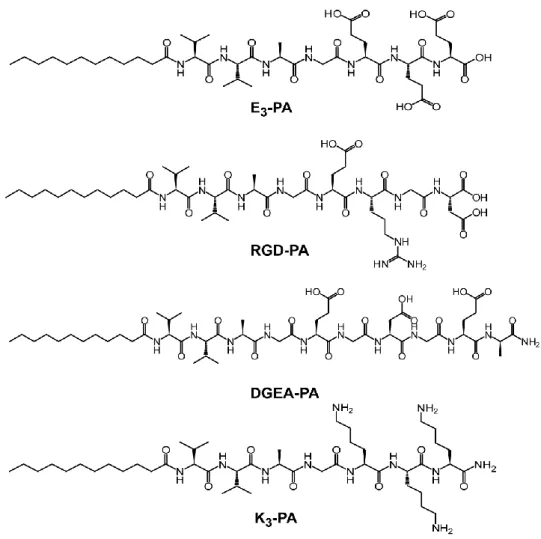
Figure 1.1. Diagram of fibronectin modular structure, structure of fibronectin modules and binding units ... 7 Figure 1.2. Molecular structure of a representative peptide amphiphile. ... 26 Figure 2.1. Chemical representations of Lauryl-VVAGEEE (E3-PA),
VVAGERGD (RGD-PA), VVAGEGDGEA-Am (DGEA-PA) and Lauryl-VVAGKKK-Am (K3-PA) ... 41
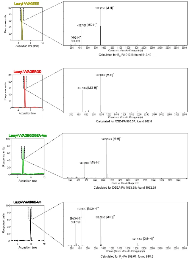
Figure 2.2. Liquid chromatography-mass spectrometry (LC-MS) analysis of the synthesized PAs ... 42 Figure 2.3. Tranmission Electron Microscopy (TEM) images of PA combinations………..45 Figure 2.4. Crosslinking mechanism of PEGDMA. ... 47 Figure 2.5. Scanning electron microscopy (SEM) images of PEG (w/o peptide nanofibers) and PEG-peptide composites ... 48 Figure 2.6. BET analysis showing the pore size distributions and cumulative pore volumes of PEG (w/o peptide nanofibers) and PEG-peptide composites ... 50 Figure 2.7. Total pore volume and specific surface area of PEG (w/o peptide nanofibers) and PEG-peptide composite scaffolds. ... 51 Figure 2.8. Storage/loss moduli of PEG (w/o peptide nanofibers) and PEG-peptide samples showing the gel character of resulting networks ... 52 Figure 2.9. Equilibrium storage moduli of PEG (w/o peptide nanofibers) and PEG-peptide composite hydrogels ... 52 Figure 2.10. Rheological characterizations of gels ... 54
xii
Figure 2.11. Photographs of A) PEG-PA (E3-PEG, 12% wt PEGDMA) and B) only
peptide gel (E3+K3) with the same storage moduli showing the increased elasticity
and stability of the composite system ... 54 Figure 2.12. Representative Calcein-AM/ethidium homodimer stained micrographs of Saos-2 cells on PEG (w/o peptide nanofibers) samples and PEG-peptide composites showing the non-toxic effect of hydrogel scaffolds. ... 56 Figure 2.13. A) Representative Calcein-AM stained micrographs and B) relative adhesion of Saos-2 cells on PEG (w/o peptide nanofibers) and PEG-peptide (E3-PA
combination) substrates at 24 h in serum free culture conditions ... 58 Figure 2.14. Representative Calcein-AM stained micrographs of Saos-2 cells on PEG (w/o peptide nanofibers) samples and PEG-peptide (E3-PEG with 12% wt
PEGDMA) composites showing the enhanced adhesion of cells with peptide incorporation. ... 59 Figure 2.15. Representative Phalloidin stained micrographs of Saos-2 cells on PEG (w/o peptide nanofibers) and PEG-peptide substrates at 72 h. ... 62 Figure 2.16. A) Projected spreading areas and B) aspect ratios of Saos-2 cells on PEG (w/o peptide nanofibers) and PEG-peptide substrates at 72 h. ... 63 Figure 2.17. Representative ICC micrographs (40X magnification) of Saos-2 cells on crosslinked PEG (w/o peptide nanofibers) and PEG-peptide composite substrates at day 7. ... 64 Figure 2.18. A,B) RUNX2 and C,D) COL1 gene expressions of Saos-2 cells on crosslinked PEG (w/o peptide nanofibers) and PEG-peptide composite substrates at day 3 and day 7 ... 68
xiii
Figure 2.19. Representative live/dead micrographs of Saos-2 cells encapsulated within three-dimensional (top) PEG (w/o peptide nanofibers) and (bottom) RGD-PEG scaffolds at day 7 ... 71
LIST OF TABLES
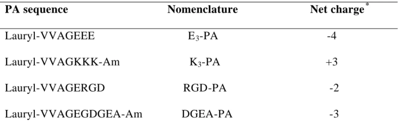
Table 2.1. Bioinspired self-assembling PA building blocks.. ... 46 Table 2.2. Nomenclature and composition of PEG and PEG-peptide composite hydrogels.. ... 46 Table 2.3. Primer list used in the qRT-PCR setups... ... 61
1
CHAPTER 1
2 CHAPTER 1
1. INTRODUCTION
1.1. MICROENVIRONMENT OF CELLS: EXTRACELLULAR MATRIX Cellular reactions are guided by the highly complex microenvironment of cells and the fate of cells is determined by information received from soluble factors, other cells, and the physical network they are encapsulated in. This physical network that provides structure and support to cells is called extracellular matrix (ECM). Cells secrete ECM molecules and maintain the matrix through continuous remodeling of its structure. ECM in turn, provides support to cells to communicate with each other and with the external environment. 1-2
For many years, ECM was known as an inert background which occupies the space between the cells to provide a physical network for structural support. However, recent investigations have clarified that ECM is much more complex than it was thought to be and acts as an active component for the control of cell behaviour.3-5 It is now accepted that, beginning with embryogenesis and continuing through adulthood, cellular development is influenced by the interaction between cells and their ECM.6 Along with its heterogeneous composition that consists of proteins, proteoglycans, and signalling molecules, ECM is a supply of complex information for cells. Information contained in the ECM provides cells temporal and positional clues such as where they are, where they should be going, how old they are (in terms of cellular differentiation), and
3
when it is time for to die (apoptosis).7 Biochemical (cell adhesion, presentation of signalling molecules) and mechanical (stiffness, remodelling) properties of ECM provided by its macromolecular components and bioactive cues can directly influence cell survival, proliferation, migration and differentiation.8-9 Thus, successful understanding of ECM structure and signals can provide us the ability to evaluate complex intracellular signalling pathways and control cellular functions.
1.2. ECM STRUCTURE AND FUNCTION
ECM consists of a great diversity of insoluble macromolecules including structural proteins such as collagens and elastin, glycoproteins including fibronectin, vitronectin and laminin, and glycosaminoglycans.10 Fibrous ECM proteins form a network of fibers and fibrils. Composition and spatial arrangement of ECM can vary from one tissue type to another. For example, bone ECM is mostly composed of collagen type I, and non-collagenous proteins including osteocalcin, fibronectin and vitronectin while cartilage ECM mostly consists of collagen type II and aggrecans.11-13 Since, different ECM macromolecules can selectively stimulate different signalling pathways through cell-ECM interactions, this tissue-specific composition of ECM might be instructive for materials science to regulate cell behaviour to obtain the desired output.
4 1.2.1. Macromolecular Components of ECM 1.2.1.1. Collagen
Collagen is the most abundant component of the ECM and it forms ~30% of the total proteins in the body.14 Collagen provides tensile strength and elasticity to tissues and organs, and it forms the structural framework of connective tissues including bone, tendons and dermis.15-16
Collagens are characterized by a distinct triplet of amino acid repeat defined as Gly-X-Y that eventually forms a triple helix structure. Gly represents glycine amino acid, while X and Y residues can be any amino acid but are commonly proline and hydroxyproline.17 Each single polypeptide chain forming the triple helical assembly is called α-chain and collagens are separated according to the composition of α-chains and their supramolecular assembly. According to the repeat length and integrity of the Gly-X-Y repeats, self-assembly of the α-chains may result in the formation of uninterrupted triple helix structure as in the case of fibrillar collagen or the presence of the non-collagenous domain can form helical interruptions. Therefore, different α-chain motifs give rise to a number of different supramolecular assemblies with various geometric networks.18 For example, in skin, tendon, bone and cartilage, the collagenous backbone of the ECM consists of crossbanded fibril-forming collagens (Type I, II, III, V, XI, XXIV, and XXVII) and the structure is supported by fibril-associated collagens with interrupted triple helices (FACIT) (Type IX, XII, XIV, XVI, XX, and f XXI) as well as microfibrillar type VI collagen.19-21 Some other collagen types like network forming collagens (Type IV, VII, and X) contain large collagenous
5
domains interrupted by short non-collagenous sequences (other than Gly-X-Y repeat). Type IV collagens which are found in the basement membrane of epithelial tissues assemble into chicken-wire-like collagenous networks, while the ones found in the Descemet’s membrane of the eye (Type VIII) and hypertrophic cartilage (Type X) forms regular hexagonal networks.22-24 This structural heterogeneity provides different organization of collagen types within the ECM of different tissues with functional diversity and contributes to a range of biological functions including cell adhesion, migration, tissue repair , molecular filtration and tumor suppression.25
1.2.1.2. Adhesive Glycoproteins
Cells adhere to ECM through interaction with adhesive glycoproteins including fibronectin, vitronectin, laminin, thrombospondins, tenascins, entactins, nephronectin, fibrinogen, and others. Adhesive glycoproteins bind to cells through cell surface integrin receptors and interact with other ECM proteins to form a complex matrix network. Interactions between the cells and ECM glycoproteins can alter many cellular responses such as survival, growth, migration and differentiation. In this section, major cell adhesion proteins namely fibronectin (which interacts with more than ten different integrin receptors), vitronectin, and laminin are discussed.
6 1.2.1.2.1. Fibronectin
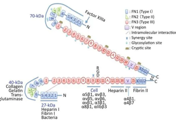
Fibronectin is a high molecular weight dimeric glycoprotein (~450 kDa per dimer) which is expressed by a variety of cells.26 Some forms of fibronectin such as the ones found in blood plasma can remain in soluble form, while the ones found in the ECM are associated into disulfide-bonded fibrillar form.7 ECM fibronectin consists of two similar subunits with a molecular weight of ∼220 to 250 kDa covalently linked through disulfide bonds near the C-terminus.27 Each fibronectin subunit contains three types of repeating modules defined as FN1 (12 type I repeats), FN2 (2 type II repeats), and FN3 (15-17 type III repeats). These modules form 90% of the total sequence. The remaining part includes a connector between 5FN1 and 6FN1 modules, a connector between 1FN3 and 2FN3 modules,
and a variable (V) sequence (Figure 1).28 Each fibronectin molecule contains binding sites of a variety of molecules including cell surface integrins (α5β1, αVβ1,
αVβ3, αVβ5, αVβ6, α3β1, α4β1, α4β7, α8β1, αIIbβ3) collagens, proteoglycans and heparin
sulfate. Therefore, fibronectins provide binding sites to cells, also serve to bind other components of the ECM together.
1.2.1.2.2. Vitronectin
Vitronectin (also known as serum spreading factor, S-protein, and epibolin) is a multifunctional glycoprotein found in blood plasma and ECM.29 It is found in the fibrillar form in ECM of a variety of tissues and colocalizes with fibronectin and elastic fibers.7 Vitronectin can also interact with a variety of ECM molecules including collagen and heparin as well as some cell surface integrins (αIIbβ3, αvβ1,
7
fibronectin, does not recognize vitronectin. Interactions between vitronectin and integrin receptors of cells activate intracellular signalling pathways to mediate cellular functions such as adhesion, spreading, migration, differentiation, growth and apoptosis.35-38
Figure 1.1. Diagram of fibronectin modular structure, structure of fibronectin modules and binding units (Reproduced with permission from ref. 28, copyright © Springer.).
1.2.1.2.3. Laminin
Laminins are large adhesive glycoproteins (400-900 kDa) that consist of three different polypeptide chains (α, β and γ) which form its heterotrimeric structure. Laminin binds to cell surface receptors such as integrins, heparins and
α-8
dystroglycan.28 Majority of the binding sites for integrin receptors are found on the long α-chain of the laminin molecule. Most of the integrin receptors that have been reported to bind laminin are found in the integrin β1 family including α1β1,
α2β1, α3β1, α6β1, α6β4, α7β1, and α9β1 integrins. Other integrins that bind to laminin
include αvβ3 and α6β4.39-40 Interaction of the laminin with integrin receptors
activates different intracellular signalling pathways involving focal adhesion kinases (FAK), mitogen-activated protein kinases (MAPK), phosphatases, and cytoskeletal components. Along with the signal transduction, cellular behaviours such as survival, adhesion, migration, proliferation and differentiation can be mediated by laminin-integrin interactions.41-44
1.2.1.3. Matricellular proteins and proteoglycans
Matricellular proteins function by binding to other matrix proteins and cell surface receptors, however they do not make any contribution to the structural integrity of the ECM.45 Members of matricellular proteins include thrombospondins, tenascins, osteonectin and osteopontin.7 Even though they are referred as “anti-adhesive proteins”, since they induce rounding and detachment of some cells in vitro, they also act as regulators of cell adhesion, migration and differentiation in various tissues.10
Proteoglycans contain a number of families of multidomain proteins that are covalently attached to glycosaminoglycan (GAG) chains. Proteoglycans are named according to the type of attached GAG chains. Large proteoglycans such as aggrecan, versican, neurocan and brevican are able to form
very-high-9
molecular-weight aggregates by interacting with hyaluronate.46 The interaction between hyaluronate and highly sulfated, negatively charged GAG side-chains of large proteoglycans provides the turgor and elasticity of many tissues.47 For example, in cartilage, large hyaluronan-aggrecan complexes are entrapped within the fibrillar collagen network and the high-content of sulfated GAGs provide the high water uptake capacity of the tissue.48 Therefore, cartilage tissue can generate enormous turgor and elasticity, and shows great mechanical resistance to pressure.46
Besides large pretoglycans, another protein family called short leucine rich proteoglycans (SLPRS), which includes decorin, biglycan, fibromodulin, chondroadherin, and aspirin, plays an important role in collagen fibril assembly as well as the storage and inhibition of transforming growth factor β and bone morphogenetic proteins.49-50 Thus, even tough proteoglycans do not support cell adhesion and growth directly, they indirectly affect cell behavior as the regulators of extracellular matrix assembly, providers of tissue resilience and modulators of growth factors.51
1.3. CELL-ECM INTERACTIONS
Several types of receptor families including integrins, syndecans and discoidin -domain tyrosine kinase receptors DDR1 and DDR2, take roles in the recognition of signals coming from the ECM.52 However, the transmission of chemical and mechanical signals from the ECM is primarily mediated by integrin receptors.53
10
Integrins are heterodimeric transmembrane receptors that provide the connection between ECM and cytoskeleton of cells. Each integrin receptor consists of α and β subunits. Up to date, 18 α and 8 β integrin subunits have been identified and various combination of these subunits were found lead to formation of 24 different heterodimers, which have unique binding characteristics determining the ligand specifity.54
Most of the integrins can bind to several types of ECM molecules, and one ECM molecule can bind to more than one integrin. Major ECM binding integrins include β1 integrin that are able to bind to fibronectin (α4β1, α5β1, α5β3, αvβ3),
collagen (α1β1, α2β1, α10β1, α11β1) and laminins (α3β1, α6β1, α7β1).55
Both α and β subunits, which pass through the cell membrane have large (700-1100 residues) extracellular domains and small (30-50 residues) cytoplasmic domains. The extracellular domains of integrins recognize their target ligand. Upon binding, conformational changes in the integrin molecules occur and their cytoplasmic domains associate with cytoskeleton and intracellular signal transduction molecules.56-58 Binding of the intracellular integrin domains to focal complex proteins including focal adhesion kinase FAKp130, integrin-linked kinase, Fyn and c-src is followed by the incorporation of intracellular proteins such as paxillin, α-actinin, vinculin, talin, and zyxin into the focal complexes.59-61
Association of the integrins with this complex signalling network activates downstream signalling cascades such as protein kinase C, Rac, Rho and MAPK pathways.61-62 Along with the signalling, clustering of ECM ligands, integrins and
11
cytoskeletal components including actin fibers lead to formation of focal adhesions.63 Depending on the regulated specific signalling pathway within the cells, integrin mediated cell-ECM interactions can alter cellular behaviours such as survival, proliferation and differentiation.64-66 Therefore, elucidation of cell-ECM interactions and utilization of integrin binding epitopes can be a useful target for biomimetic tissue engineering strategies.
1.3.1. Adhesive properties of ECM: Integrin-binding epitopes
Although ECM macromolecules such as collagens, fibronectin, vitronectin and laminin have long protein backbones, integrin binding is very specific and integrins recognize only a few short peptide sequences within the molecules. One of the most studied integrin-binding epitopes is RGD-adhesive peptide sequence found in fibronectin, vitronectin, laminin and other adhesive glycoproteins.67 Even tough it was first discovered in vitronectin, RGD sequence is well -known for its binding to αvβ3 integrins that recognize the sequence located in the 3rd
repeat ofFN3 domain in fibronectin.68-69 RGD peptide motif is also found within the typical Gly-X-Y-Gly-X-Y order of collagen molecules, however most of these sequences lack of bioactivity. One of the active forms is found in type IV collagen and the three aminoacids forming the R-G-D sequence is located in the separate α chains of the collagen molecule, which is recognized by αvβ3
integrins.70 Another well-known integrin-binding peptide sequence found in the collagens is GFOGER sequence, which has been located in type I collagen. 71 GFOGER sequence binds to β1 family of integrins, including α1β1, α2β1, α10β1,
12
Also, RGD sequences located in the α1 and α2 chains of the laminin molecule have been found to be adhesive and they are recognized by α6β1 and α7β1
integrins.72-73 Other studies have identified another short peptide sequence YIGSR located in the β1 chain of laminin responsible for integrin-mediated cell adhesion and differentiation.74-75 α1 chain of the laminin contains another adhesive sequence IKVAV which promotes cell adhesion, migration, neurite outgrowth and tumor growth.76
Apart from these extensively studied adhesive sequences, some other integrin-binding epitopes were identified in fibronectin (REDV77, LDV78 and PHSRN79), collagen (DGEA80) and laminin (PDSGR81).
1.3.2. Mechanical properties of ECM
Collagen and elastin are the two major structural proteins of the native ECM. Mechanical properties of ECM are determined by a complex structure constructed by interwoven fibers of collagen and elastin proteins in a diameter from 10 to hundreds of nanometers.82 Naturally, elastin is a highly elastic ECM protein that can stretch up to 2-3 times of its original length and turn back to its initial position with a minimum energy loss.83 On the other hand, collagen is about 100 times stiffer than elastin and it is almost inextensible.84 The amounts and organization of these two proteins within the ECM determine the mechanical stiffness of different native tissues which can vary significantly throughout the body (for example, brain: 0.2-1 kPa, muscle: 10 kPa, osteoid: 30-45 kPa).85-88
13
mechanical backbone to provide specific binding epitopes to integrin receptors of cells. These interactions make it possible for cells to sense the physical features of their microenvironment.82 Therefore, cells are not only sensitive to adhesive properties of ECM but also to its mechanical properties. They can sense the mechanical stiffness of their environment, and as a response to perceived mechanical stimuli, they generate biochemical activity through the signal tranduction mechanism called mechanotransduction.89-90 Associated with mechanical signal transduction, matrix stiffness can regulate cellular functions including adhesion91, spreading92, migration93, proliferation94 and differentiation95-96.
1.3.2.1. Cell probing of ECM stiffness: Mechanotransduction
Many of the integrins are found in focal adhesion plaques, which are sites of high concentrations of various cytoskeletal proteins, and they are involved in various aspects of cell-cell and cell-ECM interactions, which are critical for cell behavior, specifically cell adhesion, migration, survival, and differentiation. Extracellular domains of integrins bind to specific peptide sequences in ECM, while intracellular domains connect to the cytoskeleton through focal adhesions that contain actin related proteins such as talin, vinculin, paxillin, and zyxin.97 They regulate cytoskeletal organization and mediate transmembrane signal transduction. ECM-integrin interaction leads to the reorganization of the actin cytoskeleton, initiation of signal transduction cascades and coordination of responses to growth factors. The cytoplasmic domains of integrin subunits are required for these functions.98 Indeed, the β1 integrin cytoplasmic domain has
14
been shown to contain all the information required for its localization to focal adhesion plaques, 99,100,101 and for the initiation of many of the integrin-mediated signalling events,100,101 although the cytoplasmic domains of the α subunits can modify some of these parameters.102
When a mechanical stress is applied to a tissue, force is transferred over the ECM and channeled to microfilaments, microtubules and intermediate filaments of cytoskeleton through integrins.103 Resulting rearrangement of cytoskeletal filaments comprise shape changes in the molecules associated to cytoskeleton. This shape change alter the biophysical properties (thermodynamics, kinetics) and biochemistry (chemical reaction rates) of the molecules.104 Enzymes, substrates and many signal transduction molecules such as ion channels, protein kinases, G proteins, small GTPases and growth factors, oriented on the integrin binding sites of cytoskeletal backbone, regulate cellular metabolism according to these changes.105 Force tranmission through integrins and cytoskeletal filaments concentrates stress not only on focal adhesions but also organelles at the distant sites of cytoplasm and nucleus.106 Forces transferred to nucleus through the cytoskeleton may also effect gene regulation by activating stress-sensitive ion channels on nuclear membrane and altering nucleolar function, chromatin folding, and access to transcription factors. Thus, mechanotransduction at cellular level not only defines the cell morphology but also regulates gene transcription and differentiation.107
15 1.3.3. Nanostructure and porosity of ECM
In addition to its adhesive and mechanical properties, architectural cues of the native ECM are also important for the modulation of cellular behaviour. To maintain metabolic activity, cells need to receive nutrients and remove the metabolic waste. Therefore, cells require an permeable ECM environment that allows the diffusion of nutrients and waste products.6 Thus, porosity of the ECM is crucial to provide diffusion and it affects the cellular processes. A compact ECM with high cell density and dense composition can reduce the nutrient diffusion into the interior layers of tissues and ejection of the waste compounds as in the case of solid tumors, which develop necrotic cores due to poor diffusion.108
Porosity is also important for the regulation of cell function. In each individual natural tissue, porosity and permeability of the microenvironment are in an ideal arrangement for the control of cell functions such as differentiation. For example, in bone tissue, ECM consists of an interwoven fiber network of collagen and elastin including proteoglycans and inorganic hydroxyapatite content.109 During osteogenesis, cells differentiate into osteoblasts which are the primary cells responsible for bone matrix minerilization by secreting type I collagen and hydroxyapaptite. As these components are secreted into the bone ECM by osteoblasts, matrix porosity and permeability of the mineralized bone tissue as well as growth factor levels decrease. Along with these changes, within the mineralized matrix osteoclastic activity becomes predominant and osteoclasts provide destruction of bone and reabsorbtion of minerals.110 As such, regulation
16
of cell functions and reorganization within the tissues are critically linked to not only adhesive and mechanical properties of ECM but also its permeability and porosity.
1.4. HYDROGELS AS ECM MIMICS
Hydrogels are versatile biomaterial platforms for developing ECM analogs for in vitro and in vivo cell culture and tissue engineering applications. They are ideal candidates for mimics of the native ECM with their high water content, facile transport of oxygen, nutrients and wastes, and tissue-like elasticity.111 Furthermore, many hydrogels can be formed under mild and cytocompatible conditions, and easily modified with chemical functionalities, mechanical properties and degradability.112
Hydrogels can be synthesized from either naturally or synthetically derived polymer systems offering a broad spectrum of chemical and mechanical properties. Naturally derived hydrogels are typically formed of ECM components including collagen113, hyaluronic acid114, fibrin115, dextran116, and Matrigel117. Since, these hydrogels are derived from natural sources, they are inherently bioactive, biocompatible and biodegradable. They also promote cellular functions due to the numerous endogenous factors presented. However, these materials are very complex and it is challenging to determine the isolated effects of single cues on cellular behaviour.118 In addition, there is risk of contamination, and batch-to-batch variability, which can result in different effects on cells, and make tuning of the biochemical and mechanical properties difficult.
17
On the other hand, synthetic polymers which provide certainty for the exact composition, biochemical and mechanical properties of the cellular microenvironment have evolved as an attractive platform to investigate the effects of specific biochemical and biophysical signals on cellular behaviour. Many different polymeric building blocks including polyethylene glycol (PEG)119, poly(vinyl alcohol) (PVA)120, and poly(2-hydroxy ethyl methacrylate)121 can be used to form synthetic hydrogels as 2D and 3D cell culture platforms. PEG is considered as a golden standard with its bioinert nature and high hydrophilicity. PEG hydrogels are accepted as a blank state since they lack functional sites to interact directly with cells. Even though, they don’t provide any integrin mediated cell material contact, it has been shown that PEG hydrogels support the v iability of cells and allow ECM deposition as they are degraded.122 In addition, the hydroxyl end groups of PEG can be easily modified with other chemical groups such as arylates, metacrylates, maleimides, thiols and azides that can react with each other to form 3D hydrogel networks.119 These inert hydrogels are highly reproducible with their easy manufacturing process and they allow for precise control over the mechanical properties. However, they lack bioactivity to promote cell behaviour, and act just as a template to permit cellular function.118 However, some of its biochemical and biophysical cues can be integrated into these convenient hydrogel platforms to properly mimic the complex system of native ECM and bioactive matrices with controllable properties can be obtained.
18
1.4.1. Bioengineering approaches to create synthetic ECM analogues
The rapid increase in the understanding of matrix biology has provided strategies to utilize the native ECM as an ultimate model for creating functional biomimetic scaffolds.123 However, understanding the signals that guide cell fate lies at the interfaces of biology, chemisty and materials science. One should consider the biochemical, mechanical and physical properties of the natural cell microenvironment for succesful fabrication of functional tissue analogues.
1.4.1.1. ECM-mimetic bioactive modification
Cell arctitecture and function are affected by the binding of specific ligands to cell surface receptors activating specific signal transduction pathways. Modulation of biological outcomes of the interaction between a biomaterial and cells can be acquired by introducing bioactive molecules that provide signals to direct cellular behaviour.124 ECM-derived short peptides125 as well as ECM-derived proteins126-127 have been intensively used to modify PEG hydrogels to provide chemical cues that modulate cell adhesion, migration, proliferation and differentiation. Usage of the entire protein structure for incorporation of bioactivity can result in the denaturation and degredation of proteins quickly after immobilization. ECM-derived short peptide sequences have the advantage of stability, easy tunability of functions just by changing the amino acid sequences and synthesis in a large scale.128 Many bioactive short peptide sequences derived from native ECM proteins including collagen, fibronectin and laminin have been utilized to provide biochemical functionality to PEG hydrogels. Current strategies to tether bioactive epitopes to PEG hydrogel networks are mainly based on
19
covalent attachment via mono-, di-, or multivalent reactive groups such as acrylate, amine, thiol, azide, and maleimide.123
Incorporation of bioactive peptide epitopes into the crosslinked polymer matrix induces attachment of cells to the otherwise non-adhesive PEG hydrogels. Cell-adhesive peptide sequences are crucial for regulation of cell-material interactions and cellular functions.129 RGD is certainly the most widely used short peptide sequence to render PEG hydrogels bioactive.130-131
A major approach to create bulk cell-adhesive PEG hydrogels is copolymerization of PEG diacrylate (PEGDA) with monoacrylates of RGD peptide. Hern and Hubbell synthesized monoacrylated RGD monomers with (RGD-PEGMA) or without (RGD-MA) PEG spacers by functionalizing the N-terminal amines of RGD peptides with N-hydroxyl succinimide (NHS) ester of acrylic acid (AA-NHS).131 Eventually, copolymerization of RGD-MA or RGD-PEGMA monomers with PEGDA resulted in the formation of cell-adhesive photopolymerized PEG matrices. Incorporation of RGD peptide into hydrogel network provided significant increase in fibroblast adhesion and spreading. This method has been studied with various other cell adhesive peptides suchs as YIGSR, REDV, VAPG and IKVAV to incorporate bioactivity into PEG hydrogels.123
Another available approach is functionalization of short peptides with the same reactive groups that are employed in the crosslinked polymer network formation. When the functionalized peptides are mixed with polymer precursor solution, the
20
peptide sequence is distributed within the network upon gelation and can provide signalling to cells.132 With regard to this strategy, many studies in the literature used acryl-RGD monomers synthesized by coupling of monoacrylated PEG-N-hydroxysuccinimide to the N-terminal α-amino group of the RGD peptide. Along with copolymerization of acryl-PEG-RGD and PEGDA, it is possible to obtain RGD coupled photopolymerized PEG matrices.131,133 It is shown that, osteoblasts cultured on these hydrogel matrices, presented a higher degree of spreading and cytoskeletal organization. In addition, increase in the mineralization was observed along with increasing RGD epitope concentration.134
Another method for peptide coupling to PEG hydrogels is thiol-acrylate photopolymerization. Anseth and co-workers synthesized thiol-containing RGD peptide in the form of CGRGDSG and this peptide was photopolymerized with PEGDA by using UV light for 10 min135. This method was cytocompatible for encapsulation of cells within 3D PEG hydrogels to direct cellular functions. Similar to this strategy, Liu et al.136 functionalized tetrahydroxyl PEG with acrylate and then reacted with thiol-containing RGD peptide. This method was implementad as an injectable PEG/RGD hybrid hydrogel to encapsulate human mesenchymal stem cells (hMSCs) and in vitro results confirmed that hMSCs encapsulated within the PEG/RGD hydrogel undergo chondrogenic differentiation with RGD-dose dependence.
Click chemistry has also been employed to fabricate bioactive PEG hydrogels with enhanced mechanical properties. Yang et al. synthesized cell-adhesive PEG
21
hydrogels by click chemistry between 4-arm PEG acetylene (4-PEG-Ace) and RGD diazide (RGD-2N3).137 PEG networks were formed by Copper (I) catalysis
between RGD-2N3 and 4-PEG-Ace forming 1,2,3-triazoles under physiological
conditions. Primary human dermal fibroblasts encapsulated into RGD-PEG hydrogels showed significantly improved attachment and proliferation.
These affords provide fundamental knowledge to understand cell-material interactions through cell adhesion. Although these strategies are very straightforward and widely used, several challenges still remain in terms of creating precisely controlled bioactive hydrogels. Incorporation of adhesive peptides into the network requires multistep complex chemical reactions to create functionalized peptide and polymer monomers and the level of peptide incorporation directly influences the network structures and mechanical properties of the resulting covalent network. Therefore, these covalent chemistries are insufficient in terms of offering spatiocontrol over the gel’s functionalization.
1.4.1.2. Controlling the mechanical properties
In addition to chemical cues, mechanical properties of materials are also known to influence cell behaviour.138 Cells generally adhere more strongly to stiffer substrates compared to soft ones.88 When the cells are attached to surface, they spread out by forming actin-myosin fibers, therefore substrate stiffness influences the cytoskeletal organization and cell morphology. Many studies showed that stiffer substrates support extended cell spreading while the cells on soft substrates
22
preserve their rounded shape.139 These changes in cell morphology are accompanied by changes in cell behaviour including differentiation. The effect of substrate stiffness on cellular differentiation was demonstrated by Engler and co -workers.140 They showed that MSCs commit to a specific lineage with extreme sensitivity to substrate stiffness. It was indicated that soft gels that mimic elasticity of brain tissue are neurogenic, while stiffer matrices that mimic muscle tissue are myogenic, and rigid gels that mimic bone tissue are osteogenic.
The most common way to control the mechanical properties of polymeric materials is by varying the concentarions or molecular weights of polymers and crosslinkers.141 In one approach, Anseth et al. developed photocrosslinkable gels based on multi-vinyl macromers of PEG and PLA to optimize the compressive modulus of the gel, mimicking the physiological loads.142 Increasing the initial PEG macromer concentration from 10% to 20% resulted in gels with elastic moduli ranging from 60 to 500 kPa. In another approach, Healy and colleagues developed interpenetrating networks with variable moduli (vmIPN).143 For the first step of vmIPN synthesis, they polymerized acrylamide gels directly onto the glass surfaces with various amounts of N,N′-methylenebis(acrylamide) (BIS) to change mechanical stiffness. They used a second layer of PEG(NH2)2 for the
functionalization of surfaces with RGD peptide. They found that soft PEG-peptide based materials with 0.5 kPa moduli mimicking the physiological stiffness of brain promote differentiation of neural stem cell (NMCs) into neurons, while stiff gels with 1-10 kPa moduli promote differentiation into glial cells. Moreover, Gilbert et al. engineered a tunable PEG hydrogel platform by
23
using PEG-SH and PEG-VS precursors and they produced hydrogels with a range of rigidity by changing the percentage of PEG polymer in the precursor solution.144 Eventually, skeletal muscle stem cells (MuSCs) on soft PEG hydrogels that mimic muscle elasticity (12 kPa) showed self-renewal and regenerated functional muscle tissue when implanted, while the ones cultured on rigid substrates lost their ability of regeneration.
In summary, current investigations demonstrate that mechanical properties of materials affect cellular behaviour including differentiaton and the cytoskeletal regulation plays an important role in translating feedback from substrate stiffness into cell behaviour.145 However, all these strategies demonstrate a uni-functional perspective. Further research is still need to investigate the effects of mechanical properties in combination with other factor such as varied bioactive signals and scaffold nanostructure (i.e. porosity, dimensionality) similar to complex microenvironment of native ECM.
1.4.1.3. Tuning the porosity and permeability
Most important concern about the synthetic polymer scaffolds in case of three -dimensional (3D) cell culture is the fact that cells may suffer from lack of nutrients and gases within the 3D matrix. 3D matrices have physical obstacles that prevent cell proliferation, migration and morphogenesis.141 In general, chemically crosslinked polymer hydrogels form mesh-like structures with pores less than 10 nm. Eventough they provide diffusion, encapsulation of cells within the polymeric matrices prevents cellular events such as spreading, where cells
24
entrapped within the crosslinked scaffold remain in the rounded morphology and cell functions are restricted.146
Researchers have managed to improve diffusion and increase cell functions through different engineering strategies. Some physical techniques such as leaching and gas foaming have been developed to create porous PEG hydrogels. By using crystal colloids that could be further removed by solvent extraction (leaching), PEG scaffolds with pore sizes ranged between 20-60 µm were formed.147 Another approach, using CO2 as a porogen, resulted in the formation
of pores ranging in size from 100 to 600 µm and MSCs encapsulated into these PEG scaffolds showed enhanced osteogenesis.148 One recent study indicated that incorporating hydrophilic nanoparticles partially reduced the crosslinking density and improved the permeability of PEG hydrogels and viability as well as functionality of encapsulated cells was improved by this method.149
These methods provide cell functionality, transport of nutrient and removal of wastes for cell survival, however, they only allow cell seeding after fabrication process due to non-physiological fabrication conditions and it is hard to control material integrity and mechanical properties by using these strategies.
1.4.1.4. Self assembly as a strategy for structural and bioactive ECM mimics Self-assembly is the spontenous arrangement of individual building blocks into ordered and stable architectures by means of non-covalent bonds such as hydrogen bonding as well as electrostatic and hydrophobic interactions.150 The
25
most commonly investigated self-assembling material for tissue engineering applications is the peptide amphiphile (PA), which contains a hydrophilic peptide region capable of making hydrogen bonds to form β-sheet structure and a hydrophobic region usually consist of a single carbon tail (Figure 1.2. A).151 Peptide amphiphiles are known to self-assemble into one-dimensional (1D) nanostructures under physiological conditions, forming predominantly nanofibers with a cylindrical geometry (Figure 1.2. B,C,D).152 The amphiphilic peptides can form hydrogels under physiological conditions by encapsulating water. These fibrous structures closely mimic the features of native ECM with their nanofibrillar architecture and high water content.153 Furthermore, the resulting nanostructures can be highly bioactive and are of great interest in biomedical applications. Bioactive signalling epitopes derived from native ECM proteins can be easily incorporated into the peptide structure by simply changing the amino acid sequences.152 However, the nature of non-covalent assembly limits flexibility in terms of tuning the mechanical properties of the resulting PA hydrogels.154 Therefore, by using the strategies to extend the horizons of self-assembly and integrating these with bioactive manipulation and architectural features, self -assembly can be used to open an entire new chapter in the field of biomimetic scaffold design.
26
Figure 1.2. A) Molecular structure of a representative peptide amphiphile. B)
Molecular graphics illustration of a PA molecule with a bioactive epitope and its self-assembly into nanofibers. C) Scanning electron micrograph of the PA nanofiber network formed by adding cell media (DMEM) to the PA aqueous solution. D). Transmission electron micrograph of the PA nanofibers. (Reproduced with permission from ref. 152, copyright © 2010 John Wiley & Sons, Inc.).
27
CHAPTER 2
BIOACTIVE POROUS PEG-PEPTIDE COMPOSITE
HYDROGELS WITH TUNABLE MECHANICAL PROPERTIES
28 CHAPTER 2
BIOACTIVE POROUS PEG-PEPTIDE COMPOSITE HYDROGELS WITH TUNABLE MECHANICAL PROPERTIES
2.1 INTRODUCTION
Hydrogels have been intensively studied as molecularly engineered scaffolds for controlled drug delivery155, cell encapsulation156 and tissue regeneration157 applications. They mimic native extracellular matrix (ECM) in terms of its highly hydrated and porous network structure.[112,158-159] However, when the complexity of
natural ECM160 is considered, hydrophilicity and porosity are not sufficient by themselves to meet the design requirements for guiding cellular behavior. The biological outcomes of introducing a biomaterial to the cellular microenvirenment are dependent on cell-material interactions at the nanoscale level.161 Cells sense their microenvironment with receptors called integrins.162 They can sense biochemical properties of a material such as the presence of bioactive ligands130 as well as biophysical characteristics including dimensionality163 and matrix stiffness95. Along with integrin signalling, specific signal transduction mechanisms can be activated within the cells in response to different stimuli and the signalling pathways can regulate cell fate.66,162,164-166 Therefore, functionalization of hydrogels is crucial for the modulation of cellular characteristics, and plays an important role at biochemical and biophysical interfaces depending on the desired cellular outcome for a specific therapeutic application.
29
Synthetic polymers have been used as a tool for the modification of biophysical characteristics since they provide convenient control over the mechanical properties.167 Cells can sense the mechanical properties of their environment and as a response to perceived mechanical stimuli, they generate biochemical activity along with the signal tranduction mechanism called mechanotransduction.89-90 Matrix stiffness can regulate cellular functions including adhesion91, spreading92, migration93, proliferation94 and differentiation95-96. One of the most commonly used synthetic polymers to investigate the effects of mechanical stimuli on cellular behavior is polyethylene glycol (PEG), which provides precise control over material stiffness. PEG is an ideal hydrogel material with its good water solubility, biocompatibility, nonimmunogenity and resistance to protein adsorption.168 However, due to its protein-repellent property, PEG alone can not provide cell attachment and induce further cell-material interactions. Current strategies for creating functional PEG hydrogels that provide specific biochemical characteristics of native ECM, require incorporation of ECM-derived bioactive molecules via crosslinking chemistries.123,169 Short peptide sequences are major targets for addition of bioactivity. Fibronectin derived RGD is the most commonly used adhesive peptide sequence to introduce bioactivity to PEG hydrogels.123 Various strategies have been described in the literature to create RGD-coupled hydrogel networks of PEG macromers. Micheal-type addition reactions and acrylate polymerization are the most widely utilized crosslinking chemistries.131,170 Nevertheless, covalent conjugation of functional epitopes to the polymer chain requires complex chemical reactions and can result in limited mobility and accessibility of bioactive ligands.96 For example, peptide monoacrylates such as RGD-PEGMA (polyethylene glycol monacrylate) can
30
copolymerize with polyethylene glycol diacrylate (PEGDA) to create cell-adhesive PEG hydrogels with acrylate polymerization.131 However, due to the indiscriminate polymerization of modified peptide and polymer monomers, the distribution of RGD epitopes within the resulting network is random. Also, peptide incorporation into the hydrogels is limited because the acrylation of peptides affects hydrogel formation and its mechanical properties. Since, ligand presentation and convenient control over the mechanical properties play important role in controlling cell behaviour, crosslinking-chemistries stay as insufficient approaches for incorporation of bioactivity to PEG hydrogels. In addition, limited porosity of the crosslinked PEG hydrogels could prevent cell motility, cell-cell interactions and diffusion, especially in case of three dimensional (3D) culture conditions. A number of approaches have been shown to generate porous PEG networks such as salt leaching171 and gas foaming172. However, these methods require multiple steps and they still have broad pore size distributions reaching up to 600 µm with poor pore interconnectivity. Therefore these strategies are far from presenting a bioactive nanoscale architecture for mimicking the real ECM environment.
When compared to current PEG systems, supramolecular peptide networks which have fibrous structure and tailorable bioactive properties, are versatile hydrogel platforms that can eliminate the limitations of covalent crosslinking.173-174 Under physiological conditions, supramolecular peptides can self-assemble into one-dimensional nanostructures, predominantly cylindrical nanofibers.152 Through incorporation of specific amino acids into the sequence, self-assembled peptide networks allow construction of bioactive hydrogels closely imitating the nanoscale
31
architecture and function of native ECM.175 The resulting hydrogels can present a variety of bioactive signals on the nanofiber surfaces at high concentration without any limitation of ligand presentation. Current strategies for incorporation of biochemical factors to direct cellular processes, are mainly based on utilization of short peptide sequences derived from the native ECM proteins such as fibronectin
176-177
, laminin178, collagen179 etc. For instance, previously mentioned RGD epitope has been widely used to produce adhesive self-assembled peptide networks.176,180-181 It has been shown in many studies that αvβ1 integrin binding RGD sequence induce
adhesion, spreading and migration of fibroblasts182, osteoblasts134 and mesenchymal stem cells183. Another bioactive epitope of interest is α2β1 integrin binding DGEA
(Asp-Gly-Glu-Ala) derived from collagen type-1. The DGEA peptide can promote survival and osteogenic differentiation of hMSCs and mouse pre-osteoblast MC3T3 cells.184-186 Self-assembled peptides can be modified to perform a desired function by simply changing the amino acid sequence. Therefore, non-covalently assembled peptide nanofibers can be utilized as versatile ECM mimicking nanostructures displaying a variety of biologically active signals without the need of complex covalent chemistries.
In this work, we present a novel PEG-peptide nanofiber composite hydrogel system with independently tunable biochemical, mechanical and physical cues that does not require any chemical modification of polymer backbone to create synthetic ECM analogues. This approach allows non-interacting modification of multifactorial niche properties (i.e. bioactive ligands, stiffness, porosity), since no covalent conjugation method was used to modify PEG monomers for
32
incorporation of bioactivity and porosity. Combining the self-assembled peptide nanofibers with crosslinked polymer network simply by facile mixing followed by photo-polymerization resulted in formation of porous hydrogel systems. Resulting porous network can be functionalized with desired bioactive signalling epitopes by simply altering the amino acid sequence of peptide amphiphile molecules. In addition, the mechanical properties of the composite system can be precisely controlled by changing the PEG concentration. Ultimately, multifunctional PEG-peptide composite scaffolds reported in this work, can fill a critical gap in the available biomaterials as versatile synthetic mimics of ECM with independently tunable properties. Such a system could provide a useful tool allowing the investigation of how complex niche cues interplay to influence cellular behaviour and tissue formation both in 2D and 3D platforms.
33 2.2 MATERIALS & METHODS
2.2.1. Materials
All protected amino acids, lauric acid, Rink amide MBHA resin, Fmoc-Glu(OtBu)-Wang resin (100-200 mesh), Fmoc-Aps(OtBu)-Wang resin (100-200 mesh), N,N,N′,N′-Tetramethyl-O-(1H-benzotriazole-1-yl) uranium hexafluorophosphate (HBTU) and diisopropylethylamine (DIEA) were purchased from Novabiochem ABCR or Sigma-Aldrich. All other chemicals and materials used in this study were analytical grade and purchased from Invitrogen, Fisher, Merch, Alfa Aesar, and/or Sigma-Aldrich.
2.2.2. Synthesis and Characterization of Peptide Amphiphiles
Fmoc solid phase peptide synthesis method was employed to synthesize Lauryl-Val-Val-Ala-Gly-Lys-Lys-Lys-Am (K3-PA),
Lauryl-Val-Val-Ala-Gly-Glu-Glu-Glu (E3-PA), Ala-Gly-Glu-Arg-Asp (RGD-PA),
Lauryl-Val-Val-Ala-Gly-Glu-Gly-Asp-Gly-Glu-Ala-Am (DGEA-PA). For K3-PA and DGEA-PA
Rink amide MBHA resin (Novabiochem) served as the solid support while Fmoc -Glu(OtBu)-Wang resin 200 mesh) and Fmoc-Asp(OtBu)-Wang resin (100-200 mesh) were used for E3-PA and RGD-PA as solid supports. Carboxylate
group activation of 2 mole equivalents of amino acid was succeeded by 1.95 mole equivalents of HBTU, and 3 mole equivalents of DIEA for 1 mole equivalent of functional sites on the solid resin. Fmoc groups were removed at each coupling step with 20% piperidine/dimethylformamide for 20 min. Amino acid coupling time was set to be 2 h at each cycle. Lauric acid served as the source of lauryl group and its coupling mechanism was similar to amino acid coupling. 10%
34
acetic anhydride-DMF solution was used to permenantly acetylate the unreacted amine groups after each coupling step. Cleavage of protecting groups and peptide molecules from the solid support was carried out by trifluoroacetic acid (TFA) cleavage cocktail (95% TFA, 2.5% water, 2.5% triisopropylsilane) for 3 h. Excess TFA was removed by rotary evaporation. Synthesized peptides were then precipitated in diethyl ether overnight. The precipitate was collected by centrifugation and dissolved in ultra pure water. This solution was frozen at -80 °C followed by lyophilization for one week. The purity of the peptides was assessed using Agilent 6530 quadrupole time of flight (Q-TOF) mass spectrometry with electrospray ionization (ESI) source equipped with reverse -phase analytical high performance liquid chromatography (HPLC). Syntesized peptides were purified with a preparative HPLC system (Agilent 1200 series). All peptide molecules were freeze-dried and reconstituted in ultrapure water at pH 7.4 before use.
2.2.3. Transmission Electron Microscopy (TEM) Imaging of PA Nanofibers For TEM imaging the samples were prepared by mixing 1 mM PA solutions at 3:4 (E3-PA/K3-PA), 3:2 (RGD-PA/ K3-PA), and 1:1 (DGEA-PA/K3-PA) ratios on
a 200 mesh carbon TEM grid. After 5 min incubation, the unbound peptide nanofibers were rinsed off with water and the remaining peptide nanofibers were air-dried in a fume hood. Staining was performed with uranyl acetate. TEM imaging was performed with a FEI Tecnai G2 F30 transmission electron microscope at 300 kV.