Comparative Antineuroapoptotic Effects of
Dexmedetomidine and Propofol in
Cranial Injury: An Animal Study
AABBSSTTRRAACCTT OObbjjeeccttiivvee:: Traumatic brain injury (TBI) is a common consequence of accidents, and apoptosis is now recognized as one of its important pathophysiological factors. The primary hy-pothesis of this study was to show the early antineuroapoptotic effects of propofol and dexmedetomidine by showing the small number of apoptotic cells after mild TBI. MMaatteerriiaall aanndd M
Meetthhooddss:: Forty five rats, anesthetized with intraperitoneal 50mg/kg ketamine hydrocloride and 5mg/kg xylazine, were randomly assigned into 5 groups. Groups 1 (trauma) and 2 (no trauma) were applied propofol while Groups 3 (trauma) and 4 (no trauma) were applied dexmedetomidine. No ad-ditional anesthetics were applied to Group 5 (trauma). The mean arterial pressure (MAP), rectal temperature and blood glucose levels were monitored for 2 hours. Then, the brains of the rats were removed after sacrification and craniectomy, and the apoptotic cell analysis was done in midsagi-tal, parasagittal and hippocampal regions. RReessuullttss:: The median values for mean body weight, MAP, and temperature were similar(p>0.05), but glucose levels were significantly higher in Group 5 in the first 45 min (p<0.05). Among the trauma groups, the apoptotic cell number was significantly higher in Group 5 in all regions (p<0.05). In contrary, there was no significant difference in the number of apoptotic cells in any of the region in groups without trauma (Groups 2 and 4) (p>0.05). CCoonn--cclluussiioonn:: The number of apoptotic cells in rat brains with mild TBI, in which propofol and dexmedetomidine applied, was smaller. However, these two agents had no superiority to each other in terms of antineuroapoptotic effect. These agents were thought to be protective against the early phase brain damage.
KKeeyy WWoorrddss:: Brain injuries; propofol; dexmedetomidine; apoptosis Ö
ÖZZEETT AAmmaaçç:: Travmatik beyin hasarı (TBH) kazaların sık karşılaşılan bir sonucudur, ve son za-manlarda apoptozun TBH’nin önemli patofizyolojik faktörlerinden biri olduğu anlaşılmıştır. Bu çalışmanın primer hipotezi; hafif TBH sonrası propofol ve deksmedetomidinin erken antinöroa-poptik etkinliğini rat beyinlerindeki düşük apoptotik hücre sayısı ile göstermekti. GGeerreeçç vvee YYöönn--tteemmlleerr:: İntraperitoneal 50mg/kg ketamin hidroklorür ve 5 mg/kg ksilazin ile anestetize edilen 45 rat, randomize edilerek 5 gruba ayrıldı. Grup 1 (travmalı) ve Grup 2’ye (travmasız) propofol, Grup 3 (travmalı) ve Grup 4’e (travmasız) deksmedetomidin uygulandı. Grup 5 (travmalı) rat-lara ise ek anestetik kullanılmadı. Ratların iki saat süresince ortalama arter basınçları (OAB), rektal ısı ve kan glukoz seviyeleri monitörize edildi. Sakrifikasyon ve kraniektomiden sonra rat-ların beyinleri çıkartıldı ve midsagittal, parasagittal ve hipokampal bölgelerde apoptotik hücre analizleri yapıldı. BBuullgguullaarr:: Tüm ratların ortalama vücut ağırlıkları, OAB ve ısı değerleri benzer (p>0,05) iken, ilk 45 dakikada Grup 5’te glukoz seviyeleri anlamlı olarak yüksek bulundu (p<0,05). Travma grupları karşılaştırıldığında, Grup 5’te tüm bölgelerde apoptotik hücre sayısı daha yüksekti (p<0,05). Tersine, travmasız gruplarda (Grup 2 ve 4) apoptotik hücre sayıları açısından anlamlı bir fark bulunamadı (p>0,05). SSoonnuuçç:: Propofol ve deksmedetomidin kullanılan hafif TBH’li ratlarda apoptotik hücre sayısının daha düşük olduğu görüldü. Ancak, bu iki ajanın antinöroapoptotik et-kiler açısından birbirlerine üstünlüklerinin olmadığı saptandı. Bu ajanların erken dönemde beyin hasarına karşı koruyucu olabileceği düşünüldü.
AAnnaahhttaarr KKeelliimmeelleerr:: Beyin yaralanmaları; propofol; deksmedetomidin; apoptoz
TTuurrkkiiyyee KKlliinniikklleerrii JJ MMeedd SSccii 22001144;;3344((33))::331199--2277
Züleyha KAZAK BENGİSUN,a
Hakan SABUNCUOĞLU,b
Emine Aysu ŞALVIZ,c
Önder ÖNGÜRÜ,d
Tayfun İDE,e
Serdar KAHRAMANf
Departments of
aAnesthesiology and Reanimation, bNeurosurgery,
Ufuk University Faculty of Medicine, Ankara
cDepartment of
Anesthesiology and Reanimation, İstanbul University
İstanbul Faculty of Medicine, İstanbul
dDepartmen of Pathology,
Gülhane Military Medical Academy,
eGülhane Military Medical Academy
Research and Development Center, Ankara
fDepartment of Neurosurgery,
Yeni Yüzyıl University Faculty of Medicine, İstanbul
Ge liş Ta ri hi/Re ce i ved: 12.08.2013 Ka bul Ta ri hi/Ac cep ted: 01.04.2014 Ya zış ma Ad re si/Cor res pon den ce: Züleyha KAZAK BENGİSUN Ufuk University Faculty of Medicine, Department of
Anesthesiology and Reanimation, Ankara,
TÜRKİYE/TURKEY [email protected]
doi: 10.5336/medsci.2013-37319
raumatic brain injury (TBI) is a common se-quence of traffic accidents and incidents at home and work, and it is also the most com-mon cause of death especially acom-mong children and young adults.1The outcome in patients surviving
TBI is mainly determined by secondary events such as cerebral edema, ischemia or neurodegeneration. Acute and chronic neurodegeneration following TBI is characterized by programmed death (apop-tosis) of the neuronal cells.1Since underlying basic
molecular mechanisms of apoptosis resulting in neurological damage have not been well under-stood yet, several significant attempts to synthesize therapeutic drugs have met with little clinical suc-cess. However they have provided some insights for future research.1-6
Propofol (2,6 diisopropylphenol), an anes-thetic agent, is shown to be an effective neuropro-tective agent both in animal models and in clinical practice owing to its positive effects on oxidative stress, inhibition of NADPH oxidase, reduction of infarct volume, and attenuation of apoptosis.7-11
Some studies have suggested but not directly in-vestigated that dexmedetomidine is (α2-adreno-ceptor agonist) also a neuroprotective and antiapoptotic agent.12-15
In this study, our primary hypothesis was to show the early antineuroapoptotic effects of propo-fol and dexmedetomidine by indicating the small number of apoptotic cells in three different regions of rat brains after mild TBI. Our secondary hy-pothesis was having hemodynamically stable rats in propofol and dexmedetomidine groups in terms of mean arterial pressure (MAP), rectal tempera-ture, and blood glucose levels after mild TBI.
MATERIAL AND METHODS
ANIMAL PREPARATION
This study was approved by the Animal Investiga-tion Committee of Gulhane Military Medical Acad-emy, and we adhered National Institutes of Health guidelines for the use of experimental animals dur-ing the study. Forty five six-week-old male Wistar albino rats, weighed between 200.1 -240.9 g (mean 219.7±14.6 g), were housed in individual cages in a
cohorted rectal temperature-controlled room (ap-proximately 22oC) with alternating 12 hours
light-dark cycles, and the animals were acclimated for 4 days before the study. Food was removed 8 h be-fore the study, but all animals were allowed free access to water.
ANESTHESIA
All rats were given intraperitoneal 50 mg/kg kkeettaa--m
miinnee hhyyddrroocchhlloorriiddee and 5 mg/kg xylazine, as a standard anesthesia. Then, aseptic right internal jugular venous and left carotid arterial lines were inserted to maintain anesthesia, infusion of fluids and to monitor MAP. The rats were randomly as-signed into 5 groups, with 9 rats in each. Anesthe-sia was induced with 10 mg/kg propofol (Abbott Propofol; Abbott Laboratories, Chicago, IL) in Groups 1 (trauma) and 2 (no trauma), and 40 μg/kg dexmedetomidine hydrochloride (Hospira Lake Forest, IL) in Groups 3 (trauma) and 4 (no trauma). Anesthesia was maintained with 20-30 mg/kg/h propofol in Groups 1 and 2, and with 3 μg/kg/min dexmedetomidine in Groups 3 and 4 until the end of the experiment (2 hours). Group 5 (control group-trauma) had only standard anesthesia (Table 1). The MAP, rectal temperature and blood glucose levels were noted at the time of induction, and on 15, 30, 45, 90 and 120th minutes during 2-hour
ex-periment period.
SURGICAL PROCEDURE AND TRAUMATIC BRAIN INJURY Diffuse head injury was induced in 45 six-week-old male Wistar albino male rats by Marmarou method. A midline incision was done on the scalp and periosteal dissection was performed. A 10 mm metal disc was sticked between coronal and lamb-doid sutures. Metal disc (450 g) was dropped from 1 meter high, and a mild cranial injury was
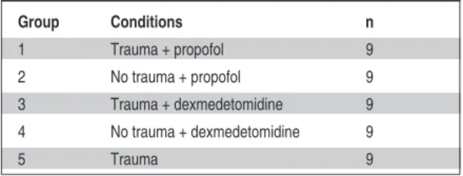
consti-Group Conditions n 1 Trauma + propofol 9 2 No trauma + propofol 9 3 Trauma + dexmedetomidine 9 4 No trauma + dexmedetomidine 9 5 Trauma 9
tuted.16The rats were sacrificed with cervical
dis-location. The rats in Groups 1, 3, 5 were sacrificed 2 hours after the mild cranial injury. The rats in Group 2 and 4 were sacrificed instantly 2 hours after anesthesia induction. After sacrification, a craniectomy was performed and then the brain was removed under the operation microscope. Brain tissue samples from three different regions (mid-sagital, parasagittal and hippocampal regions) were fixed in 10% formalin solution, and sent to pathol-ogy department for assessment of apoptotis. THE TERMINAL DEOXYNUCLEOTIDYL TRANSFERASE (dUTP) NICK-END LABELING (TUNEL) TECHNIQUE The terminal deoxynucleotidyl transferase (dUTP) nick-end labeling (TUNEL) technique was applied using “ApopTag Plus Peroxidase In Situ Apoptosis Detection Kit” (Millipore, Billerica, MA, USA), to determine the extent of neuronal cell death in tis-sue sections. The brain tistis-sue samples were put in 10% formaldehyde for fixation, and after routine tissue processing, embedded in paraffin, cut into 5 µm sections, and deparaffinized by 3 changes of xy-lene, each wash being 5 min. The sections were next dehydrated in a graded series of ethanol con-centrations (100%-70%), and washed in one change of phosphate buffered saline (PBS) for 5 min. For pretreatment, proteinase K (20 μg/mL) was applied at room temperature for 15 min, and washed in two changes of deionized water in a coplin jar, each wash being 2 min. The tissue was quenched in 3.0% hydrogen peroxide in PBS at room temperature for 5 min, and rinsed twice with PBS in a coplin jar, each wash being 5 min. After application of equilibration buffer at room temper-ature for at least 10 sec, the tissue was incubated with terminal deoxynucleotidyl transferase (TdT) enzyme in a humidified chamber at 37°C for 1 h. The sections were then placed in TdT stop buffer for 10 min, washed again with PBS, incubated at room temperature for 30 min in anti-digoxigenin conjugate solution, and washed with PBS. Peroxi-dase substrate was applied at room temperature for 5 min, monitoring the color development. After washing with deionized water, tissues were coun-terstained with methyl green. Slides were washed again with deionized water, and then 3 changes of
100% N-Butanol were done, each wash lasting ap-proximately 1 min. The sections were mounted on slides with mounting media, and coverslipped. ANALYSIS
Apoptotic cells were counted in 3 different loca-tions with the same methodology. The slides stained with TUNEL method were scanned under a light microscope, and at least 8 pictures were ob-tained at x400 high power field for saggital, parasaggital and hippocampal regions. Quantitation was achieved with image analysis using ImageJ ver-sion 1.38 program.17 It is a public domain Java
image processing program. The equipment used for morphometric analysis included a color video cam-era (Olympus DP71, Tokyo, Japan) attached to a light microscope (Olympus BX51, Tokyo, Japan), and an IBM compatible computer with Intel core2duo processor, 1 GB RAM and video monitor (BenQ G2400WA, Taipei, Taiwan). Briefly, the analysis of the procedure was as follows: initialize application, open acquired images, process binary, count, and record the measurement. After quanti-tation of apoptotic cells, a mean value was obtained for saggital, parasaggital and hippocampal regions of each case.
STATISTICAL ANALYSIS
All statistical analysis were performed with SPSS 15.0 statistical package. Data were expressed as mean±standard deviation or median [min-max] as appropriate. MAP, glucose level and rectal tem-peratures were compared within the groups using Friedman test. Since there were no within group differences according to MAP, glucose levels or temperature, no pairwise comparisons were per-formed. For glucose, MAP and temperature, we calculated the percentage changes according to the baseline values. These percentage changes were compared between the groups using Kruskal Wal-lis test. The quantity of apoptosis in groups with trauma (Groups 1,3, and 5) were compared using Kruskal Wallis test, followed by Mann Whitney test with Bonferronni adjustment. Groups without trauma (Groups 2, and 4) were compared with Mann Whitney test. P values less than 0.05 were considered as statistically significant. There are
three comparisons for one region, therefore we adapted the p value of 0.05 by Bonferroni adjust-ment, dividing 0.05 by 3 (=number of comparisons) which resulted in a p level of 0.017.
RESULTS
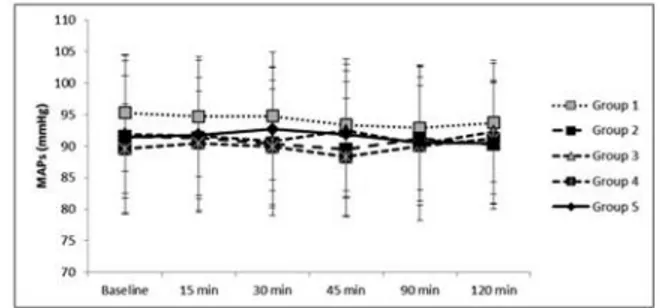
Forty five male Wistar albino rats were randomly assigned into 5 groups, with 9 rats in each. The me-dians of mean body weight of the rats in each group were 224.9, 228.9, 213.3, 216.4 and 210.6 g, re-spectively (p=0.513). At the time of anesthesia, the MAP (p>0.05) (Figure 1), and rectal temperatures (p>0.05) (Figure 2) were similar. The blood glucose levels were significantly higher in Group 5 com-pared to the other groups, just in the first 45 min-utes of the experiment (p<0.05); however there was no difference within the groups during 2-hour pe-riod (p>0.05) (Figure 3).
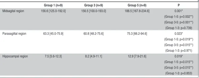
Apoptotic cells were counted in 3 different re-gions (midsagital, parasagittal, hippocampal) of rat brains. When trauma groups were compared, apop-totic cells were significantly higher in Group 5 compared to in Groups 1 and 3 (Table 2) (Figure 4). In midsagital region, Groups 1, 3 and 5 had 158.4±17.8 (median=156.6), 151.8±27.8 (median= 156.5) and 189.9±18.9 (median=188.5) apoptotic cells, respectively (p=0.001) (Figure 5). In parasagit-tal region, Group 1 had 61.7±11.5 (median=63.3), Group 3 had 61.8±11.2 (median=60.8), and Group 5 had 78.8±12.1 (median=75.3) apoptotic cells (p=0.023) (Figure 6). Additionally, in hippocampal region, Groups 1,3 and 5 had 8.0±2.2 (median=7.5), 8.1±2.1 (median=8.2) and 14.2±6.1 (median=12.9) apoptotic cells (p=0.019) (Figure 7).
In addition to the comparison of all trauma groups, the groups were also compared with each other one by one (Table 2) (Figure 4). There were statistically significant differences between Groups 1 and 5 in midsagital (p=0.002), parasagittal (p=0.019) and hippocampal regions (p=0.015). Moreover, the differences between Groups 3 and 5 in midsagital (p=0.001), parasagittal (p=0.015) and hippocampal regions (p=0.015) were also statisti-cally significant. However, the number of apoptotic cells were similar in Groups 1 and 3 (p>0.05).
The number of apoptotic cells of the groups without trauma (Groups 2 and 4) was very small, and there was no significant differences among the regions (p>0.05) (Table 3).
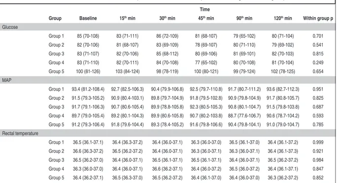
The variations of MAP, rectal temperature and glucose levels were not statistically significant in the groups (Table 4); however their calculated per-centage changes were all differed compared to baseline values in studied groups (Table 5). Ac-cordingly, the percentage changes were found to be comparable in all groups.
FIGURE 1: The mean arterial pressures (MAPs) during 2-hour experiment
pe-riod.
FIGURE 2: The rectal temperatures of the rats during 2-hour experiment
pe-riod.
FIGURE 3: The glucose levels of the rats during 2-hour experiment period. The
blood glucose levels were significantly higher in Group 5 in the first 45 minutes of the experiment (p<0.05); however there was no differences within the groups dur-ing 2-hour experiment period (p>0.05).
DISCUSSION
The outcome in patients surviving TBI is mainly de-termined by secondary events such as cerebral edema, ischemia or neurodegeneration.1Cerebral
ischemia and neurodegeneration are associated with
an increase in circulating and extracellular brain catecholamine concentrations and apoptosis.1,18-20
Interventions to reduce sympathetic tone and neu-roapoptotic effects may improve neurologic out-come, and therefore propofol and dexmedetomidine
FIGURE 4: The comparison of the quantity of the apoptotic cells in different
regions ıf the brain and the groups with trauma. Among the trauma groups (Groups 1, 3, and 5), the apoptotic cells were significantly higher in Group 5 in all regions (p<0.05).
Group 1 (n=9) Group 3 (n=9) Group 5 (n=9) P
Midsagital region 156.6 [125.0-192.0] 156.5 [100.0-193.0] 188.5 [167.8-234.6] 0.001* (Group 1-5: p=0.002**) (Group 3-5: p=0.001**) (Group 1-3: p=0.739) Parasagittal region 63.3 [45.0-75.9] 60.8 [48.2-75.6] 75.3 [66.2-94.6] 0.023* (Group 1-5: p=0.019**) (Group 3-5: p=0.015**) (Group 1-3: p=0.971) Hippocampal region 7.5 [5.6-12.3] 8.2 [4.9-11.1] 12.9 [7.9-21.6] 0.019* (Group 1-5: p=0.015**) (Group 3-5: p=0.015**) (Group 1-3: p=0.853)
TABLE 2: Comparison of the quantity of apoptotic cells in three regions of the brain in groups with trauma.
* These statistically significant values show the comparison of all groups (p<0.05).
** The groups were compared with each other one by one with Bonferronni adjustment (p<0.017).
FIGURE 5: Midsagittal region. 1A: Group 1, 1B: Group 2, 1C: Group 3, 1D:
Group 4, 1E: Group 5 (TUNEL histochemistry; x400).
(See color figure at http://www.turkiyeklinikleri.com/journal/tip-bilimleri-dergisi/1300-0292/)
FIGURE 6: Parasagittal region. 2A: Group 1, 2B: Group 2, 2C: Group 3, 2D:
Group 4, 2E: Group 5 (TUNEL histochemistry; x400).
(See color figure at http://www.turkiyeklinikleri.com/journal/tip-bilimleri-dergisi/1300-0292/)
FIGURE 7: Hippocampal region. 3A: Group 1, 3B: Group 2, 3C: Group 3, 3D:
Group 4, 3E: Group 5 (TUNEL histochemistry; x400).
(See color figure at http://www.turkiyeklinikleri.com/journal/tip-bilimleri-dergisi/1300-0292/)
Group 2 (n=9) Group 4 (n=9) p
Midsagital region 3.3 (1.1-6.1) 4.3 (1.3-6.2) 0.315 Parasagittal region 2.7 (1.2-4.6) 2.5 (1.6-4.1) 0.912 Hippocampal region 3.1 (2.3-8.1) 2.7 (1.6-5.9) 0.393
TABLE 3: Comparison of the quantity of apoptotic cells
in three regions of brain in groups without trauma.
a b c d e
a b c d e
may be used to improve survival. Accordingly, in the current study, we have two main results. First, both propofol and dexmedetomidine significantly decrease the number of apoptotic cells in different regions of the rat brains after mild TBI, and second,
these agents have no superiority to each other. Propofol (2,6-diisopropylphenol) is a short-acting, intravenous hypnotic agent that primarily acts by potentiating the function of the
gamma-TABLE 4: Glucose, mean arterial pressure and rectal temperature changes in each group. Time
Group Baseline 15thmin 30thmin 45thmin 90thmin 120thmin Within group p
Glucose Group 1 85 (70-108) 83 (71-111) 86 (72-109) 81 (68-107) 79 (65-102) 80 (71-104) 0.701 Group 2 82 (70-106) 81 (68-107) 83 (69-109) 78 (69-107) 80 (71-110) 79 (69-102) 0.541 Group 3 83 (71-107) 82 (70-106) 85 (68-112) 80 (69-106) 81 (69-101) 82 (70-103) 0.815 Group 4 83 (71-110) 82 (70-111) 84 (70-108) 77 (65-102) 80 (70-108) 81 (70-104) 0.249 Group 5 100 (81-126) 103 (84-124) 98 (78-119) 100 (80-121) 99 (79-124) 102 (78-125) 0.654 MAP Group 1 93.4 (81.2-108.4) 92.7 (82.5-106.3) 90.4 (79.9-106.8) 92.5 (79.7-110.8) 91.7 (80.7-111.2) 93.6 (82.7-112.3) 0.951 Group 2 91.5 (79.3-105.2) 90.9 (80.4-103.1) 89.8 (79.7-104.9) 91.8 (79.5-102.8) 90.9 (79.8-104.9) 91.7 (80.8-105.7) 0.825 Group 3 91.7 (79.1-106.3) 90.7 (80.6-105.4) 89.9 (78.8-105.8) 92.3 (80.5-105.3) 90.8 (80.1-104.7) 91.5 (79.8-103.8) 0.687 Group 4 89.7 (79.0-105.4) 89.2 (80.1-104.3) 89.9 (80.6-105.8) 90.7 (80.2-103.8) 88.7 (77.6-106.7) 90.6 (78.7-104.2) 0.593 Group 5 91.2 (79.3-106.4) 91.8 (79.6-104.4) 89.3 (78.4-105.2) 91.6 (79.8-106.6) 90.4 (79.8-104.1) 91.0 (79.0-104.7) 0.785 Rectal temperature Group 1 36.5 (36.1-37.1) 36.4 (36.3-37.2) 36.4 (36.0-37.1) 36.3 (36.0-37.0) 36.5 (36.1-37.0) 36.4 (36.1-37.2) 0.999 Group 2 36.6 (36.3-37.2) 36.5 (36.2-37.2) 36.4 (36.0-37.1) 36.3 (36.0-37.1) 36.3 (36.0-37.1) 36.4 (36.1-37.3) 0.921 Group 3 36.5 (36.2-37.0) 36.4 (36.0-37.1) 36.5 (36.1-37.1) 36.5 (36.1-37.1) 36.4 (36.0-37.1) 36.5 (36.2-37.2) 0.984 Group 4 36.3 (36.0-37.0) 36.4 (36.0-37.1) 36.6 (36.2-37.1) 36.4 (36.0-37.2) 36.5 (36.0-37.2) 36.4 (36.1-37.1) 0.847 Group 5 36.4 (36.2-37.1) 36.5 (36.3-37.0) 36.5 (36.2-37.2) 36.4 (36.1-37.0) 36.4 (36.0-37.0) 36.3 (36.2-37.2) 0.852
TABLE 5: Glucose, mean arterial pressure (MAP) and rectal temperature changes in percentages in each group.
Group 1 Group 2 Group 3 Group 4 Group 5 p
Glucose* Baseline-15thmin -0.02 (-0.05-0.08) -0.01 (-0.05-0.08) -0.01 (-0.04-0.09) -0.03 (-0.05-0.10) 0.03 (0.01-0.12) 0.297 Baseline-30thmin 0.02 (-0.01-0.09) 0.02 (0.00-0.08) 0.03 (-0.01-0.10) 0.02 (-0.02-0.12) 0.04 (0.01-0.11) 0.369 Baseline-45thmin 0.01 (-0.03-0.11) 0.00 (-0.03-0.11) 0.02 (-0.04-0.08) 0.04 (-0.02-0.10) 0.02 (-0.01-0.10) 0.854 Baseline-90thmin 0.01 (-0.01-0.08) 0.02 (-0.01-0.08) 0.03 (-0.02-0.11) 0.02 (-0.01-0.11) 0.02 (-0.01-0.13) 0.745 Baseline-120thmin -0.02 (-0.04-0.08) 0.01 (-0.03-0.09) -0.01 (-0.03-0.14) 0.02 (-0.05-0.11) 0.04 (0.00-0.14) 0.958 MAP* Baseline-15thmin 0.02 (-0.03-0.08) -0.01 (-0.04-0.10) -0.01 (0.00-0.09) -0.03 (-0.05-0.10) 0.04 (-0.01-0.12) 0.745 Baseline-30thmin 0.02 (-0.01-0.09) 0.03 (0.00-0.11) 0.03 (-0.01-0.10) 0.03 (-0.02-0.12) 0.03 (-0.02-0.11) 0.398 Baseline-45thmin 0.01 (-0.02-0.10) 0.00 (-0.03-0.11) 0.02 (-0.03-0.09) 0.04 (-0.03-0.10) 0.02 (-0.03-0.10) 0.785 Baseline-90thmin 0.01 (-0.03-0.12) 0.02 (-0.01-0.08) 0.03 (-0.01-0.11) 0.02 (-0.02-0.11) 0.01 (-0.01-0.11) 0.485 Baseline-120thmin -0.02 (-0.05-0.08) 0.01 (-0.03-0.09) -0.01 (-0.05-0.10) 0.02 (-0.04-0.10) 0.05 (0.01-0.16) 0.851 Rectal temperature* Baseline-15thmin 0.01 (-0.03-0.08) -0.01 (-0.04-0.10) 0.01 (-0.01-0.09) 0.02 (-0.05-0.08) 0.03 (-0.01-0.12) 0.658 Baseline-30thmin 0.02 (-0.01-0.09) 0.03 (0.00-0.11) 0.03 (-0.01-0.08) 0.02 (-0.01-0.10) 0.04 (-0.02-0.11) 0.962 Baseline-45thmin 0.01 (-0.02-0.10) 0.00 (-0.03-0.11) 0.02 (-0.03-0.10) 0.03 (-0.03-0.09) 0.05 (-0.03-0.15) 0.997 Baseline-90thmin 0.02 (-0.01-0.10) 0.02 (-0.01-0.08) 0.03 (-0.01-0.11) 0.00 (-0.01-0.10) 0.02 (-0.01-0.10) 0.896 Baseline-120thmin 0.00 (-0.05-0.08) 0.01 (-0.03-0.09) 0.01 (-0.05-0.07) 0.02 (-0.03-0.08) 0.03 (0.01-0.14) 0.924
*Glucose, MAP and rectal temperatures compared to baseline values of changes and percentage changes differed among the groups studied. These statistically significant values show the comparison of all groups (p<0.05).
aminobutyric acid (GABA)A and glycine-recep-tors.21-23Propofol’s effects on ischemia-reperfusion
injury, oxygen-glucose deprivation, apoptosis and neuroprotection have been investigated in both in vivo and in vitro models, and conflicting results were obtained.9,24-31Rossaint et al. found that
post-traumatic administration of propofol led to a dose-dependent decrease in total injury, and the combination of propofol and hypothermia were ad-ditive in regard to neuroprotection.24In another
study, propofol provided neuroprotection by re-versing the increase in glutamate extracellular con-centrations and decreasing glutamate uptake after ischemic injury.25 Adembri et al. reported that
propofol had a neuroprotective effect by demon-strating approximately 30% reduced infarct size when it was administered immediately after and up to 30 min after middle cerebral artery occlusion.9
On the other hand, dexmedetomidine is an α2-adrenoceptor agonist that acts by reducing no-radrenergic output from the locus coeruleus, de-creasing brain norepinephrine levels.32 Previous
studies have reported the ameliorative effect that α2-adrenoceptor agonists, especially dexmedeto-midine, exhibit in acute neuronal injury.18,33-37The
reason for the neuroprotective effect of dexmedeto-midine is thought to be due to its attenuating action for massive release of catecholamines occurring in cerebral hypoxic-ischemia in multiple parts of the brain.38,39The administration of dexmedetomidine
during the critical phase of synaptogenesis activates the endogenous postsynaptic norepinephrine-me-diated trophic system to produce its antiapoptotic effect.40-42Several mechanisms have been
investi-gated, however, just one study has mentioned the influence of dexmedetomidine on the number of apoptotic cells. The decreased number of apoptotic neurons with dexmedetomidine, and its neuropro-tective effect on hippocampus has been reported.43
We also obtained similar results suggesting that dexmedetomidine reduced apoptosis after mild cra-nial injury in midsagital, parasagittal and
hip-pocampal regions, and also compared its antineu-roapoptotic effect with propofol. In addition, our current study demonstrated that propofol also pre-vented apoptosis and tissue injury after mild TBI similar to dexmedetomidine.
In cell culture videomicroscopy studies, the dynamic morphologic changes at the light micro-scopic level always take place in less than 2 hours.44,45Different types of cell death share
com-mon mechanisms in the early phases, whereas ac-tivation of caspases determines the phenotype of cell death. Detection of apoptotic cells in tissue samples currently relies on the TUNEL assay.46
Therefore, we decided to investigate the tissue samples of three different regions by TUNEL method at the end of 2-hour period to understand the early apoptotic effects of TBI. Our study is the first one which compared the activities of propofol and dexmedetomidine on apoptosis. This study just intended to find the decreased quantity of apop-totic cells, and no mechanisms were taken into consideration.
Additionally, although our secondary hypoth-esis was having hemodynamically stable rats in propofol and dexmedetomidine groups in terms of MAP, rectal temperature, and blood glucose levels after mild TBI, there were no differences between the groups during the first 2 hours except for hav-ing statistically significant higher blood glucose levels in Group 5 within the first 45 minutes.
CONCLUSION
In conclusion, propofol and dexmedetomidine de-crease the formation of apoptotic cells in rats with TBI, and they are beneficial for neuroprotection. The protective effects of these two agents may be a promising for reducing damage in clinical situa-tions, however they have no superiority to each other. Although the results seem to be beneficial, the clinical significance of these results must be elucidated with further studies.
1. Plesnila N, von Baumgarten L, Retiounskaia M, Engel D, Ardeshiri A, Zimmermann R, et al. Delayed neuronal death after brain trauma involves p53-dependent inhibition of NF-kap-paB transcriptional activity. Cell Death Differ 2007;14(8):1529-41.
2. Veenith T, Goon SSh, Burnstein RM. Molecu-lar mechanisms of traumatic brain injury: the missing link in management. World J Emerg Surg 2009;4:7. doi: 10.1186/1749-7922-4-7. 3. Slemmer JE, Zhu C, Landshamer S, Trabold
R, Grohm J, Ardeshiri A, et al. Causal role of apoptosis-inducing factor for neuronal cell death following traumatic brain injury. Am J Pathol 2008;173(6):1795-805.
4. Raghupathi R. Cell death mechanisms follow-ing traumatic brain injury. Brain Pathol 2004;14(2):215-22.
5. Zweckberger K, Stoffel M, Baethmann A, Plesnila N. Effect of decompression cran-iotomy on increase of contusion volume and functional outcome after controlled cortical im-pact in mice. J Neurotrauma 2003;20(12): 1307-14.
6. Narayan RK, Michel ME, Ansell B, Baethmann A, Biegon A, Bracken MB, et al. Clinical trials in head injury. J Neurotrauma 2002;19(5):503-57.
7. Luo T, Wu J, Kabadi SV, Sabirzhanov B, Guanciale K, Hanscom M, et al. Propofol lim-its microglial activation after experimental brain trauma through inhibition of nicotinamide adenine dinucleotide phosphate oxidase. Anesthesiology 2013;119(6):1370-88. 8. Tanguy M, Seguin P, Laviolle B, Bleichner JP,
Morandi X, Malledant Y. Cerebral microdialy-sis effects of propofol versus midazolam in se-vere traumatic brain injury. J Neurotrauma 2012;29(6):1105-10.
9. Adembri C, Venturi L, Tani A, Chiarugi A, Gramigni E, Cozzi A, et al. Neuroprotective ef-fects of propofol in models of cerebral is-chemia: inhibition of mitochondrial swelling as a possible mechanism. Anesthesiology 2006;104(1):80-9.
10. Peters CE, Korcok J, Gelb AW, Wilson JX. Anesthetic concentrations of propofol protect against oxidative stress in primary astrocyte cultures: comparison with hypothermia. Anes-thesiology 2001;94(2):313-21.
11. Tawfeeq NA, Halawani MM, Al-Faridi K, Aal-Shaya WA, Taha WS. Traumatic brain injury: neuroprotective anaesthetic techniques, an update. Injury 2009;40 (Suppl 4):S75-81. 12. Sanders RD, Xu J, Shu Y, Januszewski A,
Halder S, Fidalgo A, et al. Dexmedetomidine attenuates isoflurane-induced neurocognitive impairment in neonatal rats. Anesthesiology 2009;110(5):1077-85.
13. Gertler R, Brown HC, Mitchell DH, Silvius EN. Dexmedetomidine: a novel sedative-analgesic agent. Proc (Bayl Univ Med Cent) 2001; 14(1):13-21.
14. Kose EA, Bakar B, Kasimcan O, Atilla P, Kil-inc K, Muftuoglu S, et al. Effects of intracister-nal and intravenous dexmedetomidine on ischemia-induced brain injury in rat: a com-parative study. Turk Neurosurg 2013;23(2): 208-17.
15. Schoeler M, Loetscher PD, Rossaint R, Fahlenkamp AV, Eberhardt G, Rex S, et al. Dexmedetomidine is neuroprotective in an in vitro model for traumatic brain injury. BMC Neurol 2012;12:20. doi: 10.1186/1471-2377-12-20.
16. Marmarou A, Foda MA, van den Brink W, Campbell J, Kita H, Demetriadou K. A new model of diffuse brain injury in rats. Part I: Pathophysiology and biomechanics. J Neuro-surg 1994;80(2):291-300.
17. Abramoff MD, Magelhaes PJ, Ram SJ. Image processing with image J. Biophotonics Inter-national 2004;11(7):36-42.
18. Hoffman WE, Kochs E, Werner C, Thomas C, Albrecht RF. Dexmedetomidine improves neu-rologic outcome from incomplete ischemia in the rat. Reversal by the alpha 2-adrenergic an-tagonist atipamezole. Anesthesiology 1991;75 (2):328-32.
19. Werner C, Hoffman WE, Thomas C, Miletich DJ, Albrecht RF. Ganglionic blockade im-proves neurologic outcome from incomplete ischemia in rats: partial reversal by exogenous catecholamines. Anesthesiology 1990;73(5): 923-9.
20. Unterberg AW, Stover J, Kress B, Kiening KL. Edema and brain trauma. Neuroscience 2004;129(4):1021-9.
21. Alkire MT, Haier RJ. Correlating in vivo anaes-thetic effects with ex vivo receptor density data supports a GABAergic mechanism of action for propofol, but not for isoflurane. Br J Anaesth 2001;86(5):618-26.
22. Trapani G, Altomare C, Liso G, Sanna E, Big-gio G. Propofol in anesthesia. Mechanism of action, structure-activity relationships, and drug delivery. Curr Med Chem 2000;7(2):249-71.
23. Franks NP. Molecular targets underlying gen-eral anaesthesia. Br J Pharmacol 2006;147 (Suppl 1):S72-81.
24. Rossaint J, Rossaint R, Weis J, Fries M, Rex S, Coburn M. Propofol: neuroprotection in an in vitro model of traumatic brain injury. Crit Care 2009;13(2):R61. doi: 10.1186/cc7795.
25. Velly LJ, Guillet BA, Masmejean FM, Nieoul-lon AL, Bruder NJ, Gouin FM, et al. Neuro-protective effects of propofol in a model of ischemic cortical cell cultures: role of gluta-mate and its transporters. Anesthesiology 2003;99 (2):368-75.
26. Yagmurdur H, Ayyildiz A, Karaguzel E, Akgul T, Ustun H, Germiyanoglu C. Propofol re-duces nitric oxide-induced apoptosis in testic-ular ischemia-reperfusion injury by downregu-lating the expression of inducible nitric oxide synthase. Acta Anaesthesiol Scand 2008; 52(3):350-7.
27. Jin YC, Kim W, Ha YM, Shin IW, Sohn JT, Kim HJ, et al. Propofol limits rat myocardial is-chemia and reperfusion injury with an associ-ated reduction in apoptotic cell death in vivo. Vascul Pharmacol 2009;50(1-2):71-7. 28. Chen L, Xue Z, Jiang H. Effect of propofol on
pathologic time-course and apoptosis after cerebral ischemia-reperfusion injury. Acta Anaesthesiol Scand 2008;52(3):413-9. 29. Zhu H, Cottrell JE, Kass IS. The effect of
thiopental and propofol on NMDA- and AMPA-mediated glutamate excitotoxicity. Anesthesi-ology 1997;87(4):944-51.
30. Feiner JR, Bickler PE, Estrada S, Donohoe PH, Fahlman CS, Schuyler JA. Mild hypother-mia, but not propofol, is neuroprotective in organotypic hippocampal cultures. Anesth Analg 2005;100(1):215-25.
31. Qi S, Zhan RZ, Wu C, Fujihara H, Taga K, Shi-moji K. The effects of thiopental and propofol on cell swelling induced by oxygen/glucose deprivation in the CA1 pyramidal cell layer of rat hippocampal slices. Anesth Analg 2002;94(3):655-60; table of contents. 32. Correa-Sales C, Rabin BC, Maze M. A
hyp-notic response to dexmedetomidine, an alpha 2 agonist, is mediated in the locus coeruleus in rats. Anesthesiology 1992;76(6):948-52. 33. Hoffman WE, Cheng MA, Thomas C,
Baugh-man VL, Albrecht RF. Clonidine decreases plasma catecholamines and improves out-come from incomplete ischemia in the rat. Anesth Analg 1991;73(4):460-4.
34. Maier C, Steinberg GK, Sun GH, Zhi GT, Maze M. Neuroprotection by the alpha 2-adrenoreceptor agonist dexmedetomidine in a focal model of cerebral ischemia. Anesthesi-ology 1993;79(2):306-12.
35. Halonen T, Kotti T, Tuunanen J, Toppinen A, Miettinen R, Riekkinen PJ. Alpha 2-adreno-ceptor agonist, dexmedetomidine, protects against kainic acid-induced convulsions and neuronal damage. Brain Res 1995;693(1-2):217-24.
36. Kuhmonen J, Pokorný J, Miettinen R, Haa-palinna A, Jolkkonen J, Riekkinen P Sr, et al. Neuroprotective effects of dexmedetomidine in the gerbil hippocampus after transient global ischemia. Anesthesiology 1997;87(2): 371-7.
37. Laudenbach V, Mantz J, Lagercrantz H, Desmonts JM, Evrard P, Gressens P. Effects of alpha(2)-adrenoceptor agonists on perina-tal excitotoxic brain injury: comparison of cloni-dine and dexmedetomicloni-dine. Anesthesiology 2002;96(1):134-41.
38. Globus MY, Busto R, Dietrich WD, Martinez E, Valdes I, Ginsberg MD. Intra-ischemic ex-tracellular release of dopamine and gluta-mate is associated with striatal vulnerability to ischemia. Neurosci Lett 1988;91(1):36-40.
39. Matsumoto M, Zornow MH, Rabin BC, Maze
M. The alpha 2 adrenergic agonist, dexmede-tomidine, selectively attenuates ischemia-in-duced increases in striatal norepinephrine concentrations. Brain Res 1993; 627(2):325-9.
40. Dahmani S, Paris A, Jannier V, Hein L, Rouelle D, Scholz J, et al. Dexmedetomidine increases hippocampal phosphorylated extra-cellular signal-regulated protein kinase 1 and 2 content by an alpha 2-adrenoceptor-independent mechanism: evidence for the in-volvement of imidazoline I1 receptors. Anesthesiology 2008;108(3):457-66. 41. Dahmani S, Rouelle D, Gressens P, Mantz
J. Effects of dexmedetomidine on hip-pocampal focal adhesion kinase tyrosine phosphorylation in physiologic and ischemic conditions. Anesthesiology 2005;103(5): 969-77.
42. Gähwiler BH. Organotypic monolayer cultures of nervous tissue. J Neurosci Methods 1981; 4(4):329-42.
43. Eser O, Fidan H, Sahin O, Cosar M, Yaman M, Mollaoglu H, et al. The influence of dexmedetomidine on ischemic rat hippocam-pus. Brain Res 2008;1218:250-6.
44. McCarthy NJ, Whyte MK, Gilbert CS, Evan GI. Inhibition of Ced-3/ICE-related proteases does not prevent cell death induced by oncogenes, DNA damage, or the Bcl-2 homologue Bak. J Cell Biol 1997;136(1):215-27.
45. Messam CA, Pittman RN. Asynchrony and commitment to die during apoptosis. Exp Cell Res 1998;238(2):389-98.
46. Saraste A, Pulkki K. Morphologic and bio-chemical hallmarks of apoptosis. Cardiovasc Res 2000;45(3):528-37.