Comparison of catheter‑directed
thrombolysis and anticoagulation
in intermediate‑risk pulmonary
embolism: A retrospective analysis
Derya Ozden Omaygenc, Mehmet Onur Omaygenc1ORCID:
Derya Ozden Omaygenç: 0000-0003-1037-8915 Mehmet Onur Omaygenç: 0000-0003-2995-8792
Abstract:
BACKGROUND AND OBJECTIVES: The selection of escalation of care strategies for the treatment of intermediate‑risk pulmonary embolism (PE) is a matter of debate. Here, we aimed to assess the features of our population treated either with anticoagulation (AC) alone or catheter‑directed thrombolysis (CDT). We also sought to identify a relationship between high residual systolic pulmonary artery pressure (sPAP) and demographic and clinical variables.
PATIENTS AND METHODS: The retrospective data of 30 intermediate‑high‑risk PE patients were analyzed. CDT was used in 14 (46.7%) cases. Enoxaparin (b. i. d) injections were administered in the AC group. In the CDT group, patients received 5 mg bolus dose of alteplase followed by 1 mg/h infusion for 24 h. Estimated sPAP at presentation and discharge was recorded. A value equal to or greater than 40 mmHg in the latter was accepted as a significant rise.
RESULTS: The patients in the CDT group had a lower HAS‑BLED score (2 [0–3] vs. 1 [0–3], P = 0.03). Although initial sPAP values were comparable among treatment arms, sPAP at discharge was significantly lower in the CDT group (mmHg, 42 ± 11.2 vs. 33.6 ± 9.7, P = 0.04). The reduction in sPAP at discharge was also significantly higher in this group. The degree of reduction in sPAP was considerably correlated with baseline sPAP (r: 63.2, P < 0.001). Finally, the baseline sPAP measurement and HAS‑BLED score of the patients with high residual sPAP were significantly higher (56.6 ± 13.1 vs. 67.3 ± 11.3, P = 0.02, and 1 [0–3] vs. 2 [0–3], P = 0.02, respectively). CONCLUSION: CDT was preferred over AC when lower bleeding risk was anticipated for intermediate‑high‑risk PE patients in our sample population. Eventually, CDT provided lower discharge sPAP levels and a greater reduction in sPAP. However, the factors associated with high sPAP at discharge were only high baseline sPAP measurement and HAS‑BLED score.
Keywords:
Anticoagulants, pulmonary embolism, pulmonary hypertension, thrombolytic therapy
Introduction
A
cute pulmonary embolism (PE) is one of the leading causes of inhospital mortality. It may also result in a reduction of functional capacity and quality oflife during long‑term follow‑up mainly but not invariably via development of chronic thromboembolic pulmonary hypertension (CTEPH).[1,2] Regarding the enhanced awareness of the disease, widespread access to imaging modalities in health facilities, and formation of local disease‑specific assemblies, namely
Address for correspondence:
Dr. Derya Ozden Omaygenc, Uğur Mumcu, Hastane Av. No: 1 A D: 1B, 34265 Sultangazi, Istanbul, Turkey. E-mail: drderyaozden@ yahoo.com Received: 07-06-2020 Revised:28-06-2020 Accepted: 02-02-2021 Published: 30-04-2021 Department of Anesthesiology and Critical Care, Haseki Sultangazi Education and Research Hospital, Turkey University of Health Sciences, 1Department of Cardiology, International Medical School, Istanbul Medipol University, Istanbul, Turkey
Access this article online
Quick Response Code:
Website:
www.eurasianjpulmonol.com
DOI:
10.4103/ejop.ejop_73_20
How to cite this article: Omaygenc DO,
Omaygenc MO. Comparison of catheter-directed thrombolysis and anticoagulation in intermediate-risk pulmonary embolism: A retrospective analysis. Eurasian J Pulmonol 2021;23:50-8.
This is an open access journal, and articles are distributed under the terms of the Creative Commons Attribution‑NonCommercial‑ShareAlike 4.0 License, which allows others to remix, tweak, and build upon the work non-commercially, as long as appropriate credit is given and the new creations are licensed under the identical terms.
response teams, PE has been increasingly diagnosed recently.[3‑5]
Risk modeling is the mainstay of management in PE by influencing not only the prognosis but also the level of care and selection of aggressive treatment modalities such as administration of thrombolytics, percutaneous techniques, and surgical embolectomy.[6,7] Relevant guidelines define intermediate‑risk PE as the presence of findings indicating increased right ventricular (RV) afterload in subjects without hypotension.[8,9] European guidelines further classify this group into two: intermediate‑low risk, designating the signs suggestive of RV dilation and/or dysfunction or increased biomarkers, and intermediate‑high risk, expressing the existence of both.[9] The intermediate‑risk group constitutes 20%–25% of all PE cases with a mortality rate ranging between 3% and 5%.[6,7,10,11]
CTEPH is the major long‑term complication of PE which had been reported with an estimated frequency varying between 0.5% and 4.6% in different registries related to the population selected for investigation.[7,12‑14] Currently, precise risk factors for the development of CTEPH have not been fully established.[14] However, even intermediate‑risk PE was documented to be associated with an increased incidence of CTEPH.[7,15]
In line with these data, escalation of care strategies – which was previously reserved for high‑risk population – has been more commonly used for intermediate‑risk patients in the last decade who may exhibit features of sudden decompensation.[3,4,6,16] Catheter‑directed thrombolysis (CDT) seemed a reasonable option at this setting based on the hypothesis of increased efficacy due to local administration of thrombolytics which might also be potentiated by adjunctive modalities such as suction or ultrasound‑assisted fragmentation. Moreover, a significant decline in bleeding events was expected by utilizing considerably reduced doses of these agents. In various publications, earlier improvement in RV function and less clinical deterioration were elicited by CDT use. However, a definite projection of this virtue to the clinical outcome was not observed.[2,3,6,7,17]
Here, we sought to distinguish our intermediate‑high‑risk PE patients treated either with anticoagulation (AC) alone or CDT added to AC by clinical and laboratory features. We also aimed to discriminate individuals who had higher systolic pulmonary artery pressure (sPAP) measurements at discharge from others who had relatively lower values (<40 mmHg). Estimated sPAP ≥40 mmHg at discharge was accepted as a cutoff
point which may represent a candidacy for progression to CTEPH.
Patients and Methods
Study qualification and patient selection
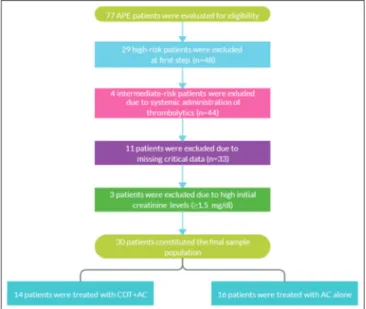
Retrospective data of 77 patients followed in the coronary care unit of a tertiary center between September 2015 and August 2019 were analyzed. After exclusion of 29 high‑risk patients treated either with systemic thrombolysis (ST) or CDT and 4 intermediate‑risk patients to whom ST was given, 44 patients were found to be eligible for inclusion. Among those, 11 cases could not be included because of lacking critical data. Finally, 3 patients whose initial creatinine levels were higher than 1.5 mg/dl were further excluded to eliminate the possibility of improper dose adjustment of anticoagulants. Eventually, 30 patients constituted the final sample population. Flow diagram is displayed in Figure 1.
All patients had a central occlusive thrombus accompanied by images compatible with increased RV afterload in CT angiography. Systolic blood pressure (SBP) was over 90 mmHg at the presentation in the entire population as required for assigning to the intermediate‑risk group. As suggested, a minimal initial troponin I level of 0.4 ng/ml was sought for confirmation of intermediate‑high‑risk status before the assignment.[8] This retrospective investigation was approved by the ethical committee of Istanbul Medipol University (Approval ID: 787, Date: 09.10.2019), hence met the global standards stated by the Declaration of Helsinki. Permission for using the data was granted by the same institution.
Figure 1: Flow diagram of the study. AC: Anticoagulation; APE: Acute pulmonary
Demographic features, diagnostic tests, treatment algorithms, and clinical endpoints
Once eligibility for inclusion was confirmed demographic features including hypertension, diabetes mellitus, obesity (body mass index [BMI] ≥30 kg/m2), and presence of malignancy were noted. In addition to underlying malignant disease, orthopedic or other major surgeries, oral contraceptive use, and immobilization were accepted as provoking factors for a PE episode and specified if existed. HAS‑BLED score was used for estimating bleeding risk during hospitalization.[18] Initial SBP, mean arterial pressure, heart rate, and oxygen saturation detected with pulse oximeter were recorded. D‑dimer, troponin I, and creatinine levels at presentation were noted. Chronic Kidney Disease Epidemiology Collaboration creatinine formula (which was automatically generated by the information management system using the equation: A × [Scr/B] C × 0.993age) was used to calculate estimated glomerular filtration rate.[19] Estimated sPAP values measured by echocardiographic examination at baseline and discharge were received from physician logs. A sPAP value equal to or higher than 40 mmHg was identified as a cutoff point in analogy to previous investigations.[20,21] Patients with and without residual high sPAP levels were also compared apart from the choice of treatment.
According to the institutional algorithms, patients treated with AC alone received subcutaneous low‑molecular‑weight heparin (100 U/kg enoxaparin, b. i. d) injections. If warfarin was chosen for long‑term AC, treatment had started the day before discharge. For non‑Vitamin K oral anticoagulating agents, treatment shift was performed on the day of discharge with the recommended dosing scheme. In the CDT group, conventional technique or ultrasound‑assisted thrombolysis (USAT) with EkoSonic Endovascular System (EKOS Corp.) was used. In conventional technique, alteplase was administered through a multi‑side hole 5F or 6F infusion catheter which had been placed in the trunk or one of the major branches (in case of unilateral dominance of central thrombus burden). After administration of 5 mg alteplase bolus at cath lab, 1 mg/h infusion was continued for 24 h. Meanwhile, a subtherapeutic dose of unfractionated heparin (400–600 U/kg) was given via intravenous route to provide an activated partial thromboplastin time prolongation between 40 and 50 s. AC was sustained by twice‑a‑day subcutaneous enoxaparin injections at the succeeding days. Oral anticoagulating agent shift was carried out as described above. If USAT was utilized, one or two dedicated catheters were advanced to right and/or left pulmonary arteries. 5 mg bolus dose of alteplase was administered at cath lab followed by 1 mg/h infusion through the catheters for 24 h. Infusion dose was
reduced to 0.5 mg/h per catheter if two catheters were used. The AC regimen was identical to that used in the conventional method.
Mortality, clinical deterioration, and major and nonmajor bleeding events according to ISTH definitions were designated as inhospital clinical endpoints.[22] The length of hospital stay was additionally noted.
Statistical analysis
The normality of continuous variables was tested with the Shapiro–Wilk test. Variables with and without normal distribution were expressed as mean ± standard deviation and median [range], respectively. Categorical variables were displayed as percentage (number of cases). Student’s t‑test was used to compare normally distributed continuous variables. Exact significance was considered in this test for distinguishing the groups, while Mann–Whitney U‑test was utilized to compare the ones which were not normally distributed. Frequencies of categorical variables among groups were distinguished via Chi‑square test. Pearson and Spearman tests were used to determine the correlation between continuous variables regarding the distribution pattern of relevant variables. A P value below 0.05 was deemed significant in all analyses. The Statistical Package for the Social Sciences (SPSS) (SPSS version 22.0, SPSS Inc., Chicago, IL, USA) was used for these assessments.
Results
Thirty patients (62.4 ± 16 years; 53.3% – female) were enrolled for the study. CDT was used in 14 (46.7%) cases. Four out of 14 cases were treated with USAT and two catheters were used in two patients. Patients in the CDT group tended to be relatively younger and had a higher BMI (P = 0.09 and P = 0.08, respectively). The patients in this group also had a lower bleeding tendency estimated by HAS‑BLED scoring (2 [0–3] vs. 1 [0–3], P = 0.03). In the entire population, three patients had malignant disease as an underlying cause and all of them were in the AC group. The remaining demographic features, baseline hemodynamic variables, and laboratory tests were comparable among groups. These data are displayed in Table 1.
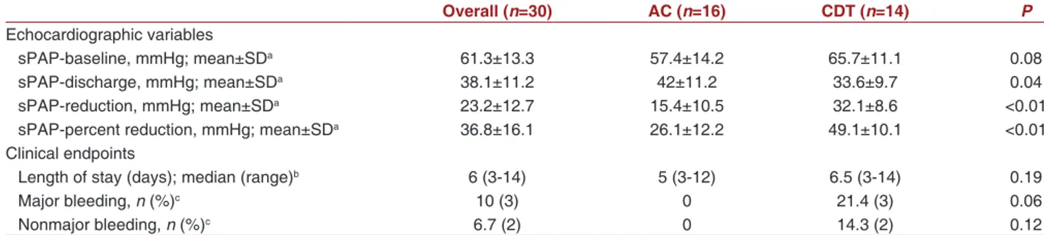
Although initial estimated sPAP values were comparable among treatment arms, sPAP at discharge was significantly lower in the CDT group. Besides, numerical and fractional reduction in sPAP at discharge was also significantly higher in this group [Table 2]. The comparison of sPAP at discharge and the degree of reduction were demonstrated with boxplots in Figure 2.
The difference in length of hospitalization was statistically indistinctive between the groups (AC: 5 [3–12] vs. CDT: 6.5 [3–14], P = 0.19). Death and clinical deterioration
were not observed in our sample population. Three patients (21.4%) had nonfatal major bleeding according to ISTH classification. One of these events was Table 2: Comparison of the echocardiographic systolic pulmonary artery pressure measurements and clinical endpoints of the study population
Overall (n=30) AC (n=16) CDT (n=14) P
Echocardiographic variables
sPAP‑baseline, mmHg; mean±SDa 61.3±13.3 57.4±14.2 65.7±11.1 0.08
sPAP‑discharge, mmHg; mean±SDa 38.1±11.2 42±11.2 33.6±9.7 0.04
sPAP‑reduction, mmHg; mean±SDa 23.2±12.7 15.4±10.5 32.1±8.6 <0.01
sPAP‑percent reduction, mmHg; mean±SDa 36.8±16.1 26.1±12.2 49.1±10.1 <0.01
Clinical endpoints
Length of stay (days); median (range)b 6 (3‑14) 5 (3‑12) 6.5 (3‑14) 0.19
Major bleeding, n (%)c 10 (3) 0 21.4 (3) 0.06
Nonmajor bleeding, n (%)c 6.7 (2) 0 14.3 (2) 0.12
aStudent’s t‑test was used, bMann‑Whitney U‑test was used, cChi‑square test was used. AC: Anticoagulation, CDT: Catheter‑directed thrombolysis, sPAP: Systolic
pulmonary artery pressure
Figure 2: Box plots displaying comparison of discharge measurements and reduction amount of systolic pulmonary artery pressures between treatment
arms. AC: Anticoagulation; CDT: Catheter-directed thrombolysis; sPAP: Systolic pulmonary artery pressure
Table 1: Demographic features, initial vital parameters, and laboratory test results of the treatment arms
Overall (n=30) AC (n=16) CDT (n=14) P
Age (years); mean±SDa 62.4±16 67.1±14.4 57.1±16.5 0.09
Gender, female, n (%)c 16 (53.3) 10 (62.5) 6 (42.9) 0.28 BMI (kg/m2); mean±SDa 26±3.6 24.9±3.8 27.2±3 0.08 Obesity, n (%)c 5 (16.7) 2 (12.5) 3 (21.4) 0.51 Hypertension, n (%)c 13 (43.3) 9 (56.3) 28.6 (4) 0.13 Diabetes mellitus, n (%)c 4 (13.3) 2 (12.5) 2 (14.3) 0.89 Malignancy, n (%)c 3 (10) 3 (18.8) 0 0.09 Provoked episode, n (%)c 10 (33.3) 5 (31.3) 5 (35.7) 0.80
HAS‑BLED score; median (range)b 1 (0‑3) 2 (0‑3) 1 (0‑3) 0,03
SBP, mmHg; mean±SDa 124.5±18.2 129.3±19 119.1±16.1 0.12
MBP, mmHg; mean±SDa 89.1±14 92±13.9 85.7±13.7 0.18
Heart rate, beats/min; mean±SDa 103.5±14.7 103.4±14.4 103.7±15.6 0.95
SaO2, %; mean±SDa 91.8±3.6 90.7±4 93±2.5 0.07
D‑Dimer, ng/ml; mean±SDa 5656.2±2987.7 5000.4±2933 6405±2975 0.20
Troponin I, ng/ml; median (range)b 0.4 (0.3‑3) 0.4 (0.3‑1.5) 0.4 (0.4‑3) 0.31
Creatinine, mg/dl; mean±SDa 1±0.22 0.99±0.24 1.03±0.20 0.62
eGFR, ml/min; mean±SDa 81.1±21.7 79.5±20.7 82.8±23.5 0.69
aStudent’s t‑test was used, bMann‑Whitney U‑test was used, cChi‑square test was used. AC: Anticoagulation, BMI: Body mass index, CDT: Catheter‑directed
retroperitoneal bleeding (not required intervention) and the others were bleeding which required transfusion with 2 units of red blood cells. There were also two nonmajor bleeding events (14.3%), a hematuria case and access‑site bleeding. All these events were observed in the CDT group [Table 2].
Cases were then regrouped according to the presence of an elevated sPAP (≥40 mmHg) at discharge. Of those treated with CDT, 5 patients (35.7%) had residually elevated sPAP at discharge while 8 patients (50%) in the AC group did so (P = 0.43). The mean HAS‑BLED score was higher in the group with high sPAP (1 [0–3] vs. 2 [0–3], P = 0.02). The baseline sPAP level was also significantly higher in this group (56.6 ± 13.1 vs.
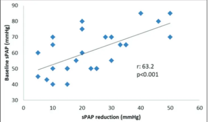
67.3 ± 11.3, P = 0.02). The remaining parameters including the use of CDT were comparable among groups [Table 3]. The degree of reduction in sPAP at discharge was considerably correlated with baseline sPAP (r: 63.2, P < 0.001). This relationship is illustrated in Figure 3. There was also a reasonable negative correlation between sPAP reduction and age (r: −42.7, P = 0.02) and sPAP reduction and HAS‑BLED score (r: −45.4, P = 0.01). Nevertheless, these correlations were presumably related to less frequent selection of CDT in older patients with an increased bleeding tendency. It should also be denoted that the correlation coefficients might not precisely interpret the relationship of these variables considering the small sample size.
Discussion
Despite having a lower mortality rate as compared to massive PE, submassive, or its correspondent intermediate risk, PE still poses a remarkable clinical significance through the likelihood of progression to shock in the acute phase and CTEPH at long term.[6,7] Recognition of RV strain findings at ECG and imaging modalities in addition to centrally located thrombus was determined as the indicator of possible deterioration.[4,23] However, only increased pulmonary artery systolic pressure was constantly found to be in a close relationship with the development of CTEPH.[7,14,15,24] In this context, low‑dose ST and catheter‑directed techniques offer prevention of clinical worsening while providing
Figure 3: Scatter plot demonstrating the correlation between baseline values and
reduction amount of systolic pulmonary artery pressure. sPAP: Systolic pulmonary artery pressure
Table 3: Comparison of the demographic and clinical features of the patients with and without high systolic pulmonary artery pressure (≥40 mmHg) at discharge
Low sPAP-d (n=17) High sPAP-d (n=13) P
Age, years; mean±SDa 58.7±16.5 67.3±14.4 0.14
Gender, female; n (%)c 10 (58.8) 6 (46.2) 0.49 BMI, kg/m2; mean±SDa 26±4 26±3.2 0.98 Obesity; n (%)c 4 (23.5) 1 (7.7) 0.25 Hypertension; n (%)c 8 (47.1) 5 (38.5) 0.64 Diabetes mellitus, n (%)c 3 (17.6) 1 (7.7) 0.43 Malignancy; n (%)c 2 (11.8) 1 (7.7) 0.71 Provoked episode; n (%)c 5 (29.4) 5 (38.5) 0.60
HAS‑BLED score; median (range)b 1 (0‑3) 2 (0‑3) 0.02
SBP, mmHg; mean±SDa 123.2±17.8 126.2±19.3 0.68
MBP, mmHg; mean±SDa 87.4±12.2 91.2±16.3 0.49
Heart rate, beats/min; mean±SDa 103.4±15.7 103.8±13.8 0,94
SaO2, %; mean±SDa 92.4±3.6 90.9±3.5 0.26
D‑Dimer, ng/ml; mean±SDa 5725.7±2718.2 5565.4±3421.2 0.89
Troponin I, ng/ml; median (range)b 0.4 (0.3‑3.0) 0.4 (0.4‑3.0) 0.81
Creatinine, mg/dl; mean±SDa 1.03±0.23 0.98±0.21 0.52
eGFR, ml/min; mean±SDa 81.9±22.1 80±22 0.81
sPAP‑Baseline, mmHg; mean±SDa 56.6±13.1 67.3±11.3 0.02
Length of stay, days; median (range)b 6 (3‑10) 6 (3‑14) 0.41
Treatment strategy, CDT; n (%)c 9 (52.9) 5 (38.5) 0.43
aStudent’s t‑test was used, bMann‑Whitney U‑test was used, cChi‑square test was used. BMI: Body mass index, CDT: Catheter‑directed thrombolysis, eGFR:
rapid improvement at exaggerated RV afterload and possibly leading to a more favorable clinical outcome at the expense of increased bleeding rates.[1,6,7,16,25,26] However, the relatively low rate of adverse events leaves the advanced treatment modalities a little room for exhibiting these advantages.[6,7,10] Hence, conflicting reports about outcome exist in the literature partly due to the high variability of study designs and the identification of endpoints. In our investigation, mortality was not observed and all bleeding events occurred in patients who were treated with CDT. This finding poses an additional significance considering the fact that common risk factors for bleeding events such as younger age, higher BMI, low HAS‑BLED score, and absence of malignant disease were in favor of this group. In a recent publication, advanced therapies (all treatment options apart from routine AC) were shown to decrease 30‑day and 90‑day mortality rates both in the massive and submassive groups. For submassive PE, advanced therapies were associated with a trend toward increased major bleeding events at 30 days, whereas this relationship lost significance at 90 days.[27] Chen et al. denoted in their meta‑analysis including 15 studies focused on moderate PE that thrombolytic treatment yielded a decline in rates of recurrent PE or death as against AC. They also reported a significant increase in nonmajor and a nonsignificant increase in major bleeding with thrombolytic therapy.[11]Another meta‑analysis performed by Chatterjee et al. also presented a similar output.[28] On the other hand, two large trials comparing ST with AC failed to confirm the mortality benefit.[26,29] In TOPCOAT, a reduction solely in mortality was not stated, but a reduction in the composite endpoint including mortality was observed.[29] In PEITHO, ST only decreased the probability of clinical worsening.[26] Bleeding rates were remarkably high in both trials. Nakamura et al. verified these data in their meta‑analysis.[30] In line with these findings, reduced dose ST and CDT garnered interest as a first‑line treatment option in intermediate‑risk PE to avoid major bleeding.[1,6]
In a retrospective registry, patients treated with low‑dose ST had 2.2% inhospital mortality and 4.4% 30‑day all‑cause mortality. Although the mean HAS‑BLED score of the patients was 0.8 in this investigation, ISTH major bleeding was observed in 11% of the cases.[31] MOPETT trial which actually focused on pulmonary artery pressure changes also compared the efficacy of ST and AC for the treatment of intermediate‑risk PE. Investigators identified acute bleeding, length of hospitalization, recurrent PE, death, and the combination of the last two as secondary endpoints. The comparison of combined secondary endpoint favored ST while no bleeding events were reported.[20] Although a low dose of
alteplase was used in a group of patients with a median HAS‑BLED score of 1, CDT was not a safe harbor for eliminating the risk of bleeding in our study. This finding was corroborating the former investigation.
Catheter‑directed techniques were tested for replacing the conventional ST in patients without hemodynamic collapse.[1,2,7] Simply inserting a multi‑side hole catheter in the pulmonary artery to provide continuous infusion of the thrombolytic agent is the most commonly used method.[5] Adjunctive modalities such as the ultrasound‑assisted disruption of the thrombus and suction thrombectomy were also used to reduce the thrombolytic dose and enhance the efficacy.[4,16,32‑34] Suction thrombectomy was positioned as a bail‑out treatment in the literature for patients at shock who had a contraindication for administration of thrombolytics.[4] USAT seemed more appropriate for the treatment of intermediate‑risk patients, particularly after the publication of first reports expressing high clinical success rates and lower major bleeding events.[33,34] Thereafter, SEATLE II raised again the questions about safety issues while revealing a major bleeding rate of 11%. It should be denoted that SEATLE II used the most stringent criteria for the definition of major bleeding among its counterparts.[25] On the other hand, investigators of these studies agreed on the rapid reversal of increased RV systolic pressure and strain pattern by utilization of CDT as compared to AC without an evident projection to clinical hard endpoints. Besides, none of these studies was powered sufficiently to establish the safety of the method.[25,33‑35] Graif et al. compared thrombolytic infusion through a simple pigtail catheter and a USAT system. In this retrospective analysis, mortality and complication rates were comparable between treatment arms with higher procedure and fluoroscopy times in the USAT group.[36] Apart from these, Avgerinos and Chaer denoted that the length of hospitalization was lower in the AC group as compared to patients treated with CDT techniques in their sample population.[35] The length of hospitalization was comparable among the AC and CDT groups in our population while the median day for stay was numerically higher in the latter (5 vs. 6.5). Patients in the CDT group tended to have higher baseline sPAP measurements. On the other hand, sPAP at discharge was significantly lower and the degree of reduction was more pronounced in this group (15.4 mmHg in AC vs. 32.1 mmHg in CDT).
Although precise evidence derived from head‑to‑head comparison of ST and CDT has been lacking in the literature, some registries and meta‑analyses were published to fill the gap about this subject. Kaymaz et al. stated that when compared to randomized ST trials, CDT showed similar mortality but reduced major bleeding rates.[37] Arora et al. compared 3107 patients treated
with ST and 1319 with CDT and reported that mortality, mortality combined with bleeding, and readmission rates were higher in the ST group.[38] Similarly, Patel et al. found that CDT was associated with lower combined inhospital mortality and intracranial bleeding.[39] Nevertheless, the current data remain inconclusive to herald the use of CDT or ST as first‑line therapy for intermediate‑risk PE. The efficacy and safety of these modalities are yet to be supported with larger evidence. Particularly, qualifying the features of catheter‑directed methods is more troublesome due to high diversities in certain issues such as the catheter selection, duration of administration, and type and dosing scheme of the thrombolytic agent.[6] Thereby, recent European Society of Cardiology guidelines recommended CDT and ST (preferably low dose) for treatment of intermediate‑risk PE only in case of rapid clinical deterioration with signs of persistent hypoxia and diminished cardiac output.[9]
In our retrospective analysis, clinicians tended to reserve CDT for younger patients with higher body mass indices ad lower HAS‑BLED scores. Although statistical significance was not observed, bleeding events identified by strict ISTH criteria and length of hospital stay were relatively higher in the CDT group. However, we need hardly mention that our study was either focused on or empowered enough to interpret the clinical outcome. It should also be denoted that the data presented here refer to a period before publication of the most recent guidelines. Vasoconstriction and acute inflammation overlapping the physical obstruction are the hallmarks of increased RV afterload in PE. In a certain fraction of these patients, pressure overload and vascular resistance augmentation persist which may contribute to the development of CTEPH.[1] Aside from CTEPH, diminished functional capacity decreased quality of life, and recurrent PE might be observed as long‑term complications of PE.[1,7,11,12] CTEPH has an estimated incidence of 3.2% in the 3rd year in intermediate‑risk PE patients treated only with AC.[12] Pengo et al. declared the 2‑year frequency of CTEPH as 3.8% in a more heterogeneous PE population.[13] Depending on the sample population, type of investigation, and definition of CTEPH, higher event rate up to 12.4% was reported.[21] MOPETT trial identified pulmonary hypertension as the detection of a pulmonary artery systolic pressure over 40 mmHg at the echocardiographic examination. In this regard, pulmonary hypertension was observed at 16% and 57% of patients treated with ST or AC, respectively.[20] Correlatively, Korkmaz et al. denoted that persistently elevated RV systolic pressure (>35 mmHg at echocardiography) was detected in 57% of their sample population. The incidence of symptomatic CTEPH was 4.6% after an episode of PE in this study which was diagnosed on an average of 9.4 months.[14] Persistently
elevated sPAP at discharge was observed in 43.3% of our sample population with a relatively lower (statistically insignificant) frequency in the CDT group (35.7%). Several factors were established as predictors of CTEPH development. High baseline sPAP values, age, and presence of intermediate‑risk features were accused in this context.[14,15,24,40] Klok et al. also suggested a prediction score that combined demographic features, CT findings, and treatment choices.[40] Although the association of CTEPH incidence and high sPAP values at presentation had been iteratively demonstrated, rapid reduction of pulmonary arterial pressure via utilization of advanced treatments (including CDT) did not transform into a clinical achievement reflected by reduced event rates.[7] In our population, HAS‑BLED score and baseline sPAP measurements were higher in patients with elevated sPAP at discharge whereas the utilization of CDT was comparable among groups. In addition, the degree of reduction in sPAP was correlated with baseline measurements.
Regarding the retrospective nature of the study, estimated sPAP values at echocardiographic examination were used instead of invasive measurements. A value ≥40 mmHg at discharge was assumed to reflect a residually high sPAP level in our study. However, the clinical consequences of this incidence by means of CTEPH occurrence and long‑term morbidity and mortality could not be specified due to lack of follow‑up data. Another limitation of our analysis was utilization of both USAT and the conventional method in the CDT group.
Conclusion
In line with previous data, CDT was preferred as principal treatment when lower bleeding risk was anticipated for intermediate‑high‑risk PE patients in our sample population. Eventually, CDT provided lower discharge sPAP levels and a greater reduction in sPAP at the expense of more bleeding events. However, the factors associated with high sPAP at discharge were only high baseline sPAP measurement and HAS‑BLED score. Ultimately, retrospective design, small sample size, absence of hard endpoints such as mortality and clinical deterioration, and utilization of two different catheters for CDT should be designated as major limitations of our study. Another consequence of the retrospective design is the heterogeneity of demographic features which may also confound the results about clinical event occurrence. Financial support and sponsorship
Nil.
Conflicts of interest
References
1. Javed QA, Sista AK. Endovascular therapy for acute severe pulmonary embolism. Int J Cardiovasc Imaging 2019;35:1443‑52. 2. Pollak JS. Catheter‑based therapies for pulmonary emboli. Clin
Chest Med 2018;39:651‑8.
3. Rothschild DP, Goldstein JA, Ciacci J, Bowers TR. Ultrasound‑accelerated thrombolysis (USAT) versus standard catheter‑directed thrombolysis (CDT) for treatment of pulmonary embolism: A retrospective analysis. Vasc Med 2019;24:234‑40. 4. Avgerinos ED, Abou Ali A, Toma C, Wu B, Saadeddin Z,
McDaniel B, et al. Catheter‑directed thrombolysis versus suction thrombectomy in the management of acute pulmonary embolism. J Vasc Surg Venous Lymphat Disord 2019;7:623‑8.
5. Xenos ES, Davis GA, He Q, Green A, Smyth SS. The implementation of a pulmonary embolism response team in the management of intermediate‑ or high‑risk pulmonary embolism. J Vasc Surg Venous Lymphat Disord 2019;7:493‑500.
6. Chodakowski JD, Courtney DM. Pulmonary embolism critical care update: Prognosis, treatment, and research gaps. Curr Opin Crit Care 2018;24:540‑6.
7. Furfaro D, Stephens RS, Streiff MB, Brower R. Catheter‑directed thrombolysis for ıntermediate‑risk pulmonary embolism. Ann Am Thorac Soc 2018;15:134‑44.
8. Jaff MR, McMurtry MS, Archer SL, Cushman M, Goldenberg N, Goldhaber SZ, et al. Management of massive and submassive pulmonary embolism, iliofemoral deep vein thrombosis, and chronic thromboembolic pulmonary hypertension: A scientific statement from the American Heart Association. Circulation 2011;123:1788‑830.
9. Konstantinides SV, Meyer G, Becattini C, Bueno H, Geersing GJ, Harjola VP, et al. 2019 ESC Guidelines for the diagnosis and management of acute pulmonary embolism developed in collaboration with the European Respiratory Society (ERS): The Task Force for the diagnosis and management of acute pulmonary embolism of the European Society of Cardiology (ESC). Eur Respir J 2019;54:1901647.
10. Dalen JE. Advanced therapies for massive pulmonary embolism. Am J Med 2018;131:1401.
11. Chen H, Ren C, Chen H. Thrombolysis versus anticoagulation for the initial treatment of moderate pulmonary embolism: A meta‑analysis of randomized controlled trials. Respir Care 2014;59:1880‑7.
12. Konstantinides SV, Vicaut E, Danays T, Becattini C, Bertoletti L, Beyer‑Westendorf J, et al. Impact of thrombolytic therapy on thelong‑term outcome of ıntermediate‑risk pulmonary embolism. J Am Coll Cardiol 2017;69:1536‑44.
13. Pengo V, Lensing AW, Prins MH, Marchiori A, Davidson BL, Tiozzo F, et al. Incidence of chronic thromboembolic pulmonary hypertension after pulmonary embolism. N Engl J Med 2004;350:2257‑64.
14. Korkmaz A, Ozlu T, Ozsu S, Kazaz Z, Bulbul Y. Long‑term outcomes in acute pulmonary thromboembolism: The incidence of chronic thromboembolic pulmonary hypertension and associated risk factors. Clin Appl Thromb Hemost 2012;18:281‑8.
15. Yang S, Yang Y, Zhai Z, Kuang T, Gong J, Zhang S, et al. Incidence and risk factors of chronic thromboembolic pulmonary hypertension in patients after acute pulmonary embolism. J Thorac Dis 2015;7:1927‑38.
16. Rao G, Xu H, Wang JJ, Galmer A, Giri J, Jaff MR, et al. Ultrasound‑assisted versus conventional catheter‑directed thrombolysis for acute pulmonary embolism: A multicenter comparison of patient‑centered outcomes. Vasc Med 2019;24:241‑7. 17. Zuin M, Rigatelli G, Roncon L. Bedside local thrombolysis in
patients with massive pulmonary embolism and no access to fluoroscopy: A new tool in the intensivist’s arsenal? Br J Anaesth 2019;122:e17‑9.
18. Pisters R, Lane DA, Nieuwlaat R, de Vos CB, Crijns HJ, Lip GY. A novel user‑friendly score (HAS‑BLED) to assess 1‑year risk of major bleeding in patients with atrial fibrillation: The Euro Heart Survey. Chest 2010;138:1093‑100.
19. Levey AS, Stevens LA, Schmid CH, Zhang YL, Castro AF 3rd,
Feldman HI, et al. A new equation to estimate glomerular filtration rate. Ann Intern Med 2009;150:604‑12.
20. Sharifi M, Bay C, Skrocki L, Rahimi F, Mehdipour M; “MOPETT” Investigators. Moderate pulmonary embolism treated with thrombolysis (from the “MOPETT” Trial). Am J Cardiol 2013;111:273‑7.
21. Barros A, Baptista R, Nogueira A, Jorge E, Teixeira R, Castro G, et al. Predictors of pulmonary hypertension after intermediate‑to‑high risk pulmonary embolism. Rev Port Cardiol 2013;32:857‑64. 22. Schulman S, Kearon C; Subcommittee on Control of
Anticoagulation of the Scientific and Standardization Committee of the International Society on Thrombosis and Haemostasis. Definition of major bleeding in clinical investigations of antihemostatic medicinal products in non‑surgical patients. J Thromb Haemost 2005;3:692‑4.
23. Mohan B, Tandon R, Bansal R, Singh M, Singh B, Goyal A,
et al. Determinants of in‑hospital clinical outcome in patients
with sub‑massive pulmonary embolism. Indian Heart J 2018;70 Suppl 3:90‑5.
24. Park JS, Ahn J, Choi JH, Lee HW, Oh JH, Lee HC, et al. The predictive value of echocardiography for chronic thromboembolic pulmonary hypertension after acute pulmonary embolism in Korea. Korean J Intern Med 2017;32:85‑94.
25. Piazza G, Hohlfelder B, Jaff MR, Ouriel K, Engelhardt TC, Sterling KM, et al. A Prospective, single‑arm, multicenter trial of ultrasound‑facilitated, catheter‑directed, low‑dose fibrinolysis for acute massive and submassive pulmonary embolism: The SEATTLE II study. JACC Cardiovasc Interv 2015;8:1382‑92. 26. Meyer G, Vicaut E, Danays T, Agnelli G, Becattini C,
Beyer‑Westendorf J, et al. Fibrinolysis for patients with intermediate‑risk pulmonary embolism. N Engl J Med 2014;370:1402‑11.
27. Secemsky E, Chang Y, Jain CC, Beckman JA, Giri J, Jaff MR,
et al. Contemporary management and outcomes of patients
with massive and submassive pulmonary embolism. Am J Med 2018;131:1506‑14.
28. Chatterjee S, Chakraborty A, Weinberg I, Kadakia M, Wilensky RL, Sardar P, et al. Thrombolysis for pulmonary embolism and risk of all‑cause mortality, major bleeding, and intracranial hemorrhage: A meta‑analysis. JAMA 2014;311:2414‑21.
29. Kline JA, Nordenholz KE, Courtney DM, Kabrhel C, Jones AE, Rondina MT, et al. Treatment of submassive pulmonary embolism with tenecteplase or placebo: Cardiopulmonary outcomes at 3 months: Multicenter double‑blind, placebo‑controlled randomized trial. J Thromb Haemost 2014;12:459‑68.
30. Nakamura S, Takano H, Kubota Y, Asai K, Shimizu W. Impact of the efficacy of thrombolytic therapy on the mortality of patients with acute submassive pulmonary embolism: A meta‑analysis. J Thromb Haemost 2014;12:1086‑95.
31. Rothschild DP, Goldstein JA, Bowers TR. Low‑dose systemic thrombolytic therapy for treatment of submassive pulmonary embolism: Clinical efficacy but attendant hemorrhagic risks. Catheter Cardiovasc Interv 2019;93:506‑10.
32. Liang NL, Avgerinos ED, Marone LK, Singh MJ, Makaroun MS, Chaer RA. Comparative outcomes of ultrasound‑assisted thrombolysis and standard catheter‑directed thrombolysis in the treatment of acute pulmonary embolism. Vasc Endovascular Surg 2016;50:405‑10.
33. Kucher N, Boekstegers P, Müller OJ, Kupatt C, Beyer‑Westendorf J, Heitzer T, et al. Randomized, controlled trial of ultrasound‑assisted catheter‑directed thrombolysis for acute intermediate‑risk pulmonary embolism. Circulation 2014;129:479‑86.
34. Kuo WT, Banerjee A, Kim PS, DeMarco FJ Jr., Levy JR, Facchini FR,
et al. Pulmonary Embolism Response to Fragmentation,
Embolectomy, and Catheter Thrombolysis (PERFECT): Initial results from a prospective multicenter registry. Chest 2015;148:667‑73.
35. Avgerinos ED, Chaer RA. Catheter‑directed interventions for acute pulmonary embolism. J Vasc Surg 2015;61:559‑65. 36. Graif A, Grilli CJ, Kimbiris G, Agriantonis DJ, Chohan OZ,
Fedele CR, et al. Comparison of ultrasound‑accelerated versus pigtail catheter‑directed thrombolysis for the treatment of acute massive and submassive pulmonary embolism. J Vasc Interv Radiol 2017;28:1339‑47.
37. Kaymaz C, Akbal OY, Tanboga IH, Hakgor A, Yilmaz F, Ozturk S,
et al. Ultrasound‑assisted catheter‑directed thrombolysis in
high‑risk and ıntermediate‑high‑risk pulmonary embolism:
A meta‑analysis. Curr Vasc Pharmacol 2018;16:179‑89.
38. Arora S, Panaich SS, Ainani N, Kumar V, Patel NJ, Tripathi B,
et al. Comparison of ın‑hospital outcomes and readmission
rates in acute pulmonary embolism between systemic and catheter‑directed thrombolysis (from the National Readmission Database). Am J Cardiol 2017;120:1653‑61.
39. Patel N, Patel NJ, Agnihotri K, Panaich SS, Thakkar B, Patel A,
et al. Utilization of catheter‑directed thrombolysis in pulmonary
embolism and outcome difference between systemic thrombolysis and catheter‑directed thrombolysis. Catheter Cardiovasc Interv 2015;86:1219‑27.
40. Klok FA, Dzikowska‑Diduch O, Kostrubiec M, Vliegen HW, Pruszczyk P, Hasenfuß G, et al. Derivation of a clinical prediction score for chronic thromboembolic pulmonary hypertension after acute pulmonary embolism. J Thromb Haemost 2016;14:121‑8.