Email: [email protected]
ORIGINAL ARTICLE
Corresponding author: Fatih Karatas, MD. Department of Medical Oncology, Karabuk University Faculty of Medicine Hospital, Karabuk, Turkey.
Tel: + 90 5057505270, Email: [email protected] Received: 14/07/2019; Accepted: 05/08/2019
Comparison of skeletal muscle mass loss in patients with
metastatic colorectal cancer treated with regorafenib or
TAS-102
Muhammet Bekir Hacioglu
1, Osman Kostek
1, Nazmi Kurt
2, Ahmet Kucukarda
1, Ali Gokyer
1,
Fethi Emre Ustabasioglu
2, Fatih Karatas
3, Nermin Tuncbilek
2, Sernaz Uzunoglu
1, Ahmet
Bilici
4, Irfan Cicin
1, Bulent Erdogan
11Department of Medical Oncology, Trakya University Faculty of Medicine, Edirne, Turkey; 2Department of Clinical Radiology, Trakya University Faculty of Medicine, Edirne, Turkey; 3Department of Medical Oncology, Karabuk University Faculty of Medicine Hospital, Karabuk, Turkey; 4Department of Medical Oncology, Medipol University Faculty of Medicine, Istanbul, Turkey.
Summary
Purpose: To assess whether regorafenib and TAS-102
treat-ments are associated with a change in Skeletal Muscle Area (SMA) as well as to compare Skeletal Muscle Mass (SMM) loss levels between regorafenib and TAS-102 treatments and prognostic significance in the patients with metastatic colo-rectal cancer (mCRC).
Methods: A total of 36 mCRC patients, who received
re-gorafenib or TAS-102 in the third-line and subsequent set-tings were assessed in the analysis. SMM changes were as-sessed with CT scans findings, and they were categorized into two groups as loss (SMM decrease ≥2%) and SMM-stable (SMM change <2%).
Results: The SMM change after regorafenib therapy was
sig-nificantly worse compared with TAS-102 therapy (p=0.001). The median overall survival (OS) was longer in SMM-stable group than in SMM-loss group (12.8 months; 95%CI:9.8-15.7) vs. 6.4 months; 95%CI:5.2-7.7, respectively;p=0.04). Cox regres-sion analysis showed that SMM loss was independent prog-nostic indicator for OS (HR, 2.87; 95%CI: 1.07-7.42, p=0.03).
Conclusion: Although patients who received regorafenib
had more SMM loss than those who received TAS-102, there was no difference in OS between drugs.
Key words: metastatic colorectal cancer, regorafenib,
TAS-102, sarcopenia, skeletal muscle mass
Introduction
Colorectal cancer (CRC) is the third most com-mon cancer worldwide and one of the leading cause of cancer-related deaths in Europe and the United States [1]. Approximately 25% of newly diagnosed CRC patients are de novo metastatic. Approximately 25-30% of patients with stages 2 and 3 disease be-come metastatic within 5 years [2,3].
Regorafenib, a new oral multi-kinase inhibitor, has shown antitumor activity in previously treated metastatic colorectal cancer (mCRC) patients
pre-viously treated with fluoropyrimidine plus oxali-platin or fluoropyrimidine and irinotecan-based chemotherapy±anti-vascular endothelial growth factor (anti-VEGF) treatment or anti-epidermal growth factor receptor (anti-EGFR) treatment [4]. The efficacy of regorafenib has been shown in two randomized placebo-controlled phase III trials, CORRECT and CONCUR, that were conducted in patients with mCRC progressing on standard treat-ments [5-7].
Trifluridine and tipiracil hydrochloride (TAS-102) is a combination of the thymidine-based nucleoside analog trifluridine and the thymidine phosphorylase inhibitor tipiracil. Efficacy and safe-ty of TAS-102 in patients with mCRC refractory or intolerant to standard therapies were evaluated in the phase 3 RECOURSE trial. Results of RECOURSE demonstrated significant improvement in overall survival (OS) and progression-free survival (PFS) with TAS-102 versus placebo [hazard ratio (HR) = 0.68 and 0.48 for OS and PFS, respectively; both p<0.001] [8].
Muscle mass loss in cancer patients, includ-ing mCRC, is common and not exclusive to un-derweight patients [9]. Muscle mass loss and low muscle mass (sarcopenia) are associated with poor treatment outcomes [10]. Skeletal muscle area pre-dicts clinical outcome independent of body weight, and objective assessment is feasible [11-15]. De-spite their prognostic utility, dynamics of the body weight and body mass index (BMI) are suscepti-ble to common conditions in mCRC, such as pe-ripheral edema and ascites, and they are poorly correlated with skeletal muscle area or adipose tissue. In contrast to visceral adipose tissue, skel-etal muscle dynamics differ considerably between regional muscle compartments, whereas the upper extremities are most susceptible to skeletal muscle loss. Estimation of skeletal muscle area from cross-sectional computed tomography (CT) scans at the level of the third lumbar vertebra is considered to be a reference method in clinical practice and was our modality of choice because of the availability of CT scans performed during the routine care of cancer patients [16,17].
Sarcopenia diagnosed with skeletal muscle area (SMA) at the level of L3 vertebra can be a negative predictor of survival outcomes for mCRC and non-metastatic CRC patients [18,19].
In this study, we aimed to investigate whether regorafenib and TAS-102 treatments are associated with a change in SMA as well as to compare skel-etal muscle mass loss levels between regorafenib and TAS-102 treatments and prognostic signifi-cance in the patients with mCRC.
Methods
Study design
The medical records of mCRC patients admitted to oncology outpatient clinics, who were treated with regorafenib monotherapy or TAS-102 for refractory mCRC as third-line and subsequent treatment settings, in 2015 through 2019, were retrospectively analyzed. Finally, 36 mCRC patients histologically confirmed, who received regorafenib or TAS-102 in the third-line and subsequent settings, who were previously treated
with fluoropyrimidine, oxaliplatin and irinotecan-based chemotherapy±anti-VEGF therapy (e.g. bevacizumab, ziv-aflibercept) or anti-EGFR [20] (e.g. panitumumab, ce-tuximab) when appropriate were included in the analysis. The baseline data of all patients were derived from file data, including disease characteristics, patient de-mographics, the Eastern Cooperative Oncology Group Performance Status (ECOG PS), treatments and response to treatments. After the failure of standard treatments, regorafenib started as monotherapy at a dose of 160 mg/ day for 21 days with a 28-day administration. At the discretion of the treating physicians, a lower initial dose was allowed depending on the clinical condition of the patient and thereafter the dose was increased 40 mg/ week up to a maximum dose of 160 mg, based on the patient’s tolerability. TAS-102 started as 35 mg/m2 for 5 days a week, with 2-day rest, for 2 weeks, followed by a 14-day rest period, thus completing one treatment cycle. The regimen was repeated every 4 weeks. The protocol allowed for a maximum of three reductions in dose in decrements of 5 mg/m2. OS was calculated as the time from the date of regorafenib and TAS-102 initiation to the date of death due to any reason or lost follow-up.
Ethical approval
This study was conducted after obtaining ethical ap-proval from the Local Research Ethics Committee, with decision number 2019/484. All procedures and stages in this multicenter retrospective study were carried out in line with the World Medical Association Declaration of Helsinki, “Ethical Principles for Medical Research In-volving Human Subjects”, amended in October 2013.
Skeletal muscle parameters
Baseline and follow-up CT studies of the patients were performed (Aquillon, 64- detector, Toshiba Medical Systems, Tokyo, Japan) and the CT parameters were as follows: gantry rotation time, 0.5 s; section collimation, 0.5 mm; helical pitch 53; 125 mAs; and 120 kVp. CT im-ages which were performed at the diagnosis and follow-up during the treatment period were used for analysis. To measure the cross-sectional areas of SMA, L3 was set as a landmark [21], and muscles were identified based on their anatomic features, and the structure of those specific muscles was quantified based on pre-established thresholds of skeletal muscle tissue [22]. Cross-sectional areas (cm2) of muscle tissues were computed from each image. The total lumbar-skeletal muscle cross-sectional area is linearly correlated to the whole-body muscle [9]. Skeletal muscle volume (SMV) (L) = 0.1 66 L∕cm2 × skel-etal muscle area in cm2 + 2.1 42 L;
Skeletal muscle mass (SMM) (kg) = Skeletal muscle vol-ume in L × 1.06 g∕cm3.
Absolute and percentage of SMM changes were calculated for any two available consecutive CT scans. A measurement error of 2% was adopted based on previ-ously reported accuracy of CT for skeletal muscle analy-sis as in the available literature [9]. SMM changes were categorized into two groups: SMM loss (SMM decrease ≥2%) and not loss (SMM change <2%). Moreover, the
images were evaluated by two radiologists who had ab-dominal imaging experience and were blind about treat-ment groups.
Statistics
Data were presented as median and 25th-75th in-terquartile range (IQR). Categorical variables were re-ported as frequencies and group percentages. Changes from baseline SMA, SMV and SMM were summarized by median (25-75th IQR). Change from baseline was
tested by signed rank test. OS values were estimated using the Kaplan-Meier method. Univariate and multi-variate analysis to determine independent risk factors on skeletal muscle loss were analyzed by the binary logistic regression method which was adjusted for age, gender and treatment choice. Cox regression method adjusted for age, presence of skeletal muscle loss and treatment choice was used to determine predictors of survival. A p value less than 0.05 was considered as statistically significant. Characteristics All (n=36) n (%) Regorafenib (n=21) n (%) TAS-102 (n=15) n (%) p value Age, years <0.001 Median 62 67 53 Interquartile range 52-69 62-72 48-69 Gender 0.73 Female 18 (50.0) 11 (52.4) 7 (46.7) Male 18 (50.0) 10 (47.6) 8 (53.3)
Primary tumor site 0.84
Right 9 (25.0) 5 (23.8) 4 (26.7)
Left 27 (75.0) 16 (76.2) 11 (73.3)
RAS status 0.46
Wild 17 (47.2) 11 (52.4) 6 (40.0)
Mutant 19 (52.8) 10 (47.6) 9 (60.0)
Primary tumor resection 26 (72.2) 13 (61.9) 13 (86.7) 0.10
Metastasis status 0.32 Synchronous 27 (75.0) 4 (19.0) 5 (33.3) Metachronous 9 (25.0) 17 (81.0) 10 (66.7) Metastasis site Liver 24 (68.6) 13 (65.0) 11 (73.3) 0.59 Lung 7 (20.6) 3 (15.8) 4 (26.7) 0.43 Peritoneal 5 (14.7) 5 (26.3) 0 (0) 0.05 Metastasectomy 13 (36.1) 5 (23.8) 8 (53.3) 0.09 First-line backbone 0.66 FOLFOX/CapeOx 25 (69.4) 14 (66.7) 11 (73.3) FOLFIRI 11 (30.6) 7 (33.3) 4 (26.7)
First-line targeted option 0.82
Anti-VEGF 20 (55.6) 12 (57.1) 8 (53.3)
Anti-EGFR 7 (19.4) 4 (19.0) 3 (20.0)
Second-line backbone
FOLFOX/CapeOx 13 (36.1) 7 (33.2) 6 (40.0) 0.68
FOLFIRI 23 (63.9) 14 (66.7) 9 (60.0)
Second-line targeted option
Anti-VEGF 18 (50.0) 10 (47.6) 8 (53.3) 0.73 Anti-EGFR 7 (19.4) 4 (19.0) 3 (20.0) 0.94 Treatment-line 0.47 3rd line 23 (63.9) 15 (71.4) 8 (53.3) 4th line 10 (27.8) 5 (23.8) 5 (33.3) 5th line 3 (8.3) 1 (4.8) 2 (13.3)
FOLFOX/CapeOX: 5-Fluorouracil-oxaliplatin-folinic acid/capecitabine-oxaliplatin, FOLFIRI: 5-Fluorouracil-irinotecan-folinic acid, Anti-VEGF: anti-vascular endothelial growth factor, Anti-EGFR: anti-epidermal growth factor receptor
Results
A total of 36 patients (21 patients in re-gorafenib and 15 patients in TAS-102) were treated at the third-, fourth- and fifth-line settings. Median age was 62 years (IQR, 52-69) and patients receiv-ing TAS-102 were younger comparreceiv-ing with those with regorafenib group (53, IQR, 48-69) vs. 67 (IQR, 62-72) years p<0.001, respectively). Table 1 dem-onstrates the demographic and clinical character-istics of the study subjects. Treatment regimens at the first- and second-line were similar between groups. Subsequently, 15 (71.4%) were treated with regorafenib at the third-line, 5 (23.8%) at the fourth-line, and 1 (4.8%) at the fifth-line. For TAS-102, 8 patients (53.3%) were treated at the third-line, 5 (33.3%) at the fourth-line and 2 (13.3%) at the fifth-line.
Median duration of treatment was comparable between groups; it was 13.2 (IQR,11.2-21.0) weeks in the regorafenib and 8.3 (IQR,7.3-23.0) weeks in the TAS-102 group (p=0.35). Table 2 shows the
skel-etal muscle measurements of the study subjects. Although baseline CT image analysis of SMA, SMV and SMM were similar between groups, these pa-rameters at progression CT analysis showed that in the sorafenib group there was a trend towards reduced skeletal muscle parameters compared with the TAS-102 therapy (p=0.06). Accordingly, skel-etal muscle mass change after regorafenib thera-py was significantly worse than TAS-102 therathera-py (p=0.001). Predictors of the skeletal muscle loss are shown in Table 3. Whereas age, gender, tumor site, RAS mutation, treatment line, liver and peritoneal metastasis were not significant predictors of skel-etal muscle loss, only regorafenib therapy was an independent predictor of the skeletal muscle loss (HR 10.0, 95%CI 1.46-68.5, p=0.01).
Median OS was 6.9 months (95%CI 4.7-9.2); it was similar in the regorafenib group (median 6.5; 95%CI 3.7-9.2 months) compared with the TAS-102 group (median 7.8; 95%CI 6.2-9.4 months) (p=0.96). On the other hand, there was a significant associa-tion between OS in the skeletal muscle groups; it
All Regorafenib TAS-102 p value
Baseline 0.34 SMA 112.8 (97.3-124.4) 108.1 (94.3-122.5) 117.1 (106.1-135.2) SMV 18.7 (16.1-20.6) 17.9 (15.6-20.3) 19.4 (17.6-22.4) SMM 19.8 (17.1-21.9) 19.0 (16.6-21.5) 20.6 (18.7-23.8) Time at progression 0.06 SMA 101.4 (85.5-118.3) 95.7 (80.5-113.7) 113.7 (102.8-134.2) SMV 17.8 (14.2-20.1) 15.9 (13.4-18.9) 18.9 (16.9-134.2) SMM 18.8 (15.1-21.4) 16.8 (14.2-20.0) 20.0 (17.8-24.4) Change 0.001 SMM -5.3 (-9.1 - -1.9) -7.8 (-13.9 - -4.8) -2.1 (-5.1 - -0.6) P value <0.001 <0.001 0.02
SMA: skeletal muscle area, SMV: skeletal muscle volume, SMM: skeletal muscle mass
Table 2. Skeletal muscle measurements of the study subjects
Variables Univariate analysis Multivariate analysis
HR 95%CI p value HR 95%CI p value
Age, >60 y 4.37 0.88-21.61 0.07 2.30 0.24-21.50 0.46
Gender, male 0.19 0.03-1.13 0.06 0.15 0.02-1.10 0.06
Right side 0.28 0.05-1.45 0.13 N/A N/A N/A
Regorafenib 8.31 1.40-49.1 0.02 10.0 1.46-68.5 0.01
RAS mutant 1.56 0.34-7.13 0.56 N/A N/A N/A
Liver metastasis 1.12 0.22-5.66 0.88 N/A N/A N/A
Peritoneal metastasis 1.52 0.14-15.78 0.72 N/A N/A N/A
Treatment line (>3) 2.40 0.41-13.8 0.32 N/A N/A N/A
N/A: no assessment
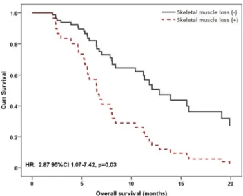
was longer (median 12.8; 95%CI 9.8-15.7 months) in the skeletal muscles stable group than those in skeletal muscle loss group (median 6.4; 95%CI 5.2-7.7 months) (p=0.04;Figure 1). Cox regression analysis showed that skeletal muscle loss was a significant prognostic indicator of OS (HR 2.87; 95%CI 1.07-7.42, p=0.03) (Table 4;Figure 1).
Discussion
There is no standard chemotherapy for re-fractory mCRC patients [23]. In the phase III tri-als (CORRECT and RESOURCE) regorafenib and TAS-102 have been shown to be superior to pla-cebo as new treatment option [5,8]. The ESMO and NCCN guidelines recommend these two agents as an additional line of therapy for mCRC refractory to chemotherapy. Regorafenib or TAS-102 can be before or after each other [24,25]. There is not any head to head study, comparing regorafenib to TAS-102 therapy. On the other hand, recent studies have shown that muscle loss is associated with progno-sis in patients with CRC. In our study, the clinical efficacy of regorafenib and TAS-102 was similar. In addition, patients who received regorafenib therapy had more muscle mass loss than TAS-102 therapy, and also muscle loss predicted mortality regardless of treatment options.
Sarcopenia has been described by Rosenberg as a systemic and progressive loss of skeletal muscle associated with age and atrophy [26]. Generally, the surface area of the muscle measured by CT at the level of L3 vertebra can be identified by SMA [27]. Sarcopenia is associated with poor prognosis in various types of cancer [28-30]. Sarcopenia accom-panied by cancer-associated cachexia is responsible for 20% of cancer-related deaths [31]. This condi-tion has also been shown to be associated with poor prognosis in patients with CRC [18]. In addition, sarcopenia has been reported to adversely affect treatment response rates, OS and PFS, independent of disease progression [32,33]. In our study, it has been shown that the OS of patients with muscle loss was worse. In the light of this information, changes in skeletal muscle mass during and after treatment may indicate prognostic significance and may be an important marker for the tumor biology.
Regorafenib inhibits tyrosine kinases involved in tumor angio- and oncogenesis and further sta-bilizes the microenvironment that is effective in tumor growth. These effects consist of multiple ki-nase inhibition (VEGF receptors 1-3, KIT, PDGFR-alpha, PDGFR-beta, RET, FGFR1 and 2, TIE2, DDR2, TrkA, Eph2A, RAF-1. BRAF, BRAFV600E, SAPK2, PTK5, and Abl). Recently, in an animal study, Huot et al reported that regorafenib has been shown to cause skeletal muscle loss by a possible mechanism including increasing levels of autophagy-depend-ent protein markers and abnormal mitochondrial homeostasis via ERK1/2 and GSK3β pathway [34]. TAS-102 is a novel oral anticancer agent consisting of trifluorothymidine as a thymidine analog which is incorporated into DNA in order to interfere with DNA synthesis and inhibit proliferation, and tip-iracil hydrochloride, which prevents the degrada-tion by inhibidegrada-tion of thymidine phosphorylase [8]. The effect of TAS-102 on skeletal muscle loss is not well known. In a recent study, regorafenib has been shown to cause more skeletal loss than TAS-102 [35]. In our study, skeletal muscle loss was seen in both TAS-102 and regorafenib patients, however in patients who received regorafenib, the skeletal muscle loss was significantly higher than in pa-tients who received TAS-102. In the sex and age
Variables HR 95% Confidential interval p value
Lower Upper
Skeletal muscle loss 2.82 1.07 7.42 0.03
Age (> 60 years) 0.58 0.22 1.55 0.28
Regorafenib 0.80 0.30 2.17 0.67
Table 4. Cox regression analysis of OS in study subjects
Figure 1. Overall survival of the patients according to
adjusted multivariate analysis, regorafenib caused more muscle loss than TAS-102.
Regorafenib and TAS-102 are the treatment op-tions in the third or the fourth lines of patients with mCRC. Median OS with regorafenib in the COR-RECT trial was 6.4 months [5] and in the CONCUR trial median OS was 8.8 months with regorafenib treatment [6]. In the RESOURCE, a prospective randomized trial of TAS-102, the median OS was 7.1 months [8]. A retrospective comparative study of real-life data showed that median OS was 6.7 months with regorafenib and 6.5 months with TAS-102. According to this study, although there was no difference in efficacy between the two drugs, different toxicity profiles were reported [36]. In our study, median OS was 7.8 months in patients who received TAS-102 and 6.5 months in patients who received regorafenib (p=0.96). Similarly, no differ-ence was observed between the efficacy of the two drugs, and skeletal muscle loss was significantly higher in the regorafenib arm. However, this differ-ence does not present a drug-related OS differdiffer-ence in patients with skeletal muscle loss.
There are some major limitations of our study. Its retrospective nature caused some disadvantages
in the assessment of treatment-associated symp-toms and toxicities. In addition, the study was made on a small number of patients. Also, SMM measurements were evaluated twice: at baseline and under treatment. Moreover, there was not any data on the change in quality of life (QoL) scores and detailed toxicity profiles at the baseline and under treatment. Despite these limitations, it has been an outstanding strength of the study to con-clude that regorafenib therapy resulted significant-ly in higher loss of SMM compared to TAS-102 therapy. We suggest that the results of this study give an opinion to treat more fragile mCRC patients to prevent muscle loss during the third- and sub-sequent lines.
In conclusion, both drugs had similar efficacy, however OS was shorter in patients with muscle loss. Although patients who received regorafenib had more muscle loss than those who received TAS-102, there was no difference in OS between the two groups.
Conflict of interests
The authors declare no conflict of interests.
References
1. Bray F, Ferlay J, Soerjomataram I, Siegel RL, Torre LA, Jemal A. Global cancer statistics 2018: GLOBOCAN estimates of incidence and mortality worldwide for 36 cancers in 185 countries. CA Cancer J Clin 2018;68:394-424.
2. Van Cutsem E, Cervantes A, Adam R et al. ESMO con-sensus guidelines for the management of patients with metastatic colorectal cancer. Ann Oncol 2016;27:1386-1422.
3. Karatas F, Sahin S. The relationship Between Primary Tumor Localization and Survival in Patients with Re-current Colorectal Cancer. Bozok Med J 2019;9:11-5. 4. Marks EI, Tan C, Zhang J, Zhou L, Yang Z, Scicchitano
A, El-Deiry WS. Regorafenib with a fluoropyrimidine for metastatic colorectal cancer after progression on multiple 5-FU-containing combination thera-pies and regorafenib monotherapy. Cancer Biol Ther 2015;16:1710-9.
5. Grothey A, Van Cutsem E, Sobrero A et al. Regorafenib monotherapy for previously treated metastatic colo-rectal cancer (CORRECT): an international, multicentre, randomised, placebo-controlled, phase 3 trial. Lancet 2013;381:303-12.
6. Li J, Qin S, Xu R, Yau TC et al. Regorafenib plus best supportive care versus placebo plus best supportive care in Asian patients with previously treated metastat-ic colorectal cancer (CONCUR): a randomised,
double-blind, placebo-controlled, phase 3 trial. Lancet Oncol 2015;16:619-29.
7. Sahin S, Hacioglu MB. Treatment Outcomes of Meta-static Colorectal Cancer Patients Treated with Re-gorafenib as Third-Line Setting-A Multicenter Study. Istanbul Med J 2019;20:82-7.
8. Mayer RJ, Van Cutsem E, Falcone A et al. Randomized trial of TAS-102 for refractory metastatic colorectal cancer. N Engl J Med 2015;372:1909-19.
9. Kurk SA, Peeters PHM, Dorresteijn B et al. Impact of different palliative systemic treatments on skeletal muscle mass in metastatic colorectal cancer patients. J Cachexia Sarcopenia Muscle 2018;9:909-19.
10. Gokyer A, Kucukarda A, Kostek O et al. Relation be-tween sarcopenia and dose-limiting toxicity in patients with metastatic colorectal cancer who received re-gorafenib. Clin Transl Oncol 2019;doi:10.1008/s12094-019-02080-4 (Epub ahead of print).
11. Martin L, Birdsell L, Macdonald N et al. Cancer ca-chexia in the age of obesity: skeletal muscle depletion is a powerful prognostic factor, independent of body mass index. J Clin Oncol 2013;31:1539-47.
12. Barret M, Berthaud C, Taieb J. [Sarcopenia: a concept of growing importance in the management of colorectal cancer]. Presse Med 2014;43:628-32.
sarcopenia, obesity and sarcopenic obesity predictive of outcome in patients with colorectal liver metastases? HPB (Oxford) 2015;17:438-46.
14. Blum D, Stene GB, Solheim TS et al. Validation of the Consensus-Definition for Cancer Cachexia and evalu-ation of a classificevalu-ation model--a study based on data from an international multicentre project (EPCRC-CSA). Ann Oncol 2014;25:1635-42.
15. Fearon K, Strasser F, Anker SD et al. Definition and classification of cancer cachexia: an international con-sensus. Lancet Oncol 2011;12:489-95.
16. Mourtzakis M, Prado CM, Lieffers JR, Reiman T, McCa-rgar LJ, Baracos VE. A practical and precise approach to quantification of body composition in cancer patients using computed tomography images acquired during routine care. Appl Physiol Nutr Metab 2008;33:997-1006.
17. Kilgour RD, Cardiff K, Rosenthall L, Lucar E, Trutschnigg B, Vigano A. Use of prediction equations to determine the accuracy of whole-body fat and fat-free mass and appendicular skeletal muscle mass measure-ments from a single abdominal image using computed tomography in advanced cancer patients. Appl Physiol Nutr Metab 2016;41:70-5.
18. Vashi PG, Gorsuch K, Wan L, Hill D, Block C, Gupta D. Sarcopenia supersedes subjective global assessment as a predictor of survival in colorectal cancer. PLoS One 2019;14:e0218761.
19. Sun G, Li Y, Peng Y et al. Can sarcopenia be a predictor of prognosis for patients with non-metastatic colorectal cancer? A systematic review and meta-analysis. Int J Colorectal Dis 2018;33:1419-27.
20. Karatas F, Sahin S, Imamoglu GI, Altinbas M. Panitu-mumab as a possible cause of hematospermia: what is the mechanism? Future Oncol 2015;11:3229-31. 21. Antoun S, Baracos VE, Birdsell L, Escudier B, Sawyer
MB: Low body mass index and sarcopenia associated with dose-limiting toxicity of sorafenib in patients with renal cell carcinoma. Ann Oncol 2010;21:1594-8. 22. Mitsiopoulos N, Baumgartner RN, Heymsfield SB,
Lyons W, Gallagher D, Ross R. Cadaver validation of skeletal muscle measurement by magnetic resonance imaging and computerized tomography. J Appl Physiol 1998;85:115-22.
23. Sahin S, Karatas F. The impact of primary tumor locali-zation on survival and treatment outcomes in patients with metastatic colorectal cancer-a multicenter study. JBUON 2019;24:479-87.
24. Benson AB, 3rd, Venook AP, Cederquist L et al. Co-lon Cancer, Version 1.2017, NCCN Clinical Practice Guidelines in Oncology. J Natl Compr Canc Netw 2017;15:370-98.
25. Yoshino T, Arnold D, Taniguchi H et al. Pan-Asian adapted ESMO consensus guidelines for the manage-ment of patients with metastatic colorectal cancer: a JSMO-ESMO initiative endorsed by CSCO, KACO, MOS, SSO and TOS. Ann Oncol 2018;29:44-70.
26. Rosenberg IH. Epidemiologic and methodologic prob-lems in determining nutritional status of older persons. Am J Clin Nutr 1989;50:1121-1235.
27. Cruz-Jentoft AJ, Baeyens JP, Bauer JM et al. Sarcopenia: European consensus on definition and diagnosis: Re-port of the European Working Group on Sarcopenia in Older People. Age Ageing 2010;39:412-23.
28. Nakanishi R, Oki E, Sasaki S et al. Sarcopenia is an independent predictor of complications after colorectal cancer surgery. Surg Today 2018;48:151-7.
29. Harimoto N, Yoshizumi T, Shimokawa M et al. Sarcope-nia is a poor prognostic factor following hepatic resec-tion in patients aged 70 years and older with hepatocel-lular carcinoma. Hepatol Res 2016;46:1247-55. 30. Takamori S, Toyokawa G, Okamoto T et al. Clinical
Impact and Risk Factors for Skeletal Muscle Loss Af-ter Complete Resection of Early Non-small Cell Lung Cancer. Ann Surg Oncol 2018;25:1229-36.
31. Skipworth RJ, Stewart GD, Dejong CH, Preston T, Fearon KC. Pathophysiology of cancer cachexia: much more than host-tumour interaction? Clin Nutr 2007;26:667-76.
32. Sasaki S, Oki E, Saeki H et al. Skeletal muscle loss during systemic chemotherapy for colorectal cancer indicates treatment response: a pooled analysis of a multicenter clinical trial (KSCC 1605-A). Int J Clin Oncol 2019.doi:10.1007/s/10147-019-01460-8 (Epub ahead of print).
33. Kostek O, Demircan NC, Gokyer A et al. Skeletal muscle loss during anti-EGFR combined chemotherapy regi-mens predicts poor prognosis in patients with RAS wild metastatic colorectal cancer. Clin Transl Oncol 2019.doi 10.1007/s12094-019-02079-x (Epub ahead of print).
34. Huot JR, Essex AL, Gutierrez M et al. Chronic Treatment with Multi-Kinase Inhibitors Causes Differential Toxic-ities on Skeletal and Cardiac Muscles. 2019;11:pii:E571. doi 10.3390/cancers 11040571.
35. Huemer F, Schlintl V, Hecht S et al. Regorafenib Is As-sociated With Increased Skeletal Muscle Loss Com-pared to TAS-102 in Metastatic Colorectal Cancer. Clin Colorectal Cancer 2019;18:159-66.
36. Masuishi T, Taniguchi H, Hamauchi S et al. Regorafenib Versus Trifluridine/Tipiracil for Refractory Metastatic Colorectal Cancer: A Retrospective Comparison. Clin Colorectal Cancer 2017;16: e15-e22.