Yunus Emre Altuntas,1 Mustafa Oncel,2 Mustafa Haksal,2 Metin Kement,1 Ersin Gundogdu,1 Nihat Aksakal,3
Fazli Cem Gezen2
1Department of General Surgery, Kartal Training and Research Hospital, Istanbul, Turkey 2Department of General Surgery, Medipol University Faculty of Medicine, Istanbul, Turkey 3Department of General Surgery, Istanbul University Faculty of Medicine, Istanbul, Turkey
ABSTRACT
OBJECTIVE: This study aimed to reveal the risk factors and outcomes of gallbladder perforation (GP) during laparoscopic cholecystectomy.
METHODS: Videotapes of all patients who underwent an elective cholecystectomy at our department were retrospectively analyzed, and the patients were divided into two groups based on the presence of GP. The possible risk factors and early outcomes were analyzed.
RESULTS: In total, 664 patients [524 (78.9%) females, 49.7±13.4 years of age] were observed, and GP occurred in 240 (36.1%) patients, mostly while dissecting the gallbladder from its bed (n=197, 82.1%). GP was not recorded in the operation notes in 177 (73.8%) cases. Among the studied parameters, there was no significant risk factor for GP, except preopera-tively elevated alanine transaminase level (p=0.005), but the sensitivity and specificity of this measure in predicting GP were 14.2% and 7.4%, respectively. The two groups had similar outcomes, but the operation time (35.4±17.5 vs 41.4±18.7 min, p=0.000) and incidence of drain use (25% vs 45.8%, p=0.000) increased in the GP group.
CONCLUSION: The present study reveals that GP occurs in 36.1% of patients who undergo laparoscopic elective cholecys-tectomy, but it may not be recorded in most cases. We did not find any reliable risk factor that increases the possibility of GP. GP causes an increase in the operation time and incidence of drain use; however, the other outcomes were found to be similar in patients with GP and those without.
Keywords: Alanine transaminase; gallbladder perforation; laparoscopic cholecystectomy.
Received: June 24, 2017 Accepted:August 22, 2017 Online: January 12, 2018
Correspondence: Dr. Yunus Emre ALTUNTAS. Kartal Egitim ve Arastirma Hastanesi, Genel Cerrahi Klinigi, Istanbul, Turkey. Tel: +90 216 441 39 00 e-mail: [email protected]
© Copyright 2018 by Istanbul Provincial Directorate of Health - Available online at www.northclinist.com
North Clin Istanb
doi: 10.14744/nci.2017.88155
Gallbladder perforation during elective laparoscopic
cholecystectomy: Incidence, risk factors, and outcomes
I
n general surgery practice, cholecystectomy is the sec-ond most commonly performed abdominal operation [1, 2]. Laparoscopic cholecystectomy has some advan-tages over the conventional technique, including better cosmetic results, lesser postoperative pain, a shorter hos-pital stay, and early return to daily activities [3]. Besides, cholecystectomy is not a risk-free procedure and may cause severe complications, including bile duct injury, bleeding, abscess, and pancreatitis.Gallbladder perforation (GP), which is a common intraoperative complication during cholecystectomy, has been reported to occur with a high incidence of 10%-33% [4]. The risk factors and consequences of GP have also been studied [4-9]. It has been advocated that male sex, a history of acute cholecystitis or previous laparo-tomies, the use of a laser, an inflamed or nonvisualized gallbladder, and a difficult operation increase the risk of GP [5-7]. In addition, bile and stone spillage have rarely
been reported to lead to severe problems [10-15]. GP does not worsen the outcomes of the procedure, but it has been stated that lost stones after GP may infre-quently cause secondary complications, including pain, fever, or intraabdominal abscesses, because they are a potential nidus of infection and bile spillage may lead to chemical peritonitis [4, 10-16]. However, most of the in-formation present in the literature may be misleading be-cause the data is mostly based on retrospective informa-tion, and it is probable that GPs were not recorded in the operation documents because GP is generally believed to be harmless, with no adverse consequences in most in-stances. Thus, to understand the incidence, risk factors, and consequences of GP, we aimed to analyze compact disc videos of the operations.
MATERIALS AND METHODS
The Institutional Review Board approved the de-sign and content of the study (Reference number: B104ISM4340029/1009/20). All the patients who underwent an elective cholecystectomy between March 2011 and March 2015 at our department were retro-spectively reviewed. The procedures were performed or supervised by one of six surgeons at our department and were performed using a four-trocar technique as de-scribed previously [17]. In case of GP during the oper-ation, the management was generally alike: free bile was aspirated, the soiled areas were irrigated with physiolog-ical saline until clear, and spilled stones were retrieved whenever possible. These patients continued to receive intravenous and peroral antibiotics for 1 week in most instances. The placement of a drain and conversion to open surgery were decided by the operating surgeon. The patients were generally discharged from the hospital on the next day, but longer hospitalization was sometimes necessary.
The primary aims of the present study were to reveal the incidence and risk factors for and outcomes after intraoperative GP during elective laparoscopic surgery. The patients for whom conversion to open surgery was undertaken were excluded from further analyses, but the reasons for conversion were stated. An experienced sur-geon (YEA) blinded to the patients, operators, and out-comes of the procedures watched the operation videos taped on compact discs (CD) of all the included patients. The patients were classified into two groups based on the presence of GP at the time of the operation (Perforation or No perforation groups). The patients were excluded
if the operation CDs were not available or defective. During this inspection, the degree of difficulty was cal-culated according to Cuschieri’s scale, which defines the complexity of the procedure in four grades [18]. Grade 1 refers to an easy cholecystectomy without any further problems. Grade 2 refers to the presence of light peri-cholecystitis or adherences or fatty tissue masking the cystic pedicle or mucocele. According to this scale, grade 3 defines severely difficult cholecystectomies in patients with gangrenous cholecystitis; shrunken fibrotic gall-bladder; intense pericholecystitis; subhepatic abscesses; or advanced hepatic diseases, such as cirrhosis or portal hypertension. In those patients, the dissections of the cystic pedicle or the body gallbladder from the hepatic bed are hard or sometimes impossible because of the ac-companying problem or adherence of Hartman’s pouch over the common bile duct. Patients with grade 4 diffi-culties were excluded because it refers to conversion to open surgery [18].
The timing of perforation was noted as follows: dur-ing traction of the gallbladder, durdur-ing the dissection of adhesions and bands, during the dissection of Callot’s triangle prior to cystic duct clipsing, during the further dissection of Callot’s triangle after cystic duct clipsing, during the dissection of the gallbladder from the hepatic fossa, and during the extraction of the gallbladder from the abdominal cavity. A chart review was completed for all patients, and computer-based data were scanned for the following information as risk factors for and out-comes of intraoperative GP: demographics; body mass index; the presence/absence of previous hospitalization for acute cholecystitis, biliary pancreatitis, or choloangi-tis; the American Society of Anesthesiology score; labo-ratory findings [alanine transaminase (ALT), aspartate transaminase (AST), gamma-glutamyl transaminase (GGT), albumin, white blood cell (WBC), hemoglobin, total bilirubin, and amylase]; the necessity and findings of magnetic resonance imaging cholangiography (MRCP) and/or endoscopic retrograde cholangiography (ERCP); ultrasound findings; the presence/absence of previous laparotomy [regional (midline or right subcostal inci-sions) or others (McBurney or Pfannenstiel)]; previous operations (operations of the gallbladder or gastroduo-denal region; or others, including appendectomy, section, or gynecological); the experience of the operator (staff or resident); the degree of difficulty of the operation as described by Cuschieri; operation time; the presence or omission of drain placement; hospitalization period; complications; and re-hospitalization and its causes,
mortality, and pathological findings. Finally, the opera-tion notes were also scanned by two investigators (EG and MH) who were blinded to the patients’ information and operation CDs, and whether or not the perforation was stated in the operation notes was assessed.
The patients with a previous hospitalization related to gallbladder stones received an interval cholecystec-tomy from 6 to 8 weeks after the initial presentation. In our routine practice, if the operation and hospitalization period are uneventful, the patients are seen at day 7 post-operatively. In case of having no complaints, further fol-low-up is not offered.
Statistical Analysis
Data were analyzed using SPSS 17.0 for Windows (SPSS Inc., Chicago, IL, USA). Results were given as percentages, mean and standard deviations, or median and ranges. Quantitative and qualitative variables were compared using Student’s t-test and chi-square (Pearson’s or Fischer’s exact) test, respectively. A Mann–Whitney U test was preferred when there was an abnormal dis-tribution of the samples confirmed by the Kolmogorov– Smirnov test. A p value <0.05 was accepted to be signif-icant.
RESULTS
In total, 737 patients [575 (78.0%) females with a mean (SD) age of 49.1±13.3 years] underwent an elective cholecystectomy at our department during the study pe-riod. However, the operation CDs were not available or defective in 60 (8.1%) cases, leaving 677 cases for further analyses. Of these, 13 (1.9%) necessitated conversion to open surgery due to severe adhesions (n=9, 1.3%) not identifying anatomical structures (n=2, 0.3%), severe bleeding (n=1, 0.1%), and injury to the transverse colon (n=1, 0.1%). Thus, current data include a total number of 664 cases [524 (78.9%) females with a mean (SD) age of 49.7±13.4 years].
Of 664 patients, an intraoperative GP was observed in 240 (36.1%) patients (Perforation group) during trac-tion of the gallbladder (n=15, 6.3%), the dissectrac-tion of adhesions and bands (n=2, 0.8%), the dissection of Cal-lot’s triangle prior to cystic duct clipsing (n=9, 3.8%), the further dissection of Callot’s triangle after cystic duct clipsing (n=6, 2.5%), the dissection of the gallbladder from its bed (n=197, 82.1%), and the extraction of the gallbladder from the abdominal cavity (n=11, 4.6%).
The remaining patients (n=424) were present in the No perforation group. Of 240 patients, GP was not stated in the operation records in 177 (73.8%) cases.
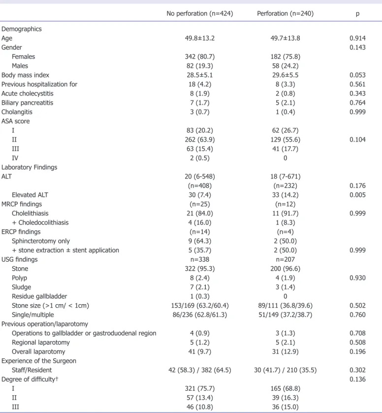
The present study evaluated the risk factors; how-ever, almost none of the analyzed parameters were found increase intraoperative GP during elective laparoscopic cholecystectomy, including patient-related aspects, pre-operative laboratory or other diagnostic findings, and operative features (Table 1). The only risk factor that increased the risk of GP was preoperative ALT level (p <0.05). Of the 63 patients with elevated ALT levels, 33 (52.4%) had GP, but the sensitivity and specificity of el-evated ALT in predicting GP were 14.2% and 7.4%, re-spectively. There were no differences between the blood tests, apart from ALP. Although some patients required MRCP or ERCP due to elevated bilirubin levels or choledocholithiasis, there were no patients who were in need of intraoperative cholangiography, exploration of the common bile duct, or laparoscopic ultrasonography in the current study.
The outcomes after the operations were also analyzed. The results showed no statistically significant differences between the groups regarding postoperative hospital-ization period, complications, and re-hospitalhospital-izations (Table 2). In addition to the median (range) operation time [32 (10-120) vs 36.5 (11-120)], the rate of drain use was significantly increased in patients with GP (p <0.001 for both). No patients required reoperation as a consequence of the complications. However, the patho-logical evaluations revealed gallbladder cancer in two pa-tients (0.8%), both in the Perforation group. Subsequent procedures were required for both the patients, and they remained alive 5 and 9 months after the operations with-out any evident tumor recurrence.
DISCUSSION
Cholecystectomy is one of the most common indications for surgery worldwide [1]. Besides, GP frequently occurs during laparoscopic cholecystectomy. A recent review on 18.280 patients has revealed that the incidence of GP is 18.3% [19]. In another analysis, the rate of GP in 1059 consecutive laparoscopic cholecystectomies was 29% [20]. However, it is possible that even this higher rate may not be the actual incidence of GP because prospec-tive information has shown that the frequency of GP during laparoscopic cholecystectomy reaches up to 33% [9]. Consequently, it may be more reasonable to con-sider the highest reported rates in the literature because
No perforation (n=424) Perforation (n=240) p Demographics Age 49.8±13.2 49.7±13.8 0.914 Gender 0.143 Females 342 (80.7) 182 (75.8) Males 82 (19.3) 58 (24.2)
Body mass index 28.5±5.1 29.6±5.5 0.053
Previous hospitalization for 18 (4.2) 8 (3.3) 0.561
Acute cholecystitis 8 (1.9) 2 (0.8) 0.343 Biliary pancreatitis 7 (1.7) 5 (2.1) 0.764 Cholangitis 3 (0.7) 1 (0.4) 0.999 ASA score I 83 (20.2) 62 (26.7) II 262 (63.9) 129 (55.6) 0.104 III 63 (15.4) 41 (17.7) IV 2 (0.5) 0 Laboratory Findings ALT 20 (6-548) 18 (7-671) (n=408) (n=232) 0.176 Elevated ALT 30 (7.4) 33 (14.2) 0.005 MRCP findings (n=25) (n=12) Cholelithiasis 21 (84.0) 11 (91.7) 0.999 + Choledocolithiasis 4 (16.0) 1 (8.3) ERCP findings (n=14) (n=4) Sphincterotomy only 9 (64.3) 2 (50.0)
+ stone extraction ± stent application 5 (35.7) 2 (50.0) 0.999
USG findings n=338 n=207 Stone 322 (95.3) 200 (96.6) Polyp 8 (2.4) 4 (1.9) 0.930 Sludge 7 (2.1) 3 (1.4) Residue gallbladder 1 (0.3) 0 Stone size (>1 cm/ < 1cm) 153/169 (63.2/60.4) 89/111 (36.8/39.6) 0.502 Single/multiple 86/236 (62.8/61.3) 51/149 (37.2/38.7) 0.760 Previous operation/laparotomy
Operations to gallbladder or gastroduodenal region 4 (0.9) 3 (1.3) 0.708
Regional laparotomy 5 (1.2) 5 (2.1) 0.508
Overall laparotomy 41 (9.7) 31 (12.9) 0.196
Experience of the Surgeon
Staff/Resident 42 (58.3) / 382 (64.5) 30 (41.7) / 210 (35.5) 0.302
Degree of difficulty† 0.136
I 321 (75.7) 165 (68.8)
II 57 (13.4) 39 (16.3)
III 46 (10.8) 36 (15.0)
(Data are presented as either median [range] or mean [±standard deviation]. Information in the parentheses indicates the percentages).
*The levels of Hgb are presented separately in male and female patients; †: Data are presented according to Cuschieri’s scale, which defines the complexity of the procedure in 4 grades; however patients with grade 4 difficulties were excluded since it refers to conversion to open surgery [18].
ASA: American Society of Anesthesiology score; AST: Alanine transaminase; ALT: Aspartate transaminase; GGT: Gamma-glutamyl transaminase; WBC: White blood cell; Hbg: Hemoglobin; MRCP: Magnetic resonance imaging cholangiography; ERCP: Endoscopic retrograde cholangiography; USG: Ultrasonography.
most perforations may not be documented in operation records. The controversy probably arises from the fact that some surgeons do not report GP in the operation records, which are the data sources of retrospective stud-ies. A recent analysis on operative notes has revealed that GP with or without bile and stone spillage was not doc-umented in some instances [21]. Thus, we believe that the rate of GP presented in the present study is more realistic because the data were obtained from operation videos. Accordingly, our data shows that GP may be more common than expected and occurs in at least one third of all patients undergoing laparoscopic cholecystec-tomy. In addition, the present study has also shown that GP is not noted in almost three-fourth of the operation records probably because most surgeons do not consider this problem a major complication in most instances and believe that GP is harmless and thus do not report it.
Several studies have evaluated the potential risk fac-tors for GP during laparoscopic cholecystectomy. A mul-tivariant logistic regression analysis has revealed male sex, a history of acute cholecystitic, the use of a laser, and the presence of a grossly inflamed gallbladder as individ-ually significant risk factors for GP [5]. Other studies have underlined some other parameters influencing the frequency of GP, for example, age; preoperative ultra-sound findings, including a thickened gallbladder wall and hydrops; the presence of a previous laparotomy; the nature of the stone (pigment stones); and the surgeon’s experience [5, 7, 9, 22]. However, these studies may be criticized to include both groups of patients who under-went emergent and elective procedures, which are
proba-bly different types of operations. Thus, the patients who underwent emergent cholecystectomies were excluded in the present study. In addition, the patients for whom conversion to open surgery was undertaken were also ex-cluded because it was generally impossible to determine whether the adverse consequences in these cases were due to GP or the conversion itself. Thus, the present study focuses on a more specific condition that only includes elective non-problematic laparoscopic cholecystectomy, which is actually the case in most instances.
Although the present study has revealed that a pre-operatively elevated ALT level may be an indicator for a possible GP at the time of the operation, we are not sure whether it is a significant factor or an incidental finding because the risk for GP was only 52.4% in patients with elevated ALT levels, which was <two-fold of that with normal ALT levels. Consequently, because the sensitiv-ity and specificsensitiv-ity were unacceptably low, in our opinion, the preoperative ALT level cannot be considered a pre-dictive factor for GP. Interestingly, a correlation between GP and the difficulty scale of the cholecystectomy was not attained within the framework of the present data. Although the statistical analysis revealed no difference, the p value of 0.136 may suggest a trend. Our data did not reveal any other risk factor that increased the pos-sibility of GP. Thus, we believe that GP may be unpre-dictable in the case of an elective cholecystectomy.
In a recent study, it has been mentioned that 69 of 131 GPs (52.7%) occurred while dissecting the gall-bladder from the hepatic fossa [6]. Because we had the same result, we can conclude that the separation of the gallbladder from its bed is probably the most risky stage of cholecystectomy for a possible GP. We believe that this information is significant, and it may warn surgeons about this particular threat. In addition, the above-men-tioned point should be emphasized during the training period of surgeons.
The consequences of GP have been extensively eval-uated previously. In animal models, it has been generally shown that GP and the consequent spillage of bile and/ or stones are harmless and do not cause any infection or mortality during the follow-up period [23, 24]. In contrast, the data derived from human studies remain controversial. Although, at least theoretically, GP leads to the contamination of the peritoneal cavity with bile, calculi, and bacteria, some believe that that the adverse consequences of spillage after GP during laparoscopic cholecystectomy may be minimized by the prompt
re-Table 2. Outcomes after gallbladder perforation
No perforation Perforation p
(n=424) (n=240)
Operation time (minutes) 35.4±17.5 41.4±18.7 0.000
Drain use 106 (25.0) 110 (45.8) 0.000
Hospitalization period (days) 1.3±1.0 1.3±0.9 0.664
Complications Bile drainage 0 2 (0.8) 0.130 Evisceration 0 1 (0.4) 0.361 Abscess 3 (0.7) 3 (1.3) 0.673 Cholangitis 4 (0.9) 1 (0.4) 0.659 Respiratory 2 (0.5) 0 0.538 Overall 9 (2.1) 7 (2.9) 0.522 Re-hospitalization 6 (1.4) 5 (2.1) 0.538
trieval of as many of the spilled stones as possible, abun-dant irrigation of the peritoneal cavity, and adequate an-tibiotic therapy [6]. It has been shown in most studies that GP does not increase the complication risk, reopera-tions, or hospital stay [6, 25]. A recent prospective study has also advocated that GP and retained gallstones do not adversely affect respiratory mechanics or alter post-operative pain [9]. Besides, other studies have reported adverse consequences of bile spillage due to GP because bile is frequently contaminated in the presence of gall-stones [26]. In an analysis of 1059 consecutive laparo-scopic cholecystectomies, increased incidences of fever and intraabdominal abscesses have been reported if GP had happened at the time of the operation [20]. In ad-dition, some case reports have advocated that retained gallstones and bile spillage cause infection or abscesses, fibrosis, adhesions, cutaneous sinuses, small-bowel ob-structions, or generalized septicemia [27, 28]. Finally, even conversion to open surgery has been recommended in a study in patients with a loss of numerous or large pigment stones that cannot be retrieved by laparoscopy [4]. However, we believe that the adverse consequences of spillage after GP during laparoscopic cholecystectomy may be minimized by the prompt retrieval of as many of the spilled stones as possible, abundant irrigation of the peritoneal cavity, and adequate prophylactic antibiotic therapy [6], as mentioned above. With this approach, we have found that GP and consequent intraabdominal contamination does not increase the risk of complica-tions or alter the outcomes during the early postopera-tive period. Consequently, the present study reveals that GP increases the incidence of drain use and lengthens the operation time, both of which are probably the con-sequences of GP because the retrieval of stones and peri-toneal irrigation are required in these cases. However, the present study found no other adverse consequence of GP in patients who underwent laparoscopic cholecystec-tomy. In contrast, similar short-term complications may be related to the treatment strategy followed in patients with GP. In addition, many of the documented complica-tions from split gallstones are long-term, and they often happen after 1 year. Therefore, based on the results of the present study, we cannot comment on any long-term complications. Thus, we believe that GP is something that may be avoided whenever possible, but in the case of GP, the outcomes do not alter if certain rules for limiting the contamination are followed.
Finally, it is necessary to detail the outcomes in two patients with gallbladder cancer in the present study.
Th-ese patients received further operations, including par-tial hepatic resections and lymph node dissection in the hepatic hilum, because their pathological results denied early T1 tumors. However, the spillage of bile has led to tumor implants over the anterior wall of the stomach in one patient; this patient required a distal gastrectomy. We believe that tumor spillage is the most significant ad-verse consequence of GP, but it also is a very exceptional problem because incidental gallbladder cancer is rare in patients undergoing elective cholecystectomy.
The present study has some limitations mostly due to its retrospective nature. Missing information, namely, the lack of operation CDs of 60 patients, limits the value of the present data. In addition, the findings of the study may be criticized in some standpoints. First, although the difference was statistically significant, we do not know whether a 6-min increase in the operation time reflects an importance in daily practice. In addition, the increased incidence of drain use in the Perforation group may be because there was no clear-cut criteria about the indications to put a drain. This may stem from the ret-rospective nature of the study. Furthermore, the increase in the operation time and the necessity of drain use may not be caused by GP, but all the three endpoints occurred due to the difficulty of the operation. The present study data may be criticized regarding the experiences of the surgeons because all staff surgeons do not have the same expertise. In addition, because the surgeons know they are being recorded, their performance may be more meticulous, which may have an impact on our results.
In conclusion, the present study reveals that GP oc-curs in 36.1% of the patients undergoing laparoscopic elective cholecystectomy but is less commonly reported in the operation notes. GP is unpredictable because there are probably no risk factors that increase the risk of GP, except a preoperatively assessed elevated ALT level; however, the sensitivity and specificity of this measure are low. The early postoperative outcomes are similar in patients with GP and those without, but the operation time lengthens and the incidence of drain use increases in patients with GP.
Conflict of Interest: No conflict of interest was declared by the au-thors.
Financial Disclosure: The authors declared that this study has re-ceived no financial support.
Authorship contributions: Concept – Y.E.A., M.O.; Design – Y.E.A., M.O.; Supervision – Y.E.A., M.O., M.K.; Materials – Y.E.A., M.H., F.C.G.; Data collection &/or processing – Y.E.A., E.G., N.A.; Analysis and/or interpretation – Y.E.A. M.K.; Writing – Y.E.A., M.O.; Critical review – Y.E.A., M.O., M.H.
REFERENCES
1. Schäfer M, Krähenbühl L, Farhadi J, Büchler MW. Cholelithiasis-la-paroscopy or laparotomy? Ther Umsch 1998;55:110–5.
2. Chekan EG, Pappas TN, Minimally invasive surgery. In: Townsend CM Jr, editor. Sabiston Textbook of Surgery: The biological basis of modern surgical practice. Philadelphia: WB Saunders; 2001. p. 292–310. 3. Memon MA, Deeik RK, Maffi TR, Fitzgibbons RJ Jr. The outcome
of unretrieved gallstones in the peritoneal cavity during laparoscopic cholecystectomy. A prospective analysis. Surg Endosc 1999;13:848–57. 4. Brockmann JG, Kocher T, Senninger NJ, Schürmann GM. Complica-tions due to gallstones lost during laparoscopic cholecystectomy. Surg Endosc 2002;16:1226–32. [CrossRef ]
5. Mohiuddin K, Nizami S, Fitzgibbons RJ Jr, Watson P, Memon B, Me-mon MA. Predicting iatrogenic gall bladder perforation during laparo-scopic cholecystectomy: a multivariate logistic regression analysis of risk factors. ANZ J Surg 2006;76:130–2. [CrossRef ]
6. Sarli L, Pietra N, Costi R, Grattarola M. Gallbladder perforation dur-ing laparoscopic cholecystectomy. World J Surg 1999;23:1186–90. 7. De Simone P, Donadio R, Urbano D. The risk of gallbladder
perfora-tion at laparoscopic cholecystectomy. Surg Endosc 1999;13:1099–102. 8. Duca S, Bãlã O, Al-Hajjar N, Lancu C, Puia IC, Munteanu D, et al. Laparoscopic cholecystectomy: incidents and complications. A retro-spective analysis of 9542 consecutive laparoscopic operations. HPB (Oxford) 2003;5:152–8. [CrossRef ]
9. Uygar Kalayci M, Veli Akin B, Alis H, Kapan S, Nuray Turhan A, Aygun E. Short-term effects of gallbladder perforations during laparo-scopic cholecystectomy on respiratory mechanics and depth of pain. Surg Endosc 2008;22:1317–20. [CrossRef ]
10. Gallinaro RN, Miller FB. The lost gallstone. Complication after laparo-scopic cholecystectomy. Surg Endosc 1994;8:913–4. [CrossRef ]
11. Golub R, Nwogu C, Cantu R, Stein H. Gallstone shrapnel contamina-tion during laparoscopic cholecystectomy. Surg Endosc 1994;8:898–900. 12. Jacob H, Rubin KP, Cohen MC, Kahn IJ, Kan P. Gallstones in a retroperitoneal abscess: a late complication of perforation of the gall-bladder. Dig Dis Sci 1979;24:964–6. [CrossRef ]
13. Kakani PR, Bhullar IS. Complications of spilled gallstones during la-paroscopic cholecystectomy. Contemp Surg 1993;43:357–61. 14. Leland DG, Dawson DL. Adhesions and experimental intraperitoneal
gallstones. Contemp Surg 1993;42:273–5.
15. Sax HC, Adams JT. The fate of the spilled gallstone. Arch Surg 1993;128:469. [CrossRef ]
16. Stewart L, Smith AL, Pellegrini CA, Motson RW, Way LW. Pigment gallstones form as a composite of bacterial microcolonies and pigment solids. Ann Surg 1987;206:242–50. [CrossRef ]
17. Zucker KA. Surgical Laparoscopy. St. Louis, Missouri: Quality Pub-lishing; 1991. p. 143–82.
18. Cuschieri A, Berci G. Laparoscopic Billiary Surgery. London: Blackwell Scientifıc Publications; 1992. p. 134–42.
19. Woodfield JC, Rodgers M, Windsor JA. Peritoneal gallstones
following laparoscopic cholecystectomy: incidence, complications, and manage-ment. Surg Endosc 2004;18:1200–7. [CrossRef ]
20. Rice DC, Memon MA, Jamison RL, Agnessi T, Ilstrup D, Bannon MB, et al. Long-term consequences of intraoperative spillage of bile and gallstones during laparoscopic cholecystectomy. J Gastrointest Surg 1997;1:85–90. [CrossRef ]
21. Wauben LS, van Grevenstein WM, Goossens RH, van der Meulen FH, Lange JF. Operative notes do not reflect reality in laparoscopic cholecys-tectomy. Br J Surg 2011;98:1431–6. [CrossRef ]
22. Kimura T, Goto H, Takeuchi Y, Yoshida M, Kobayashi T, Sakuramachi S, et al. Intraabdominal contamination after gallbladder perforation during laparoscopic cholecystectomy and its complications. Surg En-dosc 1996;10:888–91. [CrossRef ]
23. Welch N, Hinder RA, Fitzgibbons RJ Jr, Rouse JW. Gallstones in the peritoneal cavity. A clinical and experimental study. Surg Laparosc En-dosc 1991;1:246–7. [CrossRef ]
24. Zisman A, Loshkov G, Negri M, Herbert M, Halpern Z, Lin G, et al. The fate of long-standing intraperitoneal gallstone in the rat. Surg Endosc 1995;9:509–11. [CrossRef ]
25. Barrat C, Champault A, Matthyssens L, Champault G. Iatrogenic per-foration of the gallbladder during laparoscopic cholecystectomy does not influence the prognosis. Prospective study. Ann Chir 2004;129:25–9. 26. Keighley MR. Micro-organisms in the bile. A preventable cause of sepsis
after biliary surgery. Ann R Coll Surg Engl 1977;59:328–34.
27. Yamamuro M, Okamoto B, Owens B. Unusual presentations of spilled gallstones. Surg Endosc 2003;17:1498. [CrossRef ]
28. Zulfikaroglu B, Ozalp N, Mahir Ozmen M, Koc M. What happens to the lost gallstone during laparoscopic cholecystectomy? Surg Endosc 2003;17:158. [CrossRef ]