ACQUIRED TOLERANCE OF HEPATOCELLULAR CARCINOMA
CELLS TO SELENIUM DEFICIENCY:
A SELECTIVE SURVIVAL MECHANISM
A THESIS SUBMITTED TO
THE DEPARTMENT OF MOLECULAR BIOLOGY AND GENETICS
AND THE INSTITUTE OF ENGINEERING AND SCIENCE OF
BİLKENT UNIVERSITY
IN PARTIAL FULFILLMENT OF THE REQUIREMENTS
FOR THE DEGREE OF DOCTOR OF PHILOSOPHY
BY
MELİHA BURCU IRMAK
TO MY MOTHER, FATHER and
SISTER...
I certify that I have read this thesis and that in my opinion it is fully adequate, in
scope, and in quality, as a thesis for the degree of Doctor of Philosophy.
Assist. Prof. Rengül Çetin Atalay
I certify that I have read this thesis and that in my opinion it is fully adequate, in
scope, and in quality, as a thesis for the degree of Doctor of Philosophy.
Prof. Dr. Mehmet Öztürk
I certify that I have read this thesis and that in my opinion it is fully adequate, in
scope, and in quality, as a thesis for the degree of Doctor of Philosophy.
Prof. Dr. Aslıhan Tolun
I certify that I have read this thesis and that in my opinion it is fully adequate, in
scope, and in quality, as a thesis for the degree of Doctor of Philosophy.
Prof. Dr. Meral Özgüç
I certify that I have read this thesis and that in my opinion it is fully adequate, in
scope, and in quality, as a thesis for the degree of Doctor of Philosophy.
Assist. Prof. M. Cengiz Yakıcıer
Approved for the Institute of Engineering and Science
Prof. Dr. Mehmet Baray
ABSTRACT
Acquired Tolerance of Hepatocellular Carcinoma Cells to Selenium-Deficiency:
A Selective Survival Mechanism
Meliha Burcu Irmak
Ph.D. in Molecular Biology and Genetics Supervisor: Assist. Prof. Rengül Çetin Atalay
September, 2003
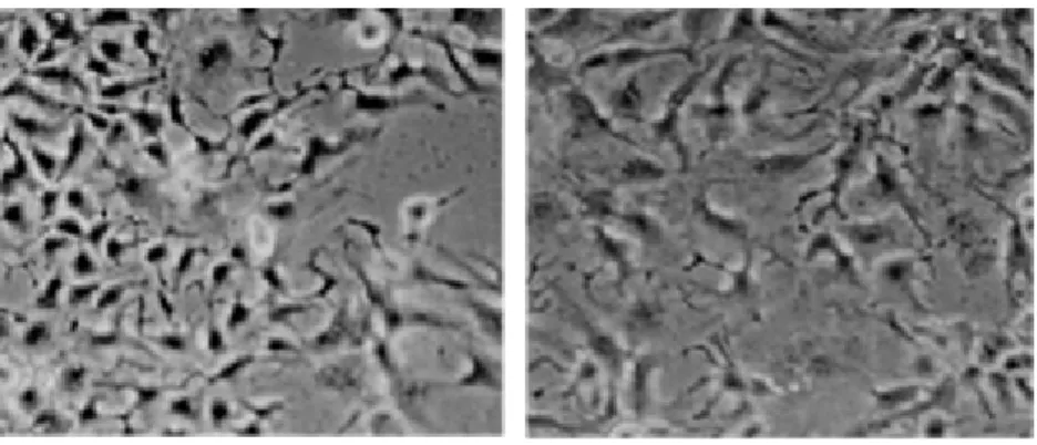
Selenium-deficiency causes liver necrosis. Selenium is protective against viral hepatitis and hepatocellular carcinoma (HCC). The underlying molecular mechanisms of selenium effects are ill-known. In this study in vitro response of hepatocellular carcinoma-derived cell lines to selenium-deficiency is examined alone or in conjunction with Vitamin E and Copper/Zinc. Here we show that in
vitro selenium-deficiency in a subset HCC-derived ‘hepatocyte-like’ cell lines
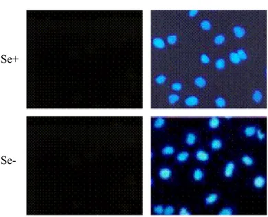
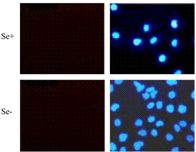
causes oxidative stress and apoptosis. The oxidative stress and consequent cell death induced by selenium-deficiency on these cells are reverted by the antioxidant effect of Vitamin E. However, ten among thirteen HCC cell lines are tolerant to selenium-deficiency and escape its deadly consequences. Nine of ten tolerant cell lines have integrated hepatitis B Virus (HBV) DNA in their genomes, and some display p53-249 mutation, indicating past exposure to HBV or aflatoxins, established factors for oxidative stress and cancer risk. Thus, as demonstrated by the gain of survival capacity of apoptosis sensitive cell lines with Vitamin E, such malignant cells have acquired a selective survival advantage that is prominent under selenium-deficient and oxidative stress conditions.
ÖZET
Karaciğer Kanser Hücrelerinin Selenyum Eksikliğine Karşı Edinilmiş Töleransı:
Seçici Bir Yaşam Mekanizması
Meliha Burcu Irmak
Doktora Tezi, Moleküler Biyoloji ve Genetik Bölümü Tez yöneticisi: Yard. Doç. Rengül Çetin-Atalay
Eylül, 2003
Selenyum eksikliği karaciğer nekrozuna sebep olmaktadır ve selenyum viral hepatit ve karaciğer kanserine karşı koruyucu etki gösterir. Selenyum etkilerinin altında yatan moleküler mekanizmalar halen açıklanamamıştır. Bu çalışmada karaciğer kanseri kökenli hücre hatlarının selenyum eksikliğine in vitro tepkisi yalnız ya da Vitamin E ve bakır/çinkonun varlığında araştırılmıştır. Araştırmamızda in vitro selenyum eksikliğinin bir grup karaciğer kanseri kökenli ‘hepatosit-benzeri’ hücre hattında oksidatif stres ve apoptoza yol açtığını gösterdik. Bu hücrelerde selenyum eksikliğinin yarattığı oksidatif stres ve buna bağlı hücre ölümü, Vitamin E’nin antioksidan etkisiyle ortadan kalkmaktadır. Ancak onüç karaciğer kanser hücre hattından on tanesi selenyum eksikliğinin ölümcül sonuçlarına karşı tolerans göstermektedir. Toleranslı on hücre hattından dokuz tanesi genomlarında hepatit B virüsü (HBV) DNA’sı bulundurmakta ve de bazıları p53-249 mutasyonu sergilemektedir. Bu da geçmişte bu hücre hatlarının, oksidatif strese ve kansere yol açtığı belirlenen, HBV veya aflatoksinlere maruz kaldıklarının göstergesidir. Sonuç olarak, bu tür malin hücrelerin selenyum eksikliği ve oksidatif stres koşullarında ortaya çıkan edinilmiş seçkin yaşama avantajları, apoptoza duyarlı hücre hatlarının Vitamin E ile yaşama kapasitelerini kazanmalarıyla da gösterilmiştir.
ACKNOWLEDGMENTS
I am thankful to Dr. Rengül Çetin-Atalay for her advices, helps, supports, friendship and contributions to my scientific background throughout my Ph.D. studies.
It is my pleasure to express my special thanks to Prof. Dr. Mehmet Öztürk for his scientific guidance and sharing his invaluable knowledge and experience when we needed. I will always be proud of having had a chance to work with him all my life.
I wish to express my appreciation to Dr. M. Cengiz Yakıcıer for all his supports, helps, understanding, friendship and warm attentions with an everlasting interest during my studies.
Special thanks to my friends….
Yeliz, Ceren, Bala, Hilal and Sevgi for their friendships and helps with the experiments.
Nuri for his helps in the laboratory related to the experiments and the computers and Mehdi for all his helps and supports during my studies.
I am thankful to all MBG family members, the technicians, secretaries and my friends. As the circumstances we shared together made us come closer. Thank you for all your efforts during the generation of this thesis.
My family… Source of my energy… Without your endless support and love, this thesis would not have reached to its aim. Thank you….
Finally, I want to express my gratitude to Prof. Dr. İhsan Doğramacı for giving me the opportunity to complete my Ph.D studies at Bilkent University in Molecular Biology and Genetics Department.
TABLE OF CONTENTS
ABSTRACT... ..II ÖZET... III ACKNOWLEDGMENTS... IV TABLE OF CONTENTS... V LIST OF FIGURES... IX LIST OF TABLES... XII ABREVIATIONS... XIIICHAPTER 1. INTRODUCTION 1
1.1 Importance of Trace Elements to Human Health 1
1.2 Selenium and Human Health 4
1.3 Intrinsic Molecular Mechanisms Involving Selenium 5
1.3.1 Gluathione Peroxidases 5
1.3.2 Selenium-Dependent Glutathione Peroxidases 6
1.4 Intracellular and Extracellular Sources of Oxidative Stress 9
1.5 Cellular ROS Toxicity 11
1.6 Signaling Molecules Targeted by ROS 14
1.7 Antioxidants against Oxidative Stress 17
1.7.1 Internal Defense Mechanisms 17
1.7.1.1 Supeoxide Dismutases 17
1.7.1.2 Catalase 18
1.7.1.3 Glutathione S Transferases 18
1.7.2 Dietary Defense Mechanisms 19
1.7.2.1 Vitamin E Supplementation 19
1.7.2.2 Selenium Supplementation 20
1.7.2.3 Copper and Zinc Supplementation 21
1.8 Oxidative Stress and Cancer 22
1.9 Selenium and Cancer 24
1.10 Signaling Molecules Targeted by Selenium Supplementation 24
1.11 Selenium-Deficiency and Liver Diseases 25 1.11.1 Selenium-Deficiency, Keshan Disease, and Liver
Disorders 25 1.11.2 Selenium-Deficiency and Alcoholism 26 1.11.3 Selenium-Deficiency and Viral Liver Diseases 27
1.12 Selenium and HCC 28
1.13 Oxidative Stress and HCC 29
1.14 Hepatocellular Carcinoma 31
1.14.1 Viral Factors 31
1.14.2 Dietary Factors 33
1.14.3 Genetic Alterations 34
CHAPTER 2. AIM and STRATEGY 37 2.1 Experimental Introduction of in vitro Selenium-Deficiency 37
2.2 Aim 46
2.3 Strategy 46
CHAPTER 3. MATERIALS and METHODS 48
3.1 Materials 48
3.1.1 General Reagents 48
3.1.2 Tissue Culture Reagents 48 3.1.3 Oxidative Stress Detection Reagents 48 3.1.4 Apoptosis Assay Reagents 49 3.1.5 Immunofluorescence Reagents 49
3.1.6 Western Blot Reagents 49
3.1.7 Cell Lines 49
3.1.8 Polymerase Chain Reaction Reagents 50
3.2 Solutions and Media 50
3.2.1 Tissue Culture Reagents 50 3.2.2 Oxidative Stress Reagents 51 3.2.3 Apoptotic Assay Reagents 51 3.2.4 Immunofluorescence Reagents 52
3.2.5 SDS-PAGE Reagents 53
3.3 Equipment 55
3.4 Methods 56
3.4.1 Tissue Culture 56
3.4.1.1 Growth Conditions 56 3.4.1.2 Thawing Cell Lines 56 3.4.1.3 Sub-culture of Cell Lines 56 3.4.1.4 Cryopreservation of Cell Lines 57 3.4.1.5 Generation of in vitro Selenium-Adequate and
Selenium-Deficient Medium 57 3.4.1.6 Culture of HCC Cell Lines Under Serum-Free
Medium 57
3.4.1.7 Treatment of HCC Cell Lines with H2O2 58
3.4.2 Detection of Oxidative Stress in HCC Cell Lines 58 3.4.3 Detection and Counting of Dead Cells 59
3.4.4 Apoptosis Assays 59 3.4.4.1 TUNEL 59 3.4.4.2 Annexin V 60 3.4.4.3 NAPO Assay 60 3.4.5 Immunofluorescence 61 3.4.6 Western Blot 62
3.4.6.1 Cell Lysis and Crude Total Protein Extraction 62
3.4.6.2 Bradford Assay 62
3.4.6.3 SDS-PAGE 63
3.4.6.4 Semi-Dry Transfer of the SDS-PAGE Separated
Proteins 64
3.4.6.5 Detection of the PVDF or Nitrocellulose
Immobilized Protein of Interest 65
3.4.7 Giemsa Staining 65
3.4.8 Polymerase Chain Reaction 65
CHAPTER 4. RESULTS 67
4.1 Analysis of Oxidative Stress in Huh-7 Cell Line 67 4.2 Analysis of Survival of Huh-7 Cells under Selenium-
Deficiency 70 4.3 Analysis of Apoptosis of Huh-7 Cells 73 4.4 Analysis of the Oxidative Stress in Hep3B-TR Cell Line 80 4.5 Analysis of Survival of Hep3B-TR Cells under Selenium-
Deficiency 83
4.6 Analysis of Apoptosis of Hep3B-TR Cells 84 4.7 Rescue of Selenium-Deficient Huh-7 Cells from Oxidative
Stress and Cell Death with Vitamin E 91 4.8 Most HCC Cell Lines are Tolerant to Selenium-Deficiency 97 4.9 HBV Confers Resistance to Selenium-Deficiency in HepG2
Cells 104
4.10 Oxidative Stress and Apoptosis in Isogenic Cell Lines 108 4.11 Response of Other Cell Lines to Selenium-Deficiency 122
CHAPTER 5. DISCUSSION and PERSPECTIVES 124
WEB SOURCES 131
LIST OF FIGURES
Figure 1.4.1: Fenton Reaction. 10 Figure 1.4.2: Haber-Weiss Reaction. 10 Figure 1.5.1: ROS Toxicity. 14 Figure 1.6.1: Signaling Molecules Targeted by ROS. 16 Figure 1.7.1: Internal Defense Mechanisms against ROS. 19 Figure 1.14.1: Molecules Targeted in HCC 36 Figure 2.1.1: In vitro response of Huh-7 cells to selenium-deficiency. 38 Figure 2.1.2: Death of Huh-7 cells under selenium-deficiency in DMEM. 38 Figure 2.1.3: TUNEL assay of HUH-7 cells cultured in DMEM. 40 Figure 2.1.4: Annexin V assay of Huh-7 cells cultured in DMEM. 41 Figure 2.1.5: In vitro response of Hep3B-TR cells to selenium-deficiency. 42 Figure 2.1.6: Survival of Hep3B-TR cells under selenium-deficiency. 43 Figure 2.1.7: TUNEL assay of Hep3B-TR cells cultured in DMEM. 44 Figure 2.1.8: Annevin V assay of Hep3B-TR cells cultured in DMEM. 45 Figure 4.1.1: Analysis of oxidative stressing Huh-7 cell line by oxidant-
sensitive fluorescent dye DCFH-DA in DMEM. 69 Figure 4.1.2: Analysis of oxidative stressing Huh-7 cell line by oxidant-
sensitive fluorescent dye DCFH-DA in HAM’s medium. 70 Figure 4.2.1: Death of Huh-7 cells under selenium-deficiency in HAM’s
Medium. 71
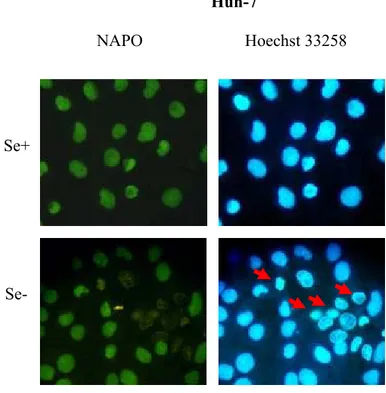
Figure 4.2.2: The percentage of death Huh-7 cells. 73 Figure 4.3.1: NAPO immunostaining of Huh-7 cells cultured in HAM’s
medium. 74
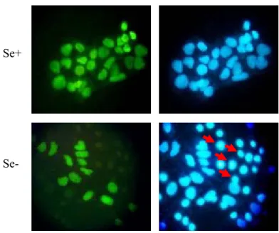
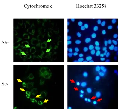
Figure 4.3.2: NAPO immunostaining of Huh-7 cells cultured in DMEM. 75 Figure 4.3.3: Cytochrome c immunostaining of Huh-7 cultured in DMEM. 77 Figure 4.3.4: Western blot analysis of PARP protein in Huh-7 cells. 78 Figure 4.3.5: Correlation of oxidative stress with apoptosis in Huh-7 cells. 79 Figure 4.4.1: Analysis of oxidative stress in Hep3B-TR cell line by
oxidant-sensitive fluorescent dye DCFH-DA in DMEM. 81 Figure 4.4.2: Analysis of oxidative stress in Hep3B-TR cell line by oxidant- sensitive fluorescent dye DCFH-DA in HAM’s medium. 82 Figure 4.5.1: Survival of Hep3B-TR cells under selenium-deficiency in
HAM’s medium. 83 Figure 4.5.2: The percentage of death Hep3B-TR cells. 84 Figure 4.6.1: NAPO immunostaining of Hep3B-TR cells cultured in
HAM’s medium. 85
Figure 4.6.2: NAPO immunostaining of Hep3B-TR cells cultured in 86 DMEM.
Figure 4.6.3: Cytochrome c immunostaining of Hep3B-TR cells cultured
in DMEM. 87
Figure 4.6.4: Western blot analysis of PARP protein in Hep3B-TR cells. 88 Figure 4.6.5: Treatment of Hep3B-TR cells with increasing doses of
exogenous H2O2. 89
Figure 4.6.6: Comparison of the survival of Huh-7 and Hep3B-TR cells. 90 Figure 4.7.1: Resistance of Huh-7 cells to selenium-deficiency induced
oxidative stress with Vitamin E. 93 Figure 4.7.2: Indifferent effect of Vitamin E on Hep3B-TR cells under
selenium-deficiency. 94 Figure 4.7.3: Conformation of resistance of Huh-7 cells to selenium-
deficiency induced oxidative stress with another source of
Vitamin E. 95
Figure 4.7.4: Indifferent effect of another source of Vitamin E on
Hep3B-TR cells under selenium-deficiency. 96 Figure 4.8.1: Survival analysis of HCC cell lines. 98 Figure 4.9.1:PCR analysis of isogenic cell lines. 104 Figure 4.9.2: Comparison of the long term survival of HepG2 and
HepG2-2.2.15 cells. 105
Figure 4.9.3: Survival of HepG2-2.2.15 cells under selenium-deficiency. 106 Figure 4.9.4: Analysis of percentage cell death of isogenic cell lines. 107 Figure 4.10.1: Analysis of oxidative stress in isogenic cell lines by oxidant- sensitive fluorescent dye DCFH-DA in DMEM. 109 Figure 4.10.2: Analysis of oxidative stress in isogenic cell lines by oxidant sensitive fluorescent dye DCFH-DA in HAM’s medium. 111 Figure 4.10.3: Correlation of oxidative stress with apoptosis in isogenic
cell lines. 114
Figure 4.10.5: NAPO immunostaining of isogenic cell lines in HAM’s
medium. 117
Figure 4.10.6: Cytochrome c immunostaining of isogenic cell lines cultured
in DMEM. 119
Figure 4.10.7: Western blot analysis of PARP protein in isogenic
LIST OF TABLES
Table 1.3.2.1: Selenium-Dependent Glutathione Peroxidases. 8 Table 3.4.8.1: Primer Sequences. 66 Table 4.8.1: Tolerance of Hepatocellular Carcinoma Cells to in vitro
Selenium-Deficiency. 103
ABREVIATIONS
A Amper
ATP Adenosin Triphosphate CaCl2 Calcium chloride
CO2 Carbondioxide
cont’ Continued
dATP Deoxyadenosine Triphosphate ddH2O Double distilled water
DMSO Dimethylsulfoxid
DNA Deoxyribonucleic Acid dNTP Deoxynucleotide Triphosphate dUTP Deoxyurasil Triphosphate
EDTA Ethylenediamine-Tetra-Acetic Acid
EtOH Ethanol g Gram Gpx-/- Gpx knockout h Hour lt Liter M Molar mg Milligram
mg/ml Milligram per milliliter mg/ml Milligram per millimeter MgCl2 Magnesium Chloride
ml Milliliter
mM Millimolar
mRNA Messenger Ribonucleic Acid
NaCl Sodium Chloride
NADPH Nicotinamide Adenine Dinucleotide Phosphate
NaOH Sodium hydroxide
ng Nanogram
nM Nanomolar
nM Nanometer
pmol Picomole
RNA Ribonucleic Acid
rpm Revolutions per minute SDS Sodium Dodecyl Sulphate
SDS-PAGE SDS-Polyacrylamide Gel Electrophoresis TEMED N,N,N,N-tetramethyl-1,2 diaminoethane Tris 2-amino-2-[hydroxymethyl]-1,3 propandiol t-RNA Transfer Ribonucleic Acid
u Unit
u/ml Unit per milliliter V Volt
w/v Weight for volume
µg Microgram
µg/ml Microgram per milliliter µl Microliter
CHAPTER 1. INTRODUCTION
1.1 Importance of Trace Elements to Human Health
Micronutrients play an important role in balancing the human health throughout their lives. Some vitamins namely, Vitamin A, Vitamin B, Vitamin D, Vitamin C, and Vitamin E; and some minerals, namely, calcium, magnesium, iodine, lithium, nickel, copper, zinc, iron, and selenium can be counted as examples.
Moreover, some of the vitamins and trace elements mentioned above have key roles in balancing the antioxidant status of the body. Among the vitamins, Vitamin C and Vitamin E, and among the trace elements, copper, zinc, iron, and selenium, are antioxidants.
Vitamin C is a water soluble vitamin. It is essential for healthy teeth and bones and helps heal wounds and scar tissue, builds resistance to infection, aids in the prevention and treatment of common cold, gives strength to blood vessels, and aids in iron absorption. This vitamin is required for the synthesis of collagen, which holds tissues together. Moreover, it has immunomodulating functions and thus influences the susceptibility of the host to infectious diseases and the course and outcome. Furthermore, its antioxidant protective role has been documented (Bhaskaram, 2002; Gaetke and Chow, 2003). Vitamin C supplementation has been shown to prevent lipid peroxidation in human fibroblast cultures (Anane and Creppy, 2001).In addition, it is involved in downmodulating human granulocyte macrophage-colony-stimulating factor (GM-CSF) signaling by suppressing GM-CSF dependent phosphorylation of the signal transducer and activator of
transcription 5 (Stat-5) and mitogen-activated protein kinase (MAPK; Juan et al., 2002).
Vitamin E, the main antioxidant scavenger, prevents the initiation and progression of oxidative damage by being incorporated into cellular membranes and acting as a potent hydroxyl radical (OH·) scavenger. The protective role of Vitamin E in oxidative stress induced DNA damage may be mediated through inhibition of free radical formation by its scavenger role. It is capable of reverting the copper induced oxidative stress; hydrogen peroxide (H2O2) induced activation of
transcription factor, nuclear factor kappaB (NFκB), and activating protein-1 (AP-1) binding activity. Individuals with impaired immune responses associated with viral infections have been reported to have low serum vitamin E levels. There is an inverse correlation between Vitamin E levels and tumor incidence. Vitamin E causes differentiation of dedifferentiated malignant cells indirectly through adenylate cyclase, which results in the release of transforming growth factor beta (TGF-β) that inhibits growth of malignant cells (Bendich, 2001; Packer et al., 2001, Bulger and Maler, 2003; Drisko et al., 2003; Fariss and Zhang, 2003; Gaetke and Chow, 2003).
Copper is an essential trace element found in a variety of cells and tissues in small amounts with highest levels in liver. It is illustrated to be associated with macromolecules such as proteins, DNA, enzymes and carbohydrates. Moreover, copper has a role in vital processes due to its function in the action of important cellular enzymes. It acts as a cofactor and is necessary for structural and catalytic properties of enzymes that are required for growth, development, and maintenance. Cytochrome c oxidase, tyrosinase, dopamine beta hydroxylase, and Copper/Zinc superoxidase dismutase (Cu/Zn SOD) are among those enzymes that require copper as a cofactor. The performance and the antioxidative and oxidative status are improved by moderate dietary copper. The level of free copper ions is important in balancing the copper-toxicity and the essentiality. Excess copper ion can lead to oxidative stress by combining with superoxide radical (-O
2·) and
neuronal degeneration similarly, copper-deficiency leads to oxidative stress due to the inactivation of Cu/Zn SOD and contribute to osteoporosis and ischemic heart disease (Gaetke and Chow, 2003; Klevay, 1998).
Zinc has been shown to be involved in growth. It is found in the biological membranes. The idea of zinc finger domains illustrates its role in gene expression and endocrine function. Moreover, zinc interacts with hormones, namely, somatomedin-c, osteocalcin, testosterone, thyroid hormones, and insulin. Bone contains high concentrations of zinc which is demonstrated to be an essential component of calcified matrix. Vitamin D effect on bone metabolism is also enhanced by zinc through stimulation of DNA synthesis in bone cells. Furthermore, zinc acts on the central nervous system and alters appetite control via altering the responsiveness of receptors to neurotransmitters. Zinc dependent enzymes participate in brain growth, and zinc finger proteins are involved in brain structure and neurotransmission. Zinc is involved in DNA and RNA synthesis, which is related to cell replication, differentiation of chondrocytes, osteoblasts, and fibroblasts, synthesis of collagen, osteocalcin, somatomedin-c, and alkaline phosphatase. Moreover, it takes a part in carbohydrate, lipid, and protein metabolism providing good food utilization. It is also found in the structure of Cu/Zn SOD that acts as an antioxidant enzyme. In addition, zinc-deficiency can lead to maternal and neonate morbidity and mortality, and diarrhea (Salguerio et
al., 2002).
Iron is essential for aerobic life. It is part of proteins required for important cellular processes. The proteins that require iron have essential roles in oxygen transport and electron transport, ATP production, DNA synthesis, and other molecular processes. Low solubility as the uncomplexed metal and its propensity to catalyze formation of toxic oxidants are the two problems associated with the utilization of iron in biological systems. Low iron status causes anemia which is a major public problem. Iron-deficiency affects more than 2 billion people and it is estimated that %50 of pregnant women in developing countries, and up to 80% in South Asia have iron-deficiency anemia. High iron ion stores leads to lipid peroxidation in the presence of OH·, induces depletion of other minerals, alters immune status, and enhances colon carcinogenesis (Quinlan et al., 2002; Tapiero
et al., 2001; Gera and Sachdev, 2002).
Selenium has been illustrated as an essential trace element due to its role in selenoproteins, namely, glutathione peroxidases (Gpxs), iodothyronine
deiodinases, thioredoxin reductases, and selenophosphate synthetases. Thioredoxin reductase has antioxidant and redox regulatory roles in conjunction with thioredoxin (Nordberg and Arner, 2001). Selenophosphate synthetases are involved in selenocysteine synthesis (Low et al., 1995). Iodothyronine deiodinases function in deiodination of thyroid hormones (Burk et al., 2003). The antioxidant role of selenium is related to its necessity for Gpxs, which are involved in removal of hydroperoxides (Flohe et al., 2000). Gpx enzymes work in the presence of selenium and protect cellular macromolecules from the damage induced by oxidative stress. Selenium-deficiency can lead to diseases such as nutritional muscle dystrophy, heart diseases, liver necrosis, and certain cancers. Selenium-toxicity, also known as selenosis, results due to excess levels of selenium. Selenium-toxicity has been documented to cause anemia, leucopenia, gastrointestinal disturbances, hair and nail changes, and neurological manifestations (Tinggi, 2003). Importance of selenium to human health will be discussed in detail in section 1.2.
1.2 Selenium and Human Health
The soil where the plants are grown is the source of selenium that is present in the food chain. Bread, cereals, fish, poultry, and meat are rich in selenium. It functions in antioxidant defense mechanism by getting incorporated into the active site of Gpxs. The National Research Council recommends 50-200 µg of daily intake of selenium for adults, depending on the geographical area. (Schwarz and Foltz, 1999). In China, two selenium-deficiency syndromes, namely, Keshan disease, which is an endemic cardiomyopathy, and Keshin-Beck disease, which is a deforming arthritis disease, have been described (see Diplock, 1994; see Rayman, 2000).
Selenium supplementation to selenium-replete individuals stimulates clonal expansion of activated T cells resulting in enhancement of immune functions (Kiremidjian-Schumacher et al., 1994). Moreover, selenium is necessary for testosterone biosynthesis, the formation and normal development of spermatozoa (Behne et al., 1996), and the regulation thyroid hormone metabolism (Arthur et
apoptosis resulting from ultraviolet radiation (UVR; Rafferty et al., 2003). Selenium has been shown to sustain the growth of selected human hepatocellular carcinoma cell lines, namely, Hep3B, HepG2, and Huh-7 under serum-free conditions, but the detailed mechanism remained undetermined (Nakabayashi et
al., 1982; Baker et al., 1993).
Besides, occurrence, virulence, and disease progression of some viral infections are related to selenium-deficiency (Rayman, 2000). For example, CVB3/0, a normally benign strain of amyocarditic Coxackievirus, becomes virulent under deficiency. Coxsackievirus recovered from the hearts of selenium-deficient mice and inoculated into selenium-adequate mice caused significant heart damage giving evidence that amyocarditic Coxsackie virus had mutated to a virulent phenotype (Beck et al., 1995). In addition, the discovery that the replication of Human Immunodeficiency virus (HIV) is hindered under selenium supplemented conditions supports the importance of selenium in viral infections (Sappey et al., 1994). In addition to these, protective role of selenium supplementation against hepatitis B and C virus (HBV, HCV) infection, progression, and primary liver cancer has been reported (Yu et al., 1997; Yu et al., 1999).
Inverse relationship has been suggested by epidemiological studies between selenium levels and different cancers including prostate cancer (Duffield-Lillico et
al., 2003; Yoshizawa et al., 1998; Willett et al., 1983), lung cancer (Knekt et al.,
1998; Kneckt et al., 1990), gastrointestinal cancer (Willett et al., 1983), stomach cancer (Kneckt et al., 1990), squamous esophageal and gastric cardia cancers (Mark et al., 2000), and liver cancer (Corrocher et al., 1986; Yu et al., 1999; Buljevac et al., 1996).
1.3 Intrinsic Molecular Mechanisms Involving Selenium 1.3.1 Glutahione Peroxidases
Elementary selenium and inorganic or organic selenocompounds cannot prevent oxidative stress. Thus, the function of selenium becomes evident when incorporated into enzymes. In eukaryotes four groups of selenoenzymes have been described so far. These are deiodinases, thioredoxin reductases, selenophosphate
synthetase, and Gpxs (Flohe et al., 2000). Gpxs can be grouped into two categories as selenium-independent and selenium-dependent.
Selenium-independent epididymis-restricted glutathione peroxidase 5 protein (Gpx5) lacks selenium in the active site of this enzyme. Thus, unlike the other Gpxs characterized to date, it was suspected that Gpx5 can back up inactive selenium-dependent Gpxs in mice subjected to selenium-deficiency (Vernet et al., 1999). Another similar selenium-independent Gpx activity is observed in Glutathione S Transferase (GST) due to selenium-deficiency (Masukawa et al., 1984; Yang et al., 2002).
Selenium-dependent Gpxs contain selenium at their active site and function only in the presence of selenium. Gpxs catalyze the reduction of hydroperoxides (R: side chain; ROOH) and H2O2 to water (H2O) and alcohol (ROH). Meanwhile,
Gpxs oxidizes two molecules of reduced glutathione (GSH) to oxidized form (GSSG), and finally glutathione reductase (Glu-Reductase) uses NADPH as an electron donor for converting GSSH back to GSH as shown in Figure 1.7.1.
1.3.2 Selenium-Dependent Glutathione Peroxidases
Mammalian cells are protected against low levels of oxidative stress by selenium-dependent Gpxs that decompose hydroperoxides and H2O2 by using GSH as a
hydrogen donor and NADPH as an electron donor (Yan and Harding, 1997; Kinnula et al., 1992) as shown in Figure 1.7.1.
These enzymes have 4 protein subunits (tetrameric), each having one atom of selenium. Selenium is incorporated into the polypeptide chain via a co-translational mechanism as the 21st amino-acid, selenocystein (Bock et al., 1991). Specific anti-codon for selenocysteyl- tRNA(ser)sec recognizes UGA codon in the
corresponding mRNA. Contrasting to usual protein synthesis, tRNA is first charged with serine forming seryl-tRNA(ser)sec, which is then transformed into selenocysteyl-tRNA(ser)sec by means of selenophosphate (Li et al., 1990).
Selenium availability is necessary for Gpx enzymatic activity and protein synthesis. In HL-60 cells grown in the absence of selenium and in erythrocytes from a selenium-deficient patient, selenium treatment restored Gpx activity and protein levels pointing out a direct relationship between selenium availability, the Gpx enzymatic activity and the Gpx protein levels (Takahashi et al., 1986). The effect of dietary selenium on Gpx is at the level of regulation of the stability of cytosolic mRNA in rat liver (Christensen and Burgener, 1992). Moreover, tissue specific differential regulation of Gpx by selenium depletion and repletion has been described (Bermano et al., 1995). Differential control of different isoforms of Gpx in hepatoma cell line, H4, at the mRNA stability level has been illustrated (Bermano et al., 1996). Human hepatoma-derived Hep3B cells and hepatoblastoma HepG2 cells exhibited time-dependent decrease and total loss in Gpx activity upon culture in selenium-deficient medium for 10 days (Baker et al., 1993). A co- and/or post-translational control mechanism in addition to the effect on mRNA stability has been suggested under selenium-depleted and -repleted conditions (Toyoda et al., 1989; Baker et al., 1993).
In mammals, at least four isoenzymes of selenium-dependent Gpxs, namely, cytosolic or mitochondrial, phospholipid hydroperoxide, and extracellular Gpx, all with ubiquitous expression pattern have been identified. Table 1.3.2.1 summarizes the selenium-dependent Gpxs, their cellular locations, and their functions. Gpx1/cGpx is cytosolic or mitochondrial and is involved in the reduction of fatty acid hydroperoxides and H2O2. If these molecules are not eliminated by Gpxs, a
chain reaction of lipid peroxidation can start and damage cellular membranes (Chambers et al., 1986; Esworthy et al., 1997). Overexpression of Gpx in cell lines confers resistancy to oxidative stress both in vitro and in vivo (Geiger et al., 1991; Mirault et al., 1991; Doroshow, 1995; Cheng et al., 1998). Mice with a null Gpx gene were demonstrated to be sensitive to oxidative stress exposure when compared to the wild type controls (de Haan et al., 1998). Fatty acid hydroperoxides, phospholipid hydroperoxides, and cholestrol hydroperoxides, that are produced in peroxidized membranes and oxidized lipoproteins, can be decomposed by phospholipid hydroperoxide Gpx (Gpx4/PHGpx; Maiorino et al., 1991; Schuckelt et al., 1991). PHGpx is localized in both membrane and cytosolic fractions (Imai et al., 1998; Flohe et al., 2000). Gpx1 and PHGpx are found in
nearly all tissues of mammalians, the former being mainly in erythroctes, kidney, and liver and the latter being mainly in renal epithelial cells and testis. On the other hand, cytosolic Gpx (Gpx2 or Gpx-G1; Chu et al., 1993; Chu and Esworthy, 1995) and extracellular Gpx (Gpx3 or Gpx-P; Takahashi et al., 1987; Yoshimura
et al., 1991; Chu et al., 1992) are weakly detected in various tissues excluding
gastrointestinal tract and kidney, respectively.
Table 1.3.2.1: Selenium-Dependent Glutathione Peroxidases. Name Abbreviation Body
Localization Cellular Localization Function Cytosolic or classical Gpx cGpx or Gpx1 Ertythrocytes, kidney, liver Cytosolic, mitochondrial Reduces fatty acid hydroperoxides, H2O2 Cytosolic Gpx Gpx2 or Gpx-G1 Gastrointestinal tract Cytosolic Reduces H2O2 Extracellular Gpx
Gpx3 or Gpx-P Kidney Extracellular Reduces H2O2
Phospholipid hydroperoxide Gpx PHGpx or Gpx4 Renal Epithelial cells, testes Cytosolic, membrane Reduces fatty acid, phospholipid, cholesterol hydroperoxides
1.4 Intracellular and Extracellular Sources of Oxidative Stress
Several environmental stresses can influence the natural balance between life and death during the lifetime of an organism. Reactive oxygen species (ROS), that are the reactive byproducts of oxygen, are amongst the most threatening and most potent ones that organisms face. ROS are continually generated under normal physiological conditions due to aerobic metabolism. Also, pathobiochemical toxic insults can lead to ROS generation. ROS consists of free radicals such as -O2·,
OH·, and peroxyl radical (ROO·) and nonradical H2O2 and singlet oxygen (1O2).
These species are transient with half-lives ranging from nanoseconds to hours depending on the species. They can react with cellular macromolecules such as proteins, lipids, nucleic acids, and carbohydrates due to their high chemical reactivity (Stahl and Sies, 2002). ROS can be eliminated by internal defense mechanisms such as Gpx, superoxide dismutase (SOD), catalase (CAT), and GST, and also by dietary defense mechanisms such as Vitamin E, selenium, and copper, zinc supplementation. Gpx activity has been explained in section 1.3 in detail. SOD, CAT and GST will be explained in section 1.7.
Both enzymatic and non-enzymatic sources can be involved in ROS generation. Mitochondria, endoplasmic reticulum (ER), membranes with ROS generating enzymes, and peroxisomes are considered to be cellular sources of ROS (See Freeman and Crapo, 1982).
Autooxidation of the reduced components of the electron transport chain that is localized to inner mitochondrial membrane is an active and continuous source of ROS during cellular respiration. -O2· and H2O2 are the free radical intermediates
formed during this process (Fernandez-Checa et al., 1998). If these are not eliminated by antioxidant mechanisms, in the presence of ferrous iron (Fe2+) or
cuprous ion (Cu+), H
2O2 is rapidly reduced to OH·, the most reactive oxygen
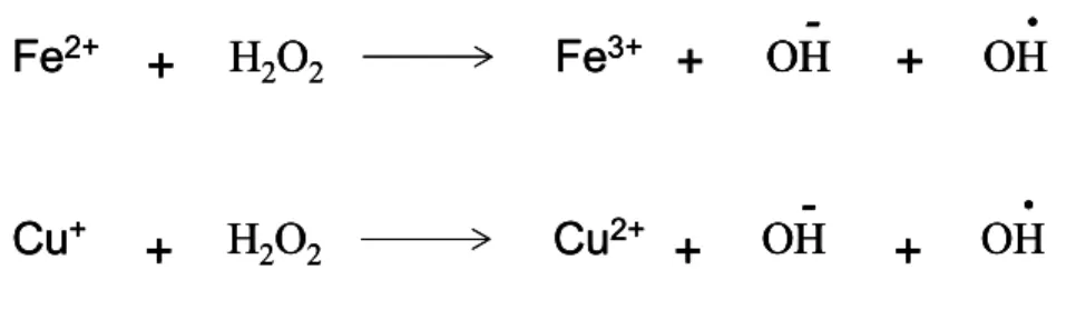
species. Fe2+ and Cu+ are oxidized to ferric iron (Fe+3) and cupric ion (Cu2+), respectively, as shown Figure 1.4.1. OH· can directly attack nucleic acids leading to DNA modifications (Zigler et al., 1985).
Moreover, as in the case of Fenton reaction, if -O
2· and H2O2 are not eliminated by
reduced and they catalyze the formation of OH·. The so called metal catalyzed Haber-Weiss reaction is shown in Figure 1.4.2 (Rigo et al., 1977).
Smooth ER contains detoxifying enzymes involved in detoxification of lipid-soluble drugs and other toxic metabolic products. Oxidization of unsaturated fatty acids and xenobiotics and finally reduction of molecular oxygen (O2) to -O2·
and/or H2O2 are catalyzed by cytochrome P-450 and b5 (Aust et al., 1972;
Capdevila et al., 1981; See Freeman and Crapo, 1982). Growth factor and/or cytokine stimulated oxidant generation may be due to the oxidases localized to the plasma membranes. NADPH oxidase system has been identified in fibroblasts and leukocytes (Meier et al., 1991). NADPH-dependent H2O2 generation in plasma
membranes is the most characterized oxidation and is involved in host defense against invading microorganisms (Meier et al., 1989; Krieger-Brauer and Kather, 1995).
Figure 1.4.1: Fenton Reaction. Metal catalysts iron (upper) or copper (lower) catalyzes the
conversion of H2O2 to OH· if H2O2 is not eliminated by defense mechanisms.
+
H
2O
2Fe
3++
+
Fe
2+OH
-
OH
.
H
2O
2Cu
2+Cu
++
+
OH
-
+
OH
.
+
H
2O
2Fe
3++
+
Fe
2+OH
-
OH
.
+
H
2O
2Fe
3++
+
Fe
2+OH
OH
-
-
OH
.
H
2O
2Cu
2+Cu
++
H
2O
2Cu
2++
OH
-
+
OH
.
Cu
++
+
OH
OH
-
-
+
OH
.
Figure 1.4.2: Haber-Weiss Reaction. Metal ions catalyze the formation of OH· if -O
2· and H2O2are not eliminated.
.
-O
2.
H
2O
2+
OH
+
OH
.
+
O
2.
-O
2.
H
2O
2+
OH
+
OH
.
+
O
2H2O2 generating enzymes in peroxisomes include glycolate oxidase, D-amino acid
oxidase, urate oxidase, L-α-hydroxyacid oxidase, and fatty acyl-CoA oxidase. Peroxisomal reduction reactions are important mainly for liver and kidney cells since toxic molecules such as ethanol are detoxified in the peroxisomes of these organelles. β-oxidation of fatty acids is also carried out in peroxisomes and mitochondria.
As well as membrane-associated oxidases, soluble enzymes such as xanthine oxidase, aldehyde oxidase, dihydroorotate dehydrogenase, flavoprotin dioxygenase are sourses of ROS generation. Moreover, ROS can be produced by autooxidation of small molecules such as dopamine, flavins, epinephrine, and hydroquinones (See Thannickal and Fanburg, 2000).
In addition to all above, ligand-induced ROS production by a variety of cytokines and growth factors in nonphagocytic cells has been illustrated (Thannickal and Fanburg, 2000). Intracellular concentration of H2O2 has been shown to increase
transiently following stimulation of rat vascular smooth muscle cells and primary lung fibroblasts (Sundaresan et al., 1995; Thannickal and Fanburg, 1995). Also, myofibroblast differentiation upon ROS generation due to TGF-β stimulation in primary lung fibroblasts has been demonstrated (Thannickal and Fanburg, 1995; Thannickal et al., 2000).
Moreover, aflatoxinB1 (AFB1) contaminated foods (Jayashree and Subramanyam, 2000), alcohol consumption, viral infections (Perez and Cederbaum, 2003; Rigamonti et al., 2003), and exposure to metal ions such as copper, iron, zinc (discussed in section 1.1), UVR (Butts et al., 2003) can be counted among the extracellular sources of ROS. There is indirect evidence that oxidative stress may play a role in genesis of hepatitis B virus (HBV) induced hepatocellular carcinoma (HCC; Hagen et al., 1994). Exposure to such external factors leads to alterations in DNA.
1.5 Cellular ROS Toxicity
Oxidative stress is associated with several disease conditions such as aging, inflammation, carcinogenesis, ischemia-reperfusion, AIDS, Parkinson’s
Huntington’s, Alzheimer, familial amyotrophic lateral sclerosis, and cataract formation in the eye (Chandra et al., 2000).
Cellular macromolecules such as proteins, DNA, and lipids are targets of free radicals, which are not eliminated by cellular antioxidant defense mechanisms. Free radicals alter cellular functions of the macromolecules permanently, leading to severe consequences that are harmful to the organism (Wells and Winn, 1996). DNA modifications such as altered purine and pyrimidine bases represent the main class of OH· mediated DNA damage that includes oligonucleotide strand breaks, DNA-protein cross-links and abasic sites generated in mammalian chromatin in vitro and in vivo (Dizdaroglu, 1992; Cadet et al., 1999). 8-hydroxy-deoxyguanosine (8-OH-dG) is formed upon oxidation of guanine by OH· consequent to X-ray and gamma irradiation (Kasai et al., 1986). These alterations in DNA lead to mutagenesis (See Ames, 1983) and carcinogenesis (See Floyd, 1990). Formation of 8-OH-dG is involved in aging (Fraga et al., 1990), diabetes mellitus (Dandona et al., 1996), inflammatory diseases (See Ames, 1983), and progression of a severe chronic hepatitis in liver cancer (Sipowicz et al., 1997). This alteration can be repaired specifically with endonuclease DNA glycosylase (Chung et al., 1991).
Polyunsaturated fatty acids are targets for lipid peroxidation. The peroxidation is induced when OH· captures a hydrogen atom from methylene carbon in the polyalkyl chain of fatty acid (R-CH2-) resulting in an alkali radical (R-C·H-). This
radical can react with O2 to create peroxyl radical (ROO·). Membrane proteins can
be altered upon further reaction of peroxyl radicals with other peroxyl radicals. Furthermore, a chain reaction occurs if they capture hydrogen molecule from adjacent fatty acids leading to the formation of more lipid peroxides as shown in Figure 1.5.1 and Figure 1.7.1 (Cheeseman, 1993; Gate et al., 1999). Malondialdehyde (MDA) is a highly reactive product of lipid peroxidation (Draper and Hadley, 1990). It can attack free amino-group of phospholipids, proteins, and lipids and consequently, form inter- and intra-molecular 1-amino-3-iminopropene bridges. By this way, cellular macromolecules are structurally altered (Halliwell and Gutteridge, 1999), leading to autoimmune response (Kergonou et al., 1987). Increased levels of lipid peroxidation have been reported
in diabetes (Sato et al., 1979), apoplexy (Mori et al., 1990), hyperlipemia (Esterbauer et al., 1990), atherosclerosis (Plachta et al., 1992), and liver diseases (Rouach et al., 1997). Besides, during inflammation, enzymes such as lipooxygenases and cyclooxygenases produce specific fatty acyl peroxides (Miller
et al., 1985) and cholesterol and fatty acid moieties of the plasmatic low-density
lipoproteins (LDL). This lipid structures can also be oxidized in the presence of oxidative stress. The oxidation of LDL is observed to be involved in atherosclerosis (Steinberg et al., 1989; Galle et al., 1995).
Like nucleic acids and lipids, proteins are also attacked by free radicals. Oxidative modification of proteins can be toxic to the cells if the ROS generated overrides the physiological levels of ROS required for intracellular ROS mediated signaling.
1O
2 and OH· attacks are common in vivo mechanisms that contribute to damage on
proteins by protein crosslinkings and cleavage of the peptide bonds to macromolecules (Cohen et al., 1998; Prinsze et al., 1990). There are studies demonstrating that via metal-catalyzed oxidative reactions that can occur in metal binding sites of the proteins, histidine can be oxidized to 2-oxohistidine (Lewisch and Levine, 1995). Also hypochlorous acid causes oxidation of thyrosine to 3-chlorotyrosine (Domigan et al., 1995). Modification of proteins by oxidation of cystein residues, formation of intra-molecular disulfide linkages, dithyrosine formation, and metal catalyzed oxidation of the proteins are the mechanisms that result in oxidative modification of proteins (Thannickal and Fanburg, 2000). Disorders associated with protein oxidation reported until now are atherosclerosis, ischemia-reperfusion injury, and aging (See Stadtman, 1992; Berliner and Heinecke, 1996).
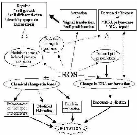
A general overview of ROS toxicity on cellular macromolecules is given in Figure 1.5.1. The figure summarizes the effects of ROS on DNA, lipids, and proteins (Mates and Sanchez-Jimenez, 1999).
Figure 1.5.1: ROS Toxicity (Mates and Sanchez-Jimenez, 1999).
1.6 Signaling Molecules Targeted by ROS
Despite the mutagenic and carcinogenic effects of ROS in case of excess, there is a huge number of signal transduction pathways regulated by ROS, but the specific molecules targeted by ROS are not yet very apparent (Thannickal and Fanburg, 2000).
Receptor kinases and phosphatases may be targets for oxidative stress. Ligand independent activation of growth factor receptors in response to UV has been documented and related to ROS. Another study recently demonstrated that exposure of human keratinocytes to 1O2 resulted in rapid loss of EGF receptor,
which indicates that oxidative stress produced by 1O
2 rapidly disrupts EGF
phosphoinositide 3-kinase (PI3K) and Akt (also known as protein kinase B) activation has been reported to be achieved through PI3K membrane recruitment to its substrate site, thereby enabling PI3K to maximize its catalytic efficiency (Qin and Chock, 2003). Another recent work illustrates that Akt activation by H2O2 is dependent on the activation of EGF receptor signaling (Wang et al.,
2000).
Due to the fact that activation of mitogen activated protein kinase (MAPK) pathways depends on mitogen and stress activated signals, redox regulation of these pathways is not surprising. There are studies showing exogeneous ROS-mediated ERK activation to be an upstream event at the level of growth factor receptors and Src kinases, there are some others suggesting oxidant induced inactivation of protein tyrosine phosphatases (PTPs) or protein phosphatase A. There are limited studies showing the endogenous ROS dependent activation of ERK MAPK pathway (Thannickal and Fanburg, 2000). Many studies demonstrate c-Jun N-terminal kinases (JNK) activation to be linked to cell death or apoptosis, while others suggest pro-survival function for JNK upon oxidative stress (Martindale and Holbrook, 2002). Pro-survival and apoptotic functions of protein kinase C (PKC) have been documented (Martindale and Holbrook, 2002).
P53 activation through environmental stress depends largely on the posttranslational mechanisms that enhance its stability and increase DNA binding activity. Oxidative stress dependent direct DNA damage activates p53. NFκB dependent upregulation of p53 upon H2O2 exposure has been documented.
Furthermore, p53 protein has been illustrated to be phosphorylated by both JNK and p38 and stabilized under oxidative stress conditions (Fuchs et al., 1998; Bulavin et al., 1999). P53 also represses the expression of anti-apoptotic factor, Bcl-2, and enhances the expression of proapoptotic factor, Bax and antioxidant enzyme Gpx upon oxidative stress (Martindale and Holdbook, 2002).
NFκB (Nuclear factor kappa B) is an oxidant responsive transcription factor that is involved in the expression of genes that have roles in immune and inflammatory responses. It is suggested that redox-regulated effect occurs downstream from inhibitor of kappaB (IκB) kinases at the level of degradation of IκB. Also AP-1 has also been considered as an oxidant responsive transcriptional complex. Both
exogenous ROS and ligand induced ROS have been implicated in AP-1 activation (Thannickal and Fanburg, 2000).
Non-receptor tyrosine kinases such as Src kinases and Janus kinases (JAK) have been reported to be activated by oxidative stress. As in the case of receptor tyrosine kinases, mostly the studies are performed by exogenously added oxidants (Thannickal and Fanburg, 2000).
The molecules targeted by ROS are summarized in Figure 1.6.1.
1.7 Antioxidants against Oxidative Stress
ROS are generated in a stepwise manner by the reduction of one electron of oxygen each time. Oxidative stress results following disturbance of the balance between intracellular pro-oxidant to anti-oxidant status of the cell consequent to the inefficiency of cellular anti-oxidant defense mechanisms to cope with ROS (Dizdaroğlu, 1992).
Antioxidants are directly or indirectly involved in maintaining intracellular balance between pro-oxidant and anti-oxidant levels. These antioxidants that favor the anti-oxidant levels in the cell are biologically important molecules. They include vitamin C (ascorbic acid), vitamin E (α-tocopherol), vitamin A, β-carotene, metallothionein, polyamines, melatonin, NADPH, adenosine, coenzyme Q-10, urate, ubiquinol, polyphenols, flavonoids, phytoestrogens, cystein, homocysteine, taurine, methionine, S-adenosyl-L-methionine, resveratrol, nitroxides, thioreductase, nitric oxide synthase, heme oxygenase-1, Gpxs, superoxide dismutases (SODs), catalase (CAT) (Matés, 2000), and GST (Arthur, 2000). The detailed explanation for Gpxs was given in 1.3.2. SOD, CAT and GST will be discussed in section 1.7.1 and the dietary defense mechanisms including Vitamin E, selenium, and Copper/Zinc in section 1.7.2.
1.7.1 Internal Defense Mechanisms
1.7.1.1 Superoxide Dismutases
The -O2· radical generated during cellular respiration is dismutated rapidly with
SOD enzymes generating H2O2 as shown in Figure 1.7.1. Four classes of SOD,
namely, manganese SOD (Mn-SOD), Cu/Zn SOD, extracellular SOD (EC-SOD), and nickel SOD (Ni-SOD) have been identified so far (McCord and Fridovich, 1969; Weisiger and Fridovich, 1973; Yost and Fridovich, 1976; Youn et al., 1996). The function of SOD is to convert -O2· to H2O2 by catalyzing one-electron
redox cycle of superoxide. Mn-SOD is localized in mitochondrial matrix while Cu/Zn-SOD is located into intracellular cytoplasmic compartments and also associated with mitochondrial and peroxisomal membranes (Kira et al., 2002; Zelko et al., 2002). Mn-SOD is a nuclear-encoded main antioxidant enzyme that
removes -O2· produced in mitochondria during respiration (Guan et al., 1998) and
has been reported to be a tumor suppressor gene (Mates, 2000). Studies with SOD knockout mice revealed that Mn-SOD was essential for life while copper/zinc SOD was not (Reaume et al., 1996). EC-SOD is a secretory Cu/Zn containing SOD that is the primary antioxidant in blood vessel interstitium and the only extracellular enzyme that reduces -O2· ( See Sentman et al., 1999; Enghild et al.,
1999).
1.7.1.2 Catalase
Catalase is a ubiquitous anti-oxidant enzyme that is predominantly localized to peroxisomes of the cells and decomposes H2O2 to H2O and O2 as shown in Figure
1.7.1. CAT confers protection against severe oxidative stress with a high affinity to H2O2. Due to its efficiency towards H2O2, it cannot be saturated by at any
concentration of H2O2 (Kinnula et al., 1992; Yan and Harding, 1997; Mates,
2000). The cells are protected against H2O2 generated within them by this enzyme.
1.7.1.3 Gulutathione S Transferases
GSTs play an important role in the detoxification and elimination of xenobiotics. It has been demonstrated that these enzymes have an additional ability to reduce lipid hydroperoxides (ROOH) by independent means via their selenium-independent Gpx activity using GSH as a hydrogen donor as shown in Figure 1.7.1. It has been shown that GST contributes to selenium-independent Gpx activity in the liver of selenium-deficient rats (Lawrence et al., 1978). In addition, dietary selenium-deficiency produced increased activity of GST suggesting that GST activity is regulated by dietary selenium (Masukawa et al., 1984). Moreover, increased resistance to oxidative stress has been documented in cells transfected with (Glutathione S transferase alpha) GSTα, which is among one of the five GST gene families (Zimniak et al., 1997). Previously it has been reported that patients with primary biliary cirrhosis, primary sclerosing cholongitis, and chronic hepatitis B had significantly elevated levels of GST-alpha (Mulder et al., 1997).
1.7.2 Dietary Defense Mechanisms
1.7.2.1 Vitamin E Supplementation
The importance of Vitamin E for human health has been described in section 1.1. Vitamin E supplementation has been suggested a valuable therapy in case of its deficiency. Vitamin E supplementation could prevent free radical induced damages to the tissues in the patients due to the radical scavenger role. Vitamin E
Figure 1.7.1: Internal Defense Mechanisms against ROS. SOD is involved in the elimination of -O 2·.
Gpx and CAT are involved in the elimination of H2O2, and Gpx and GST are involved in the
elimination of lipid peroxides (ROOH) avoiding a chain reaction (Halliwell and Gutteridge, 1999). Vitamin E prevents lipid peroxidation by scavenging OH· (Claycombe and Meydani, 2001).
VitE
H
2O
2 OHO
2 Respiration (Mitochondria) 2GSH GSSG NADPH NADP+Gpx
H
2O
SOD Glu-Reductase O2_.
.
R-CH
2-
R-CH-H2O.
O
2ROO
.
R-CH-ROOH
R-C-
.
GSHRSG
2GSH GSSGGpx
GST
ROH
H2O H2OCA
T
VitE
VitE
H
2O
2H
2O
2 OHO
2 Respiration (Mitochondria) 2GSH GSSG NADPH NADP+Gpx
H
2O
SOD Glu-Reductase O2_ O2_.
.
R-CH
2-
R-CH-H2O.
O
2ROO
.
R-CH-ROOH
R-C-
.
GSHRSG
2GSH GSSGGpx
GST
ROH
H2O H2OCA
T
supplementation to healthy elderly people has been illustrated to enhance their immune responses (Meydani et al., 1997; Meydani et al., 1998). Vitamin E supplementation has been documented to delay early progression of arteriosclerosis in heart transplant patients (Liu and Meydani, 2002). Pathogenesis of rheumatoid arthritis and systemic lupus erythematosus has been found to be associated with low serum Vitamin E concentrations (Comstock et al., 1997). Vitamin E supplemented type I diabetic children have been documented to restore GSH and MDA to normal levels (Jain et al., 2000). Vitamin E protects humans against atherosclerosis, ischemic heart disease, and development of some type of cancers such as breast, prostate, and leukemia (Ricciarelli et al., 2001). The lowered vitamin E levels in patients with viral hepatitis have been documented (von Herbay et al., 1996; von Herbay et al., 1997). High-dose supplementation of Vitamin E has been used as a safety therapy. Vitamin E is absorbed in gastrointestinal tract similar to other lipids. The primary reported side effects of high dose of Vitamin E supplementation includes breast soreness, thrombophlebitis, gastrointestinal disturbances, and depression of Vitamin K dependent coagulation factors when used with other anticoagulation agents (Bulger and Maier, 2003).
1.7.2.2 Selenium Supplementation
The importance of selenium for human health and supplementation has been described in section 1.2. In addition to those, more clinical examples related to selenium-deficiency will be provided in this section. Lowered selenium status has been observed in asthma patients and it is suggested that selenium supplementation might be beneficial to such patients, which may be at risk of selenium-deficiency (Kadrabova et al., 1996). Moreover, in patients with viral infections, selenium-deficiency is associated with skeletal muscle disorders manifested by muscle pain, fatigue, and proximal weakness (Chariot and Bignani, 2003). Severe selenium-deficiency has been reported to result in a high incidence of thyroiditis due to a decreased activity of selenium-dependent Gpx activity within thyroid cells. In those cases, selenium substitution may improve the inflammatory activity in patients with autoimmune thyroiditis, especially in those with high activity (Gartner et al., 2002). Decreased plasma selenium and Gpx
activity has been reported in patients with systemic inflammatory response syndrome and sepsis. Selenium supplementation seems to improve the outcome of those patients, but the results should be improved (Berger et al., 2001). Intensive oxidative stress is observed in the blood of patients with multiple sclerosis. Supplementation of patients with selenium improves the health of patients (Syburra and Passi, 1999).Moreover, selenium supplementation has been reported to have immunoenhancing effects in humans by stimulating cytotoxic lymphocytes and natural killer cells (Kiremidjian-Schumacher et al., 1994). In addition to these, protective role of selenium supplementation against hepatitis B and C virus (HBV, HCV) infection, progression, and primary liver cancer has been reported (Yu et al., 1997; Yu et al., 1999). In contrast to its advantages following supplementation, high doses of selenium has been reported to cause gastrointestinal tract disorders, weakness, neurological manifestations, hypochromic anemia, and leucopenia (Tinggi, 2003).
1.7.2.3 Copper and Zinc Supplementation
Safe and adequate daily intake of dietary copper has been reported to be 1.5 to 3.1 mg for adults. Individuals with copper-deficiency show hypercholesterolemia, abnormal electrocardiograms, and hypertension. The copper is required by obese women during weight loss (Klevay, 1998). Dietary copper-deficiency contributes to high blood pressure, enhancement of inflammation, anemia, reduced blood clotting and arteriosclerosis and consequently may impair cardiovascular health (Saari and Schuschke, 1999). Documentations reveal that copper-deficiency occurs secondary to gastric resection, unsupplemented total parenteral nutrition, high levels of zinc intake, or general malnutrition. Although copper has significant roles in the organism and in the immune system, the molecular mechanism for copper-deficient neutropenia is not known (Percival, 1995). Other groups demonstrated that early clinical sign of copper-deficiency is a reduction in the number of circulating neutrophils. Moreover, the growth of copper-deficient infants recovering from malnutrition is improved by copper-supplementation (Castillo-Duran and Uauy, 1988). Copper-deficiency can cause an X-linked Menkes syndrome, which is related to the deficiency of copper-dependent enzymes (Camakaris et al., 1999). In contrast to these, elevated concentrations of
copper are harmful for health. Copper-toxicity results in liver cirrhosis, damage to renal tubes, and the brain. Chronic copper-toxicity was observed in patients receiving dialysis via copper tubing. Copper-contaminated foods and water lead to gastrointestinal symptoms (Gaetke and Chow, 2003). Moreover, free copper ions take part in the formation of ROS as explained in section 1.4.
Zinc-deficiency leads to anorexia (loss of epitetite), hypogeusia (loss of taste), poor growth, alopecia, and delayed sexual maturation. Zinc supplementation has been reported to improve gastrointestinal tract diseases. A recent study demonstrated that zinc-supplementation in children with acute diarrhea had a reduction of continuing diarrhea. Zinc is essential for growth and development, DNA synthesis, neurosensory functions, and cell-mediated immunity (Duggan et
al., 2002). Another recent work showed that zinc treatment of children with
growth dysfunction appeared to ameliorate the Cu/Zn-SOD activity which was increased significantly during growth retardation (Kocaturk et al., 2002). Zinc-deficiency is a common nutritional disorder in the elderly. In those people, copper, iron, and lipoprotein status should be monitored if long-term zinc supplementation is required, since they may be affected by the zinc supplementation (Stiles and Boosalis, 1995). Zinc intake is reduced in elderly people, however, its deficiency and effects on cell-mediated immunity of the elderly have not been well documented. Zinc supplementation has been shown to overcome zinc-deficiency and normalize plasma copper levels. Moreover, serum thymulin activity, Interleukine-1 (IL-1) production significantly increases after supplementation. Improvement in response to skin-test antigens and taste acuity was observed after zinc supplementation (Prasad et al., 1993).
1.8 Oxidative Stress and Cancer
A crucial step in carcinogenesis is DNA mutation which is one of the outcomes of oxidative base lesions. Elevated levels of oxidative DNA lesions have been noted in many tumors, suggesting oxidative stress induced damage in the etiology of cancer. Oxidative mechanisms have been demonstrated to possess a potential role in the initiation, promotion, and malignant conversion (progression) stages of carcinogenesis. Given that cancer risk increases with age and is associated with an
accumulation of DNA damage, oxidative DNA damage has been investigated in cancer.
Lesions such as 8-OH-dG are established biomarkers of oxidative stress. The potential mutagenic capacity of such lesions in mammalian cells suggested them as intermediate markers of cancer. G:C → T:A transversions that are potentially derived from 8-OH-dG have been observed in vivo in the ras oncogene and the p53 tumor suppressor gene in lung and liver cancer. Also C:C → T:T substitutions in the absence of UV in tumors have been identified as signature mutations for ROS. These findings support the proposal of implication of oxidative damage in carcinogenesis (Cooke et al., 2003).
Due to elevated ROS levels, transcription factors and their corresponding genes are permanently activated, which, coupled with increased DNA damage, creates a selection survival advantage for a malignant phenotype seen in cancer (Toyokuni,
et al., 1995). It is important to note that the nuclei of undifferentiated proliferating
stem cells must be affected by oxidative stress for the initiation and progression of cancer. Current analytical procedures will not reflect lesion levels in the most important target cells due to the fact that tissue samples from tumors and normal cells will represent a heterogeneous mixture of differentiated and undifferentiated cells with the former likely to predominate (Cooke et al., 2003).
The implication of ROS in tumor formation has been mostly studied indirectly. For example, chemical promoters can generate oxidative stress and antioxidants can inhibit promotion; therefore, ROS are involved in promotion. It is possible that the antioxidants themselves may allow clonal expansion and tumor promotion by protecting initiated cells from excessive oxidant toxicity and apoptosis that would otherwise kill them (Cooke et al., 2003). Finally, in linking oxidative stress with cancer promotion, it should be kept in mind that biomolecules other than DNA may be oxidatively modified and that these may have a significant effect on the carcinogenesis as mentioned in section 1.5 and 1.6.
1.9 Selenium and Cancer
There is growing evidence supporting the association between low dietary intakes of selenium with an increased risk of certain cancers. Moreover, protective effect of selenium supplementation has been observed on the overall incidence of prostate cancer (Yoshizawa et al., 1998; Duffield-Lillico et al., 2003). The strong association between low serum selenium levels and cancer was evident for gastrointestinal and prostatic cancers (Willett et al., 1983). The association between the serum selenium level and the subsequent incidence of cancer was investigated in a group of Finnish people. Low serum selenium levels were associated with an increased risk for cancers, especially stomach and lung, among men (Knekt et al., 1990). Also another study demonstrated that very low selenium status may contribute to the risk of lung cancer (Knekt et al., 1998).Populations with low selenium intake have been documented to have low serum selenium levels and are at a high risk for squamous esophageal and gastric cancers (Mark et
al., 2000). Serum selenium concentration and the Gpx activity were positively
correlated with the incidence of liver cancer (Corrocher et al., 1986). The inverse association between plasma selenium levels and HCC has been shown to appear in a group of patients especially among cigarette smokers (Yu et al., 1999). Another research also points out the protective role of selenium in the patients with liver cirrhosis and hepatocellular carcinoma, and the potential need of selenium supplementation in these patients (Buljevac et al., 1996).
1.10 Signaling Molecules Targeted by Selenium Supplementation
In addition to the importance of selenium in certain diseases, the essentiality of selenium in the clonal growth of human fibroblasts (McKeehan et al., 1976), human hepatoma cells lines with differentiated functions (Nakabayashi et al., 1982), and mammary tumor cell lines (Barnes et al., 1979) has been clearly demonstrated.
Until recently, the signaling targets that are regulated by selenium availability were largely unknown. There is accumulating data related to the target proteins of selenium, which take part in important signaling pathways.
It has been recently documented that selenite suppresses both the JNK and stress-activated protein kinase (SAPK) as well as the p38 mitogen-stress-activated protein kinase pathway in human embryonic T cells, 293T (Park et al., 2000a). Furthermore, selenium also inhibited caspase 3 activity in embryonic kidney cells (Park et al., 2000b) and activated MAPK in human hepatocytes and adipocytes acting as an insulin-mimetic agent (Stapleton et al., 1997). These proteins are regulated through the redox regulation of their active site cysteine residues. A very recent paper described the activation of PI3K/Akt kinase pathway and inactivation of caspase 3 and apoptosis signal regulating kinase 1 (ASK1) in selenium supplemented Huh-7 cells, which blocked apoptosis and promoted cell survival. The activation of focal adhesion kinase (FAK), and Rac1, upstream inducers of PI3K, have been suggested to have a role in the activation of PI3K pathway in this study (Lee et al., 2003).
1.11 Selenium-Deficiency and Liver Diseases
Selenium has been reported to be protective against dietary necrotic liver degeneration in rats long ago (Schwarz and Foltz, 1999). Since then, the reports pointing out the importance of selenium in prevention of hepatic disease conditions with different etiologies have been accumulating.
1.11.1 Selenium-Deficiency, Keshan Disease and Liver Disorders
Animal and human studies have revealed the existence of an association between certain liver diseases and selenium-deficiency. In addition, it is well documented that in Asia and Africa, dietary selenium-deficiency is associated with a cardiomyopathy known as Keshan disease. Adults dying of Keshan disease had diagnostic lesions not only in the cardiovascular system and skeletal muscle but also in the liver. Varying degrees of focal biliary cirrhosis were identified in 50%
of the Keshan disease autopsies, and 5% developed severe lobular cirrhosis (Wallach et al., 1990).
In animal studies, pigs that suffer from Hepatosis Dietetica have been documented to have low hepatic selenium concentrations (Moir and Masters, 1979). In addition, pigs with mulberry heart disease showed low hepatic selenium concentration. It is suggested that the low selenium status together with vitamin E-deficiency increases oxidative stress and thus contributes to the development of oxidative damage in pigs (Korpela, 1990). Besides, in selenium and vitamin E-deficient pigs with microangiopathy, hepatic iron concentration has been significantly increased. Increase in iron concentration might have promoted oxidative stress and thus contributed to the development of oxidative damage (Korpela, 1990).
The importance of selenium in antioxidant defense has been shown via in vivo experiments on rats that have been put on a selenium-deficient-diet. Rats under oxidative stress due to selenium-deficiency display hepatic necrosis owing to selenium-deficiency associated with the loss of Gpx activity while selenium supplementation prevents this disease condition (Schwarz and Foltz, 1999).
Chronic selenium-deficiency may also occur in individuals with malabsorption and long term selenium-deficient parenteral nutrition. Selenium-deficiency causes myopathy as a result of the depletion of selenium-associated enzymes which protect cell membranes from damage by free radicals (See Diplock, 1994; See Rayman 2000).
1.11.2 Selenium-Deficiency and Alcoholism
One of the etiologies of HCC development is alcohol abuse. Alcohol is a source of oxidative stress. In alcohol-related liver damage, there is consistent evidence of enhanced production of free radicals and significant decrease of antioxidant defense. Alcohol-exposed cells are selectively depleted of GSH in mitochondria due to a defective functioning of the carrier responsible for the transport of GSH from cytosol into the mitochondrial matrix. This impaired transport sensitizes