Review Article
Does sleep disturbance affect the amyloid clearance
mechanisms in Alzheimer
’s disease?
Burak Yulug,
MD,
1* Lutfu Hanoglu,
MD1and Ertugrul Kilic,
PhD21
Department of Neurology,2Restorative and Regenerative Medicine Center, Istanbul Medipol University, Istanbul, Turkey
Sleep is an important factor that plays a key role in Alzheimer’s disease pathogenesis. However, it is still unclear whether poor-quality sleep may overlap with sleep disturbances in the underlying dysfunc-tional mechanisms of amyloid beta (Aβ) clearance metabolism. Here, we aimed to evaluate the current evidence on the role of sleep deprivation in Aβ
clearance metabolism. To that end, we discuss possi-ble mechanisms underlying the bidirectional interac-tion between the sleep deprivainterac-tion and Aβ clearance pathways.
Key words: Alzheimer’s disease, amyloid beta clear-ance, glymphatic system, sleep, sleep deprivation.
A
LZHEIMER’S DISEASE (AD) IS a progressive, neurodegenerative disorder that gradually dis-rupts neural circuits that are responsible for underly-ing neurocognitive symptoms. There is rapidly replicating evidence indicating that the key factor in AD pathogenesis is the aggregation of amyloid beta (Aβ) protein within critical regions of the brain. The principal underlying mechanism for the aggregation is the imbalance between clearance and production of Aβ that results in synaptic and neuronal loss due to the accumulation of toxic amyloid aggregates.1,2 It has been shown previously that individuals with AD may present with sleep and circadian rhythm disturbances. However, it is still unclear whether there is a dual interaction implying that the sleep– wake cycle itself may directly play a key role in the pathogenesis of AD.EVIDENCE FROM HUMAN STUDIES
Sleep is an important factor that plays a key role in AD pathogenesis. Apart from studies showing that individuals with AD may present with sleep and
circadian rhythm disturbances,1,3 it is still unclear whether poor-quality sleep may overlap with sleep and circadian rhythm disturbances in the underlying dysfunctional mechanisms of Aβ metabolism. There are various theories hypothesizing that sleep distur-bances might worsen the AD pathology through increased wakefulness and related excitotoxicity.4,5 Moreover, recent studies indicate that altered func-tion of brainstem neurotransmitter pathways sec-ondary to sleep impairment might lead to the impairment of the ‘default mode’ brain network, which is a key pathophysiological mechanism in AD.6,7 In recent years, human Aβ metabolism has received considerable critical attention in regards to clarifying the pathophysiological relation between sleep and AD pathogenesis. Furthermore, under-standing human Aβ physiology may also help us to establish the utility of cerebrospinal fluid (CSF) Aβ42 levels as a surrogate marker for amyloid pla-que deposition. In this context, Bateman et al. have already shown that CSF and blood Aβ metabolism are regulated in a circadian manner in healthy adults.8 These results were further confirmed by the following study indicating that age-related impair-ment in physiological CSF dynamics was strongly correlated with the amyloidosis.9
There are rapidly increasing data showing deleteri-ous effects of sleep disruption on cognition and amyloid burden in normative populations.2,3,10–14
*Correspondence: Burak Yulug, MD, Department of Neurology, Istanbul Medipol University, Regenerative and Restorative Medical Research Center, Ekinciler cad. 19, TR-34810 Istanbul, Turkey. Email: [email protected]
Received 20 April 2017; revised 28 April 2017; accepted 15 May 2017.
Although it is hard to validate the causal relation-ship between disturbed sleep and the pathogenesis of AD in humans, short-term prospective studies have already provided fruitful data revealing that sleep disruption increases the risk of incident dementia.2,3,10,11 In their prospective cohort study of older adults without dementia, Lim et al. revealed that sleep fragmentation was correlated with increased risk of AD and cognitive decline, which was abolished with improved sleep consolida-tion.10,11Subsequent longitudinal studies have indi-cated that insomnia, prolonged sleep duration, and excessive daytime sleepiness are also associated with risk of developing cognitive decline.15–17 These clinical study results were confirmed by a recent long-term follow-up study showing that there is a significant relation between insomnia and AD in cognitively normal adults.18In evaluating the under-lying pathophysiological mechanisms, Ju et al. estab-lished that poor sleep quality increased the brain amyloid aggregation in cognitively normal adults.14 These results were in line with the following study of 26 cognitively healthy adults showing that short-term sleep deprivation significantly elevated the CSF amyloid-β levels, which were reversed with a night of good sleep.12 Consistent with thisfinding, recent amyloid–positron emission tomography studies of cognitively normal populations indicated that there is a strong association between longer sleep latency, self-reported sleep quality, and brain Aβ burden19,20 that was independent of apolipoprotein E (ApoE) ε4 genotype.20
EVIDENCE FROM ANIMAL STUDIES
There are strong preclinical data indicating that sleep–wake dysregulation leads to a slight increase in extracellular Aβ in Tg2576 mice during wakefulness, which was persistently increased after sleep depriva-tion. In their interesting study, Kang et al. revealed that prolonged sleep deprivation was associated with significant increase in Aβ levels.1 These results were also confirmed with an amyloid precursor protein (APP)/PS1 mutant line, indicating that the duration of sleep deprivation may play a critical role in deter-mining the amyloid plaque load within the brain.1 These data suggest that pharmacological interven-tions aimed to increase the total sleep time may help to reduce the AD pathology. In this context, inhibit-ing the orexin receptors through almorexant resulted in significant increases in total sleep time, which was
associated with reduced Aβ plaque load in mice.1 Following transgenic mice, studies focusing on the role of amyloid plaques and tau tangles have also revealed interesting results. Di Meco et al. showed that impaired circadian response to light showed sig-nificantly increased insoluble tau that was associated with synaptic and cognitive impairment compared with normal circadian controls.21 Although the role of sleep deprivation was not directly examined in this study, these data are important in suggesting the role of impaired circadian rhythm in the pathophysi-ology of AD. From another point of view, recent transgenic mice studies have revealed that locus coer-uleus (LC) degeneration and related loss of cortical noradrenergic innervation resulted in increased neu-roinflammatory response that was correlated with increased amyloid plaques and memory deficits.22,23 As LC is a well-known region affecting the sleep– wake cycle,22,23 these preclinical data suggest that sleep disorders might contribute to amyloid pathol-ogy. Despite these promising preclinical data, it should also be noted that the results of transgenic animal species should be interpreted cautiously because most transgenic mice models are using the human-derived APP instead of a murine APP.
A POSSIBLE ROLE OF TRANSPORT
MECHANISMS?
Studies have already indicated that a significant por-tion of Aβ clearance has been attributed to the blood–brain barrier (BBB) transport of Aβ, which is mediated by multi-drug resistant proteins (i.e., ATP-binding cassette transporters, such as p-glycoprotein, and low-density lipoprotein receptor family mem-bers) in an ApoE-dependent and -independent man-ner.24,25 Beyond its critical role in BBB, ApoE also has a special role in the elimination of Aβ through its interaction with astrocytes, which play a critical role in sleep regulation. Recent studies have indicated that sleep disturbances might affect astrocyte–ApoE Aβ clearance pathways in the manner of a feed-forward mechanism that results in increased Aβ accumula-tion.2,3This is in agreement with recent data showing that sleep deprivation suppresses the glymphatic dis-tribution of CSF-derived ApoE into the brain and its elimination.26Several reports indicate that sleep dep-rivation also leads to systemic inflammation and sec-ondary release of inflammatory mediators, which promote significant alterations in the molecular com-ponents of the BBB.27 However, there is no evidence
in the literature showing that inflammatory mole-cules exert a local effect on specific BBB transport mechanisms that would increase the risk of develop-ing neurodegenerative diseases. Although there is rapidly replicating evidence indicating the role of p-glycoprotein to Aβ clearance across the BBB,28there is only one molecular positron emission tomography study in the literature that has indicated that there is no association between sleep disturbance and p-glycoprotein expression in rodents.29 Considering all of this evidence, special interest has been given to molecular transport mechanisms by which sleep dep-rivation might affect the clearance of Aβ through the interstitial fluid (ISF) bulk-flow-dependent process. Compared to peripheral tissue where lymph vessels drain the excess interstitial products to the general circulation,30 the brain, which has a high metabolic rate and related toxic byproducts, does not have a conventional lymphatic system. Instead of this, there is an interesting glymphatic clearance mechanism of the brain that enables the removal of interstitial toxic proteins, such as Aβ, through a dynamic interaction between the CSF and ISF, which is located around the cerebral vasculature.31–33 This ISF bulk- flow-dependent process drains Aβ containing ISF from the interstitium via ISF bulkflow into the CSF and peri-vascular space through the periperi-vascular and glympha-tic pathways.24,34,35 According to this hypothesis, toxic chemicals are cleared from the ISF by a complex process that requires astrocytic water-permeant chan-nel aquaporin-4 (Aqp4) during sleep. Aqp4 is a well-known water transporter that is majorly expressed on the astrocytic endfeet.36 Studies in the last decade have already shown the pivotal role of astrocytes in ‘cleaning the brain’ through the glymphatic pathway and indicate the specific modulatory role of astrocytic Aqp4 expression during the sleep.36Moreover, there have been interesting studies during recent years eval-uating the distribution and expression of Aqp4 in preclinical and clinical AD studies.37 In this context, experimental studies have already shown the role of Aqp4-dependent trans-astrocytic ISF bulkflow in the clearance of Aβ through the glymphatic sys-tem.24,37,38These results were confirmed with subse-quent studies using Aqp4-knockout mice showing reduced A 40 clearance that resulted in Aβ deposition in the cortex and hippocampus.32Although there are no actual data indicating the modulatory role of sleep–wake cycles on the Aqp4 expression, awakened mice showed much-decreased clearance of exoge-nously applied Aβ from the interstitial space that was
significantly reversed during sleep.31 Subsequent studies have confirmed the key role of the glymphatic system in Aβ clearance and have revealed that the degradation of Aβ is minimal during these transport clearance measurements.31,34,39 Additional evidence for the transport failure during sleep–wake impair-ment was provided from Xie et al., who revealed that the sleep–wake state rather than the circadian rhythm enhanced the glymphatic-system-dependent clear-ance of the exogenously applied Aβ.31 In evaluating the underlying mechanisms, they underlined the role of adrenergic signaling during wakefulness, which was responsible for increased tissue resistance to the interstitialfluid flux that resulted in decreased size of the interstitial space that was reversed with adrenergic receptor blockage. Taken together, all of these studies suggest that the astrocytic, sleep-dependent contribu-tion to the glymphatic pathway is a locus of clearance impairment in AD. This also supports that the impairment of the efflux of Ab1-42 from the glym-phatic pathway to the circulation is a critical step.
CONCLUSION
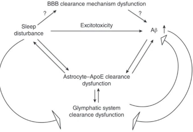
Because Aβ accumulation in the affected regions of the AD brain begins years before any signs of the disease are prominent, a therapeutic approach effec-tively enhancing clearance mechanisms would exert huge benefits in the progress of this disease. Among the many transport mechanisms that have been mentioned above, the most specific transport mech-anism that is tightly linked to sleep disturbance seems to be the glymphatic system (Fig. 1). This
BBB clearance mechanism dysfunction
Astrocyte–ApoE clearance dysfunction Glymphatic system clearance dysfunction Sleep disturbance ? ? Aβ Excitotoxicity
Figure 1. Possible role of sleep deprivation on amyloid-beta (Aβ) clearance mechanisms in the brain. ApoE, apolipopro-tein E; BBB, blood–brain barrier.
system is characterized by astrocytic Aqp4, which plays an important role in the elimination of Aβ. Future studies evaluating the effects of sleep depriva-tion on Aqp4 expression would be the logical future steps to be taken in thefield of AD research.
DISCLOSURE STATEMENT
There is no conflict of interest in this study.
AUTHOR CONTRIBUTIONS
B.Y.: Conception and design of the study, acquisition and analysis of data. L.H.: Drafting the manuscript orfigures. E.K.: Acquisition and analysis of data.
REFERENCES
1. Kang JE, Lim MM, Bateman RJ et al. Amyloid-beta dynamics are regulated by orexin and the sleep–wake cycle. Science 2009; 326: 1005–1007.
2. Gerstner JR, Perron IJ, Pack AI. The nexus of Aβ, aging, and sleep. Sci. Transl. Med. 2012; 4: 150fs134.
3. Lim MM, Gerstner JR, Holtzman DM. The sleep-wake cycle and Alzheimer’s disease: What do we know? Neuro-degener. Dis. Manag. 2014; 4: 351–362.
4. Bero AW, Yan P, Roh JH et al. Neuronal activity regulates the regional vulnerability to amyloid-β deposition. Nat. Neurosci. 2011; 14: 750–756.
5. Cirrito JR, Yamada KA, Finn MB et al. Synaptic activity regulates interstitial fluid amyloid-beta levels in vivo. Neuron 2005; 48: 913–922.
6. Bero AW, Bauer AQ, Stewart FR et al. Bidirectional rela-tionship between functional connectivity and amyloid-β deposition in mouse brain. J. Neurosci. 2012; 32: 4334–4340.
7. Clark CN, Warren JD. A hypnic hypothesis of Alzheimer’s disease. Neurodegener. Dis. 2013; 12: 165–176.
8. Bateman RJ, Wen G, Morris JC, Holtzman DM. Fluctua-tions of CSF amyloid-beta levels: ImplicaFluctua-tions for a diag-nostic and therapeutic biomarker. Neurology 2007; 68: 666–669.
9. Dobrowolska JA, Kasten T, Huang Y et al. Diurnal pat-terns of soluble amyloid precursor protein metabolites in the human central nervous system. PLoS ONE 2014; 9: e89998.
10. Lim AS, Kowgier M, Yu L, Buchman AS, Bennett DA. Sleep fragmentation and the risk of incident Alzheimer’s disease and cognitive decline in older persons. Sleep 2013; 36: 1027–1032.
11. Lim AS, Yu L, Kowgier M, Schneider JA, Buchman AS, Bennett DA. Modification of the relationship of the apoli-poprotein Eε4 allele to the risk of Alzheimer disease and
neurofibrillary tangle density by sleep. JAMA Neurol. 2013; 70: 1544–1551.
12. Ooms S, Overeem S, Besse K, Rikkert MO, Verbeek M, Claassen JA. Effect of 1 night of total sleep deprivation on cerebrospinalfluid β-amyloid 42 in healthy middle-aged men: A randomized clinical trial. JAMA Neurol. 2014; 71: 971–977.
13. Basner M, Rao H, Goel N, Dinges DF. Sleep deprivation and neurobehavioral dynamics. Curr. Opin. Neurobiol. 2013; 23: 854–863.
14. Ju YE, Mcleland JS, Toedebusch CD et al. Sleep quality and preclinical Alzheimer disease. JAMA Neurol. 2013; 70: 587–593.
15. Lobo A, Lopez-Anton R, de la Camara C et al. Non-cognitive psychopathological symptoms associated with incident mild cognitive impairment and dementia, Alz-heimer’s type. Neurotox Res. 2008; 14: 263–272.
16. Cricco M, Simonsick EM, Foley DJ. The impact of insom-nia on cognitive functioning in older adults. J. Am. Ger-iatr. Soc. 2001; 49: 1185–1189.
17. Benito-Leon J, Bermejo-Pareja F, Vega S et al. Total daily sleep duration and the risk of dementia: A pro-spective population-based study. Eur. J. Neurol. 2009; 16: 990–997.
18. Osorio RS, Pirraglia E, Agüera-Ortiz LF et al. Greater risk of Alzheimer’s disease in older adults with insomnia. J. Am. Geriatr. Soc. 2011; 59: 559–562.
19. Sprecher KE, Bendlin BB, Racine AM et al. Amyloid bur-den is associated with self-reported sleep in nondemen-ted late middle-aged adults. Neurobiol. Aging 2015; 36: 2568–2576.
20. Brown BM, Rainey-Smith SR, Villemagne VL et al. The relationship between sleep quality and brain amyloid burden. Sleep 2016; 39: 1063–1068.
21. Di Meco A, Joshi YB, Pratico D. Sleep deprivation impairs memory, tau metabolism, and synaptic integrity of a mouse model of Alzheimer’s disease with plaques and tangles. Neurobiol. Aging 2014; 35: 1813–1820.
22. Heneka MT, Ramanathan M, Jacobs AH et al. Locus ceru-leus degeneration promotes Alzheimer pathogenesis in amyloid precursor protein 23 transgenic mice. J. Neurosci. 2006; 26: 1343–1354.
23. Bondareff W, Mountjoy CQ, Roth M. Selective loss of neurones of origin of adrenergic projection to cerebral cortex (nucleus locus coeruleus) in senile dementia. Lan-cet 1981; 1: 783–784.
24. Iliff JJ, Wang M, Liao Y et al. A paravascular pathway facil-itates CSF flow through the brain parenchyma and the clearance of interstitial solutes, including amyloid beta. Sci. Transl. Med. 2012; 4: 147ra111.
25. Pascale CL, Miller MC, Chiu C et al. Amyloid-beta trans-porter expression at the blood–CSF barrier is age-depend-ent. Fluids Barriers CNS 2011; 8: 21.
26. Achariyar TM, Li B, Peng W et al. Glymphatic distribution of CSF-derived apoE into brain is isoform specific and
suppressed during sleep deprivation. Mol. Neurodegener. 2016; 11: 74.
27. Hurtado-Alvarado G, Domínguez-Salazar E, Pavon L, Velázquez-Moctezuma J, Gómez-González B. Blood-brain barrier disruption induced by chronic sleep loss: Low-grade inflammation may be the link. J. Immunol. Res. 2016; 2016: 4576012.
28. Wang W, Bodles-Brakhop AM, Barger SW. A role for P-glycoprotein in clearance of Alzheimer amyloidβ-peptide from the brain. Curr. Alzheimer Res. 2016; 13: 615–620. 29. Savolainen H, Meerlo P, Elsinga PH et al. P-glycoprotein
function in the rodent brain displays a daily rhythm, a quantitative in vivo PET study. AAPS J. 2016; 18: 1524–1531.
30. Aukland K, Reed RK. Interstitial-lymphatic mechanisms in the control of extracellularfluid volume. Physiol. Rev. 1993; 73: 1–78.
31. Xie L, Kang H, Xu Q et al. Sleep drives metabolite clear-ance from the adult brain. Science 2013; 342: 373–377. 32. Xu Z, Xiao N, Chen Y et al. Deletion of aquaporin-4 in
APP/PS1 mice exacerbates brain Aβ accumulation and memory deficits. Mol. Neurodegener. 2015; 10: 58.
33. Iliff JJ, Lee H, Yu M et al. Brain-wide pathway for waste clearance captured by contrast-enhanced MRI. J. Clin. Invest. 2013; 123: 1299–1309.
34. Tarasoff-Conway JM, Carare RO, Osorio RS et al. Clear-ance systems in the brain: Implications for Alzheimer dis-ease. Nat. Rev. Neurol. 2015; 11: 457–470.
35. Weller RO, Subash M, Preston SD, Mazanti I, Carare RO. Perivascular drainage of amyloid-β peptides from the brain and its failure in cerebral amyloid angiopathy and Alzheimer’s disease. Brain Pathol. 2008; 18: 253–266. 36. Haydon PG. Astrocytes and the modulation of sleep.
Curr. Opin. Neurobiol. 2017; 44: 28–33.
37. Yang C, Huang X, Huang X et al. Aquaporin-4 and Alzhei-mer’s disease. J. Alzheimers Dis. 2016; 52: 391–402. 38. Strickland DK, Ghiso J, Zlokovic BV. Clearance of
Alzhei-mer’s amyloid- (1-40) peptide from brain by LDL receptor-related protein-1 at the blood-brain barrier. J. Clin. Invest. 2000; 106: 1489–1499.
39. Ren Z, Iliff JJ, Yang L et al.‘Hit & Run’ model of closed-skull traumatic brain injury (TBI) reveals complex pat-terns of post-traumatic AQP4 dysregulation. J. Cereb. Blood Flow Metab. 2013; 33: 834–845.