. . . .
. . . .
Intravascular fibrin molecular imaging improves
the detection of unhealed stents assessed by
optical coherence tomography in vivo
Tetsuya Hara
1†‡, Giovanni J. Ughi
2‡, Jason R. McCarthy
3, S. Sibel Erdem
3†,
Adam Mauskapf
1, Samantha C. Lyon
2, Ali M. Fard
2, Elazer R. Edelman
4,
Guillermo J. Tearney
2§*
, and Farouc A. Jaffer
1,2§*
1Cardiovascular Research Center, Massachusetts General Hospital, Harvard Medical School, Boston, MA, USA;2
Wellman Center for Photomedicine, Massachusetts General Hospital,
Harvard Medical School, Boston, MA, USA;3
Center for System Biology, Massachusetts General Hospital, Harvard Medical School, Boston, MA, USA; and4
Massachusetts Institute of Technology, Cambridge, MA, USA
Aims Fibrin deposition and absent endothelium characterize unhealed stents that are at heightened risk of stent
thrombosis. Optical coherence tomography (OCT) is increasingly used for assessing stent tissue coverage as a measure of healed stents, but cannot precisely identify whether overlying tissue represents physiological neointima. Here we assessed and compared fibrin deposition and persistence on bare metal stent (BMS) and drug-eluting stent (DES) using near-infrared fluorescence (NIRF) molecular imaging in vivo, in combination with simultaneous OCT stent coverage.
Methods and results
Rabbits underwent implantation of one BMS and one DES without overlap in the infrarenal aorta (N ¼ 20
3.5× 12 mm). At Days 7 and/or 28, intravascular NIRF-OCT was performed following the injection of fibrin-targeted
NIRF molecular imaging agent FTP11-CyAm7. Intravascular NIRF-OCT enabled high-resolution imaging of fibrin overlying stent struts in vivo, as validated by histopathology. Compared with BMS, DES showed greater fibrin deposition and fibrin persistence at Days 7 and 28 (P , 0.01 vs. BMS). Notably, for edge stent struts identified as covered by OCT on Day 7, 92.8 + 9.5% of DES and 55.8 + 23.6% of BMS struts were NIRF fibrin positive (P , 0.001). At Day 28, 18.6 + 10.6% (DES) and 5.1 + 8.7% (BMS) of OCT-covered struts remained fibrin positive (P , 0.001).
Conclusion Intravascular NIRF fibrin molecular imaging improves the detection of unhealed stents, using clinically translatable tech-nology that complements OCT. A sizeable percentage of struts deemed covered by OCT are actually covered by fibrin, particularly in DES, and therefore such stents might remain prothrombotic. These findings have implications for the specificity of standalone clinical OCT assessments of stent healing.
-Keywords Stent thrombosis † Fibrin † Molecular imaging † Optical coherence tomography
*Corresponding author. Tel:+1 617 724 9353, Fax: +1 617 860 3180, Email:[email protected](F.A.J);[email protected](G.J.T)
†Present address: Division of Cardiology, Kobe University Graduate School of Medicine, Kobe, Japan; Dr. Erdem: I˙stanbul Medipol University, International School of Medicine and
Regenerative and Restorative Medicine Research Center (REMER), Istanbul, Turkey.
‡These authors contributed equally to this work.
§
These authors share senior authorship.
Published on behalf of the European Society of Cardiology. All rights reserved.&The Author 2015. For permissions please email: [email protected].
Received 15 June 2015; revised 14 September 2015; accepted 21 November 2015; online publish-ahead-of-print 18 December 2015
See page 456 for the editorial comment on this article (doi:1093/eurheartj/ehv768)
Translational perspective
Optical coherence tomography (OCT) is increasingly used to understand coronary stent coverage and healing, and inform mechanisms and risks regarding stent thrombosis. Here we demonstrate, via in vivo NIRF fibrin molecular imaging agent and catheter technology, that stent coverage by OCT may actually reflect coverage by fibrin, a pro-thrombotic material, particularly at the edges of drug-eluting stents. Translationally, an MRI version of the NIRF fibrin-imaging agent has already been tested in Phase II trials, and near-infrared fluorophores such as indocyanine green are clinically approved. From a catheter standpoint, clinical intracoronary testing of a NIRF-OCT catheter is underway. Therefore, NIRF-OCT fibrin imaging has translational potential and could help assess the healing of implanted coronary stents in preclinical and clinical use.
Introduction
Stent thrombosis is a life-threatening complication of coronary artery stents that occurs when a blood clot forms acutely within a stent. Patients remain at risk of stent thrombosis from both bare metal stents (BMS) and drug-eluting stents (DES), despite dual
anti-platelet therapy (DAPT).1Seminal pathology studies have revealed
that stents with delayed healing, characterized by incomplete endothelialization and pro-thrombotic fibrin deposition, are at
heightened risk of stent thrombosis.2,3Furthermore, a recent
land-mark clinical study shows that the risk of stent thrombosis persists
for years, and is reduced, but not eliminated, by prolonged DAPT.4
Therefore, the questions of which subjects remain at risk for stent thrombosis, and which subjects can safely stop DAPT, remain of paramount importance.
Assessment of stent endothelialization and fibrin deposition has
historically been performed at autopsy.2,3Recently however,
inves-tigators have applied high-resolution optical coherence tomography (OCT) to assess tissue overlying stent struts, termed ‘OCT strut coverage’, as a surrogate for coronary stent healing, and to poten-tially inform the risk of stent thrombosis. However, limitations of OCT including insufficient resolution and a lack of fibrin-specific
tis-sue contrast5,6might limit the ability of OCT to accurately identify
healed stents and to predict clinical stent thrombosis. These limita-tions might underlie the unclear association between measures of OCT strut coverage and subsequent clinical events. Therefore, a strategy to rapidly, comprehensively, and quantitatively assess fibrin deposition on coronary stents in vivo, in combination with simultan-eous tissue coverage assessment by OCT, could significantly advance our knowledge regarding stent healing, and potentially improve the prediction of stent thrombosis.
Molecular imaging offers the potential to detect fibrin at high resolution using intravascular near-infrared fluorescence (NIRF)-OCT imaging in vivo. This concept was recently introduced in a single timepoint study that imaged stents that were soaked ex vivo with high concentrations of NIR fluorescent plasma, and then implanted
in rabbit arteries and immediately imaged.7This preliminary concept
however was not informative for clinically translatable imaging, as it (i) did not demonstrate that an intravenously injectable fibrin-targeted agent could stably bind implanted stents under conditions of arterial blood flow; (ii) did not assess whether physiological levels of fibrin following stent implantation could be detected in vivo, espe-cially in subacutely placed stents with lower fibrin levels; and (iii)
uti-lized a nanomaterial imaging agent with limited translatability.8
In this translational study, we harnessed intravascular NIRF-OCT fibrin molecular-structural imaging to identify the healing status of implanted DES and BMS, compared with standalone OCT-tissue
coverage assessment, and further utilized serial NIRF-OCT to assess fibrin persistence over time in single stents in vivo.
Methods
The Institutional Animal Use and Care Committee at Massachusetts General Hospital approved all animal studies (#2004P001401). Dis-carded human blood products were obtained using a Partners institu-tional review board-approved protocol (#2013N000015). Full details are described in Supplementary material online.
Coronary stent implantation in rabbits
Coronary BMS (ML VISION, 3.5× 12 mm, Abbott Vascular) or everolimus-eluting DES (XIENCE V, 3.5× 12 mm, Abbott Vascular) was implanted into the infra-renal aorta in New Zealand white rabbits, which is the same calibre as a human coronary artery. The orientation of BMS and DES (proximal or distal segments of the abdominal aorta) was chosen randomly.
Intravascular near-infrared
fluorescence-optical coherence tomography
molecular-structural imaging in vivo
The intravascular NIRF-OCT imaging system and catheter have been pre-viously described.7See Supplementary material online, Figures S1–S2 for details on the imaging system, catheter, NIRF, and OCT image analysis, and distance-correction of the fluorescence signal to permit quantitative NIRF comparison of stents at different time points and among animals.9
Statistical analysis
Results are expressed as mean + SD. A value of P , 0.05 was consid-ered statistically significant for two and multiple groups comparison. Two-sided P-values are reported. For multiple testing, adjusted P-values are reported.
Results
Synthesis, binding, and blood half-life
of the fibrin-targeted near-infrared
fluorescence agent FTP11-CyAm7
We recently developed a fibrin-targeted NIRF imaging agent
(FTP11-Cy7) and validated it in murine thrombosis.10For the
scale-up synthesis to image fibrin in rabbit, we conjugated an in-house NIR fluorophore (CyAm7, ex/em 744/769 nm) to a validated
fibrin-binding peptide10,11(Supplementary material online, Figure S3A).
In vitro, FTP11-CyAm7 enhanced fibrin-rich clots from both rabbit and human plasma, significantly above free CyAm7 control [tar-get-to-background ratio (TBR) ¼ 20.5 + 3.5 vs. 3.4 + 1.4 for rabbit clots, 25.1 + 5.2 vs. 2.1 + 0.2 for human clots, P , 0.0001,
respectively, Supplementary material online, Figure S3B]. The blood half-life of FTP11-CyAm7 was 8.1 min in rabbits (95% CI, 6.5 – 10.9, Supplementary material online, Figure S3C).
In vivo near-infrared fluorescence-optical
coherence tomography of fibrin
deposition on coronary stent struts
A total of 20 stents (n ¼ 13 BMS, n ¼ 7 DES) were implanted and 28 NIRF-OCT imaging pullbacks of coronary stents (17 BMS, 11 DES) including serial imaging were analysed (Supplementary material on-line, Figure S1). Intravascular NIRF-OCT imaging was performed prior to and then 2 h after intravenous injection of FTP11-CyAm7. Minimal background NIRF signal was observed prior to FTP11-CyAm7 injection (data not shown), consistent with low autofluorescence in
the NIR window.12In contrast, the post-injection images revealed
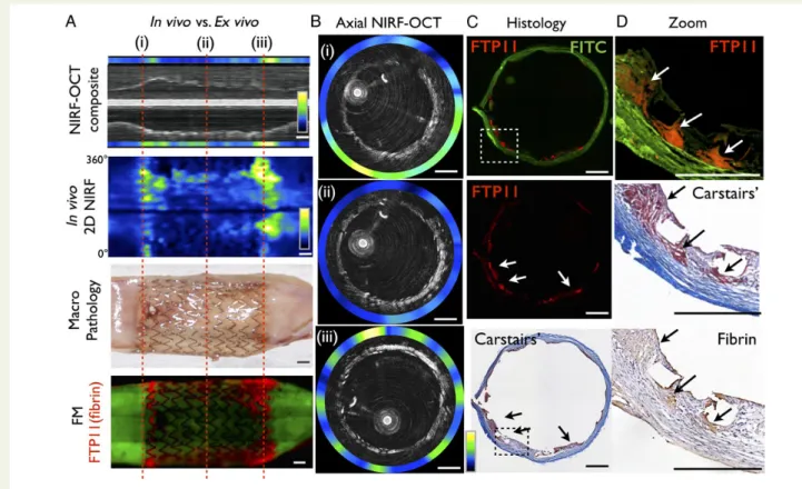
elevated NIRF signal surrounding stent struts (Figure1). Higher NIRF
fibrin signal was evident at stent edges. Axial NIRF-OCT fusion images demonstrated fibrin deposition on and around stent struts
(Figure1A and B).
Fluorescence microscopy of longitudinally opened stents con-firmed similar localization of NIRF-fibrin signal within the stent
(Figure1A). Histological evaluation demonstrated that the
FTP11-CyAm7 signal colocalized with fibrin deposition identified by
Carstairs’ stain and fibrin immunostaining (Figure1C and D).
Near-infrared fluorescence fibrin signal localized specifically near stent struts, extending prior findings of fibrin deposition patterns in a
flow loop model13and histopathological studies.2,3,14,15These
re-sults indicate that NIRF-OCT fibrin molecular imaging can accurate-ly assess fibrin deposition in stents in vivo.
Optical coherence tomography
assessment of tissue coverage on coronary
stent struts in vivo
The OCT per strut analyses of coverage and malapposition are
sum-marized and shown in Table1. Optical coherence tomography
revealed that DES exhibited greater rates of uncovered stent struts compared with BMS at both Day 7 (P , 0.001) and Day 28
(P , 0.05), consistent with prior studies.2,3,14–16
Bare metal stent fibrin dissipates from
Days 7 to 28, as assessed by near-infrared
fluorescence-optical coherence
tomography molecular-structural imaging
To assess the temporal dynamics of fibrin deposition on BMS in vivo, we next imaged BMS of two different healing stages (Days 7 and 28)Figure 1 Intravascular near-infrared fluorescence-optical coherence tomography imaging of fibrin deposition on stents. Rabbits underwent im-plantation of a 3.5 mm diameter bare metal stent in the aorta. At Day 7, the fibrin molecular imaging agent FTP11-CyAm7 was i.v. injected, fol-lowed by near-infrared fluorescence-optical coherence tomography. (A) In vivo 2D near-infrared fluorescence map of stent (upper two rows) and corresponding ex vivo imaging after longitudinally opened (lower two rows). (B) Representative cross-sectional near-infrared fluorescence-optical coherence tomography images at the stent distal edge (i), middle (ii), and proximal edge (iii). (C and D) Histological sections of the proximal stent edge (dotted line in A (i) are shown. Near-infrared fluorescence signal of FTP11-CyAm7 (red) co-localized with fibrin (bright red in Carstairs’ staining) and fibrin immunostaining. All scale bars ¼ 1 mm.
implanted in the infrarenal aorta of rabbits (N ¼ 3). We selected the Days 7 and 28 time points in rabbits to correspond to human time-points for stent healing at 1 month (subacute) and 6 months (late),
respectively.16,17In vivo NIRF-OCT molecular imaging of fibrin in
Day 7 BMS showed elevated NIRF signal, primarily at the stent edges. In contrast, little NIRF fibrin signal was observed in Day 28 BMS (P , 0.0001, Supplementary material online, Figure S4).
Greater fibrin deposition is evident in
drug-eluting stent compared with bare
metal stent at Day 7 in vivo
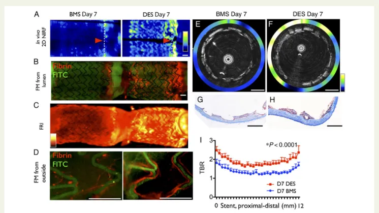
It remains unclear whether there are differences in the subacute stent thrombosis rates between BMS and DES. We compared the fibrin deposition between BMS and DES 7 days after implantation (n ¼ 7 BMS, n ¼ 7 DES), where Day 7 in the rabbit corresponds
to 1 month in humans.16We observed significantly greater NIRF
fibrin signal in DES than BMS at Day 7 after stent implantation
(P , 0.0001, Figure2). Drug-eluting stent at Day 7 also
demon-strated an edge-enhanced NIRF distribution pattern similar to BMS, but greater in magnitude. In addition, we also observed rela-tively higher NIRF fibrin signal throughout the mid stent segment
in DES compared with BMS (Figure2I).
. . . .
. . . . Table 1 Optical coherence tomography stent whole struts analyses
BMS DES P-value
Day 7 N ¼ 10 N ¼ 7
Total struts analysed 3675 2369
% covered struts 33.3 + 1.79 8.39 + 1.32 *0.0006 % malapposed struts 1.34 + 0.578 4.07 + 1.77 0.06
Day 28 N ¼ 4 N ¼ 4
Total struts analysed 2539 1375
% covered struts 92.8 + 2.98 85.8 + 2.79 *0.04 % malapposed struts 0.138 + 0.102 0.350 + 0.260 0.42
BMS, bare metal stent; DES, drug-eluting stent.
*P , 0.05 indicates significant differences between stent groups.
Figure 2 Drug-eluting stent exhibit greater fibrin deposition than bare metal stent at Day 7 in vivo. Rabbits were implanted with a nonoverlap-ping bare metal stent and drug-eluting stent, and then imaged with near-infrared fluorescence-optical coherence tomography after FTP11-CyAm7 fibrin agent injection on Day 7. (A) In vivo 2D near-infrared fluorescence map with bare metal stent (left) and drug-eluting stent (right), correspond-ing (B) fluorescence microscopy after longitudinally opencorrespond-ing stents, (C ) fluorescence reflectance imagcorrespond-ing and (D) fluorescence microscopy from the outside. Representative in vivo axial near-infrared fluorescence-optical coherence tomography images of (E) Day 7 bare metal stent and (F ) Day 7 drug-eluting stent at the distal edge (arrowhead in A). Carstairs’ stain (G and H ) demonstrates that some areas of bare metal stent tissue coverage are fibrin-negative, but almost all areas of drug-eluting stent tissue coverage are fibrin-positive. (I ) In vivo near-infrared fluorescence-fibrin signal from the proximal to distal stent edge of bare metal stent and drug-eluting stent (n ¼ 7 BMS, n ¼ 7 drug-eluting stent). FRI, fluorescence reflectance imaging; FITC, fluorescein isothiocyanate autofluorescence. Scale bars ¼ 1 mm.
Comparison of fibrin dissipation rates
in bare metal stent and drug-eluting
stent in vivo
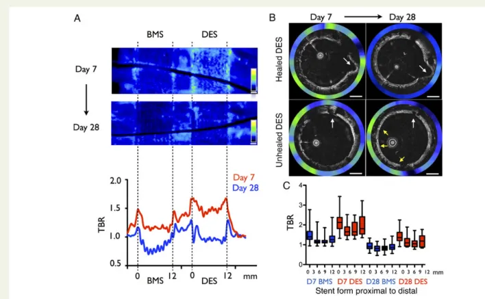
The rate of fibrin dissipation in coronary stents in vivo is unknown. To assess temporal changes in fibrin deposition in individual BMS and DES in vivo, a subset of rabbits (N ¼ 4) underwent serial NIRF-OCT fibrin imaging at Day 7 and then again at Day 28 post-stent implantation. We first confirmed that at Day 28, minimal residual NIRF signal was remnant from the initial Day 7 FTP11-CyAm7 injection (Supplementary material online, Figure S5). Co-registration of serially obtained Days 7 and 28 NIRF-OCT data-sets revealed that both BMS and DES showed reductions in
fibrin-NIRF signal (Figure3A and B). Day 28 DES exhibited quantitatively
greater fibrin deposition at all zones of the stent (edges, mid-section) compared with matched Day 28 BMS (P , 0.0001,
Figure3C). En face whole mount fluorescence microscopy
corrobo-rated higher fibrin-NIRF signal in Day 28 DES (Supplementary material online, Figure S6). Furthermore, en face microscopy of DAPI-stained tissue revealed less cellularity in Day 28 DES vs. Day 28 BMS. Carstairs’ fibrin staining also confirmed sustained peri-strut
fibrin deposition (Supplementary material online, Figure S6C and D),
as previously reported.14
A portion of optical coherence
tomography-covered edge stent struts is
near-infrared fluorescence-fibrin positive,
particularly in drug-eluting stent
While OCT-determined stent strut coverage is presumed to indi-cate a healed stent strut, ex vivo studies reveal that OCT tissue coverage can in fact represent coverage by fibrin or inflammatory cells, rather than physiological tissue, leading to an uncertainty
regarding the healing state of stents.6Although strut coverage was
highest at the stent edges as previously reported,14the NIRF fibrin
signal was also highest at the stent edges (outer 2 mm). Therefore, we analysed the extent of fibrin-rich neointima at Days 7 and 28 at the stent edges (n ¼ 8 for Day 7 stents, n ¼ 8 for Day 28 stents). Interestingly, most OCT-covered tissue on Day 7 DES struts was fibrin-rich (92.8 + 9.5% of edge struts), and significantly higher than Day 7 BMS (fibrin-positive struts 55.8 + 23.6%, P ¼ 0.0017).
Figure 3 Fibrin deposition and persistence is higher in drug-eluting stent than bare metal stent. Rabbits underwent serial near-infrared fluorescence-optical coherence tomography fibrin imaging at Days 7 and 28 (n ¼ 4 rabbits). (A) Representative 2D near-infrared fluorescence images at Day 7 (top row) and Day 28 (middle row). The lower graph shows the corresponding near-infrared fluorescence TBR signal across each stent. (B) Representative cross-sectional near-infrared fluorescence-optical coherence tomography images of a Day 28 healed drug-eluting stent (upper row) and a Day 28 unhealed drug-eluting stent (lower row). In the Day 28 unhealed case, struts are optical coherence tomography-covered but remain near-infrared fluorescence fibrin positive (e.g. 6 – 12 o’clock, yellow arrows). Co-registration was facilitated using side branches (white arrows). (C ) The near-infrared fluorescence fibrin signal decreased from Days 7 to 28 in both bare metal stent and drug-eluting stent (P , 0.001), however, fibrin persistence remained higher in drug-eluting stent (P , 0.001). Scale bars ¼ 1 mm.
At Day 28, fibrin on DES edge struts was three-fold more prevalent than on BMS (18.6 + 10.6% of DES vs. 5.1 + 8.7% of
BMS, P ¼ 0.0156, Figure4A). These findings were supported by
histological analyses (Figure4D). The functional endothelial cell
marker endothelial nitric oxide synthase was well expressed in areas of BMS neointima, in contrast to DES neointima, consistent with
prior studies.15
Optical coherence tomography grayscale
signal intensity and near-infrared
fluorescence-fibrin relationships
While differences in OCT grayscale signal intensity (GSI) of stent
strut tissue coverage might distinguish fibrin,18we found that the
OCT GSI was similar between Day 7 BMS and DES (BMS 0.50 + 0.11 vs. DES 0.51 + 0.08, P . 0.99), and between Day 28 BMS
and DES (0.59 + 0.07 vs. 0.58 + 0.07, P . 0.99, Figure4C),
des-pite higher NIRF-fibrin in DES . BMS at each time point. Signifi-cantly higher GSI was observed in Day 28 mature neointima compared with Day 7 immature neointima (P , 0.0001 for
BMS and P ¼ 0.0011 for DES, Figure4C), consistent with prior
studies.17,18
Quantitative assessment of near-infrared
fluorescence-fibrin and optical coherence
tomography coverage relationships in
bare metal stent and drug-eluting stent
healing
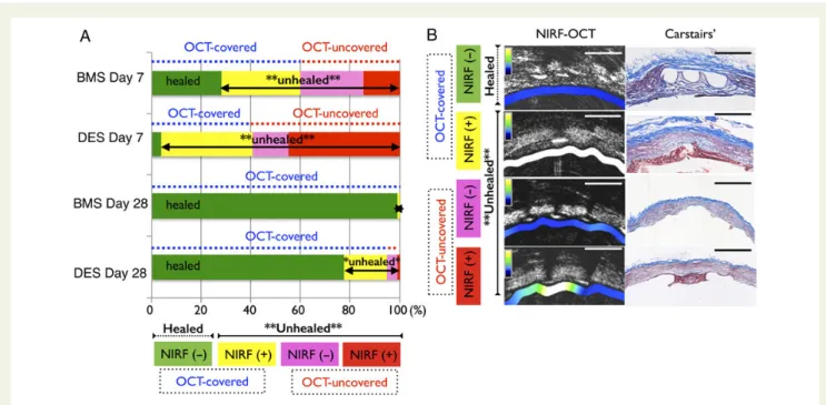
Near-infrared fluorescence-fibrin and OCT-coverage stent strut data of Days 7 and 28 BMS and DES at edges were analysed and further classified into one of four groups: NIRF-fibrin negative and OCT-covered, identifying healed stent struts; or NIRF-fibrin positive, OCT-uncovered; NIRF-fibrin positive, OCT-covered; or NIRF-fibrin negative, OCT-uncovered; these latter three groups
identifying unhealed stent struts. The data presented in Figure5
demonstrates that in subacute and late timepoints after stent implantation, a significant percentage of OCT-covered stent struts is NIRF fibrin positive and thus can be considered to
be unhealed (Figure5, yellow groups). In addition, the proportion
Figure 4 Near-infrared fluorescence molecular imaging identifies optical coherence tomography-covered, but unhealed fibrin-rich struts in vivo. (A) The percentage of fibrin-rich neointimal tissue at stent edges was assessed by near-infrared fluorescence-optical coherence tomography. Drug-eluting stent showed significantly higher rate of fibrin-rich covering tissue than bare metal stent. (B) Near-infrared fluorescence-optical co-herence tomography images of bare metal stent and drug-eluting stent at Day 7 revealed a diversity of rich (yellow arrows) and fibrin-negative (white arrows) struts, even within a single cross-section. (C ) Grayscale optical coherence tomography intensity was similar between bare metal stent and drug-eluting stent at both Days 7 and 28 timepoints (n ¼ 4 stents in each group), despite higher fibrin signal in DES . BMS at both Days 7 and 28 at the stent edges. (D) Fibrin immunostaining and endothelial nitric oxide synthase immunostaining (magnified images at bottom) are shown. Most Day 7 drug-eluting stent edge tissue coverage is fibrin-rich and lacks endothelial nitric oxide synthase expression, in contradistinction to Day 7 bare metal stent.#P , 0.05 vs. respective Day 7.
of unhealed stent struts remained significantly higher in DES compared with BMS at both Day 7 (P , 0.001) and Day 28 (P , 0.05).
Discussion
A key finding of this study is that a significant portion of OCT-covered stent struts may be actually OCT-covered by fibrin and are therefore unhealed, particularly earlier on after stenting. Specifically, over 90% of OCT-covered edge DES struts at Day 7, corresponding to the 1 month timepoint in humans, were positive for fibrin, a
pro-thrombotic interface that mediates stent thrombosis.1–3At Day 28,
corresponding to the 6 – 12 months timepoint in humans, combined NIRF-OCT determined that 23% of DES edge stents struts were unhealed—four times the percentage identified by standalone
OCT (Figure5A). The overall results demonstrate that intravascular
NIRF fibrin molecular imaging can improve the identification of unhealed clinical coronary stents by clarifying whether OCT tissue coverage represents fibrin in vivo.
Optical coherence tomography is a promising approach to iden-tify unhealed stents lacking tissue coverage, however, standalone OCT does not accurately distinguish between stent coverage by endothelium/smooth muscle cells (physiological stent healing) from coverage by prothrombotic fibrin, which indicates an unhealed
stent.6We observed a substantial proportion of OCT-covered
stent struts are actually covered by fibrin (Figure5). Therefore, a
subset of OCT-covered stent struts is actually unhealed, rather than healed. These in vivo findings extend the ex vivo study by Nakano et al. that demonstrated standalone OCT misclassification of healed stents occurs due to coverage by fibrin and inflammatory
cells.6Moreover, the observed mismatches between OCT and
NIRF fibrin imaging fundamentally changes the perspective on whether such an OCT-covered stent could be prone to stent thrombosis. This finding is of substantial importance for ongoing clinical standalone OCT-based trials—which are predicated on the concept that tissue coverage of a stent means that a stent is healed and carries a low risk of stent thrombosis. The NIRF findings herein demonstrate that caution may be needed in interpreting the healing state of stents by standalone OCT, particularly in the subacute post-implantation period where unhealed stents are more prevalent.
While OCT grayscale intensity differences might identify fibrin,18
grayscale intensity was similar between DES and BMS in our study, despite DES having significantly higher fibrin content than BMS, fur-ther strengthening the value of NIRF molecular imaging to identify fibrin-rich unhealed stent struts. In addition, OCT stent strut cover-age analysis is highly time-consuming (hours per individual stent),
limiting its application in point-of-care analysis.19In contrast, 2D
NIRF fibrin maps (e.g. Figure1A) readily display fibrin-rich areas on
stents. By specifically and rapidly assessing fibrin deposition on stents in vivo, NIRF molecular imaging powerfully complements standalone OCT structural imaging.
Figure 5 Fibrin molecular near-infrared fluorescence-optical coherence tomography imaging assessment of the healing status of edge stent struts, by stent age (Day 7 or 28), and by stent type (bare metal stent or drug-eluting stent). At both Days 7 and 28, drug-eluting stent shows a higher percentage of unhealed stent struts compared with bare metal stent. At Day 7, 59.8% of bare metal stent struts and 40.7% of drug-eluting stent struts were classified as optical coherence tomography-covered (green+ yellow groups) at stent edges, however, drug-eluting stent dis-played only a small percentage of stent struts (3.7%) that were truly healed (near-infrared fluorescence-fibrin negative, optical coherence tomography-covered, green group), in contrast to bare metal stent demonstrating 28.1% of struts as healed. At Day 28, both bare metal stent and drug-eluting stent demonstrated substantially improved stent strut healing compared with their respective Day 7 timepoints, however 22.6% of Day 28 drug-eluting stent struts still remained unhealed. (B) Representative near-infrared fluorescence-optical coherence tomography and matched Carstairs’ microscopy for the four groups. Arrow indicates thin fibrin layer over the strut.
Pathophysiologically, NIRF-OCT fibrin-structural imaging pro-vided new insights into differences in DES and BMS healing. Serial NIRF-OCT at subacute timepoints demonstrated that DES exhibited higher fibrin persistence than BMS out to Day 28 in
rab-bits, consistent with prior histopathological data.15These findings
need to be taken into context with studies of acute thrombus formation. We and others have shown, using these very same stents, that the coatings associated with DES are not inherently thrombogenic when evaluated in the acute setting associated with
implantation and before drug release.13This still remains the case.
What we now show is that as DES elute their drug, there is a signal of increased subacute fibrin deposition that diminishes over time. Collectively, these data reinforce three timeperiods where implanted stents may be susceptible to thrombosis: an acute period related to surface effects and strut dimensions; a subacute period related to concentration of drug in local vicinities; and a third period related to a stunted or incomplete vascular healing response.
Although extrapolating healing rates from rabbits to humans is approximate, prior reports demonstrate that vascular healing in rab-bits occurs 5 – 6 times faster than humans, and propose that Days 7 and 28 in rabbits correspond to 1 month and 6 – 12 months in
humans, respectively.16While current clinical guidelines indicate
that 6 – 12 months of DAPT may be sufficient for prevention of
DES thrombosis,20the recent DAPT study suggests that extended
anti-platelet therapy of 30 months is beneficial.4Therefore, the
op-timal duration of DAPT for coronary stents still remains unknown. Accordingly, the accurate identification of unhealed coronary stents could offer substantial value in guiding the duration of DAPT.
Clinical translation
The current study significantly advances the potential for intracor-onary molecular imaging of fibrin deposition on stents. While an earlier study described the feasibility of imaging fluorescently coated
clots placed on stents ex vivo,7the present study provides the first
demonstration that stent fibrin deposition can be imaged in vivo using an injectable NIRF molecular imaging agent, FTP11-CyAm7. As an MRI version of FTP11-CyAm7 has already been tested in
Phase II trials,11and NIR fluorophores such as indocyanine green
are clinically approved, the fibrin NIRF molecular imaging agent has substantial translational potential. From a catheter standpoint, clinical intracoronary testing of an NIRF-OCT catheter has already
been initiated.21These encouraging developments suggest that
clin-ical NIRF molecular imaging of fibrin deposition on stents may be feasible in the near future.
Stent thrombosis remains a highly morbid, persistent complica-tion of BMS and DES, and while despite long-term DAPT reduces stent thrombosis, thrombosis risk remains, and moreover appears
to increase quickly after cessation of DAPT.1,4The ability to image
fibrin deposition on stents could therefore have several important applications. First, identifying fibrin-bearing stents might better pre-dict the risk of stent thrombosis, motivating a re-review of optimal DAPT choices and duration, and selection for novel stent healing approaches. Second, fibrin imaging might prove to be a valuable surrogate endpoint for clinical trials evaluating new pro-healing stents and pharmacotherapies designed to reduce stent thrombosis.
Study limitations
As with other studies of rabbit stent healing,15coronary stents were
implanted into the normal aorta in rabbits without atherosclerotic plaque or curvature, as opposed to human coronary arteries. Iliac
arteries, generally used in preclinical stent healing studies,15were
not stented due to limited repeated vascular access from femoral arteries, and to allow direct comparison between two stents within one pullback. While determining the precise mechanisms underlying enhanced fibrin deposition at the stent edges is beyond the scope of this manuscript, future studies will utilize computational fluid
dynamic models22to explore this issue. As light cannot penetrate
through metallic stent struts, NIR fluorophores beneath metallic stent struts will not be detected by intravascular fluorescence reflectance imaging. Finally, delayed healing is only one feature underlying stent thrombosis, and other factors such as blood thrombogenicity and neoatherosclerosis will need to be integrated with NIRF-OCT fibrin imaging.
Conclusions
Intravascular NIRF fibrin molecular imaging improves the de-tection of unhealed stents by specifically determining whether OCT tissue coverage reflects fibrin deposition. Compared with NIRF-OCT, standalone OCT over-classifies stents as healed, even at subacute timepoints after stent implantation. Intravascular fibrin NIRF-OCT may ultimately help better identify stents at risk for stent thrombosis.
Supplementary material
Supplementary Material is available at European Heart Journal online.
Authors’ contributions
T.H. performed statistical analysis; F.A.J., G.J.T., J.M. handled funding and supervision; T.H., G.U. acquired the data; F.A.J., G.J.T. conceived and designed the research; T.H., G.U., F.J. drafted the manuscript; E.R.E., G.J.T., F.A.J. made critical revision of the manuscript for key intellectual content.
Acknowledgements
We acknowledge Hongki Yoo, PhD for assistance with NIRF-OCT imaging, and Ashok Khatri, PhD and the MGH Peptide Core for the support of synthesis of imaging agent.
Funding
NIH R01HL108229 and R01HL122388 (F.A.J), American Heart Associ-ation (#13POST14640021 to T.H.; #13GRNT17060040 to F.A.J.), MGH ECOR Support Fund (F.A.J.), and the Kanae Foundation for Research Abroad (T.H.). E.R.E. was supported in part by NIH R01GM49039. NIH R01HL093717 (G.J.T., for development of the imaging devices); CIMIT (F.A.J. and G.J.T., for development of the imaging devices).
Conflict of interest: F.A.J. receives sponsored research from Merck, Kowa, and Siemens, and nonfinancial research support from Boston Scientific. Massachusetts General Hospital has a patent licensing
arrangement with Terumo and Canon Corporations. G.J.T. (Terumo, Canon) and F.A.J. (Canon) have the right to receive licensing royalties through this licensing arrangement. G.J.T. receives consulting income from Samsung. G.J.T.’s lab receives sponsored research from Canon and InfraRedx and catheter components from Terumo.
References
1. Claessen BE, Henriques JP, Jaffer FA, Mehran R, Piek JJ, Dangas GD. Stent Throm-bosis: a clinical perspective. JACC Cardiovasc Interv 2014;7:1081 – 1092.
2. Joner M, Finn AV, Farb A, Mont EK, Kolodgie FD, Ladich E, Kutys R, Skorija K, Gold HK, Virmani R. Pathology of drug-eluting stents in humans: delayed healing and late thrombotic risk. J Am Coll Cardiol 2006;48:193 – 202.
3. Finn AV, Joner M, Nakazawa G, Kolodgie F, Newell J, John MC, Gold HK, Virmani R. Pathological correlates of late drug-eluting stent thrombosis: strut coverage as a marker of endothelialization. Circulation 2007;115:2435 – 2441.
4. Mauri L, Kereiakes DJ, Yeh RW, Driscoll-Shempp P, Cutlip DE, Steg PG, Normand SL, Braunwald E, Wiviott SD, Cohen DJ, Holmes DR Jr, Krucoff MW, Hermiller J, Dauerman HL, Simon DI, Kandzari DE, Garratt KN, Lee DP, Pow TK, Ver Lee P, Rinaldi MJ, Massaro JM, Investigators DS. Twelve or 30 months of dual antiplatelet therapy after drug-eluting stents. N Engl J Med 2014;371: 2155 – 2166.
5. Murata A, Wallace-Bradley D, Tellez A, Alviar C, Aboodi M, Sheehy A, Coleman L, Perkins L, Nakazawa G, Mintz G, Kaluza GL, Virmani R, Granada JF. Accuracy of optical coherence tomography in the evaluation of neointimal coverage after stent implantation. JACC Cardiovasc Imaging 2010;3:76 – 84.
6. Nakano M, Vorpahl M, Otsuka F, Taniwaki M, Yazdani SK, Finn AV, Ladich ER, Kolodgie FD, Virmani R. Ex vivo assessment of vascular response to coro-nary stents by optical frequency domain imaging. JACC Cardiovasc Imaging 2012;5: 71 – 82.
7. Yoo H, Kim JW, Shishkov M, Namati E, Morse T, Shubochkin R, McCarthy JR, Ntziachristos V, Bouma BE, Jaffer FA, Tearney GJ. Intra-arterial catheter for simul-taneous microstructural and molecular imaging in vivo. Nat Med 2011;17: 1680 – 1684.
8. McCarthy JR, Patel P, Botnaru I, Haghayeghi P, Weissleder R, Jaffer FA. Multimodal nanoagents for the detection of intravascular thrombi. Bioconjugate Chemistry 2009; 20:1251 – 1255.
9. Ughi GJ, Verjans J, Fard AM, Wang H, Osborn E, Hara T, Mauskapf A, Jaffer FA, Tearney GJ. Dual modality intravascular optical coherence tomography (OCT) and near-infrared fluorescence (NIRF) imaging: a fully automated algorithm for the distance-calibration of NIRF signal intensity for quantitative molecular imaging. Int J Cardiovasc Imaging 2015;31:259 – 268.
10. Hara T, Bhayana B, Thompson B, Kessinger CW, Khatri A, McCarthy JR, Weissleder R, Lin CP, Tearney GJ, Jaffer FA. Molecular imaging of fibrin deposition in deep vein thrombosis using fibrin-targeted near-infrared fluorescence. JACC Cardiovasc Imaging 2012;5:607 – 615.
11. Vymazal J, Spuentrup E, Cardenas-Molina G, Wiethoff AJ, Hartmann MG, Caravan P, Parsons EC Jr. Thrombus imaging with fibrin-specific gadolinium-based
MR contrast agent EP-2104R: results of a phase II clinical study of feasibility. Invest Radiol 2009;44:697 – 704.
12. Jaffer FA, Verjans JW. Molecular imaging of atherosclerosis: clinical state-of-the-art. Heart 2014;100:1469 – 1477.
13. Kolandaivelu K, Swaminathan R, Gibson WJ, Kolachalama VB, Nguyen-Ehrenreich KL, Giddings VL, Coleman L, Wong GK, Edelman ER. Stent thrombo-genicity early in high-risk interventional settings is driven by stent design and deployment and protected by polymer-drug coatings. Circulation 2011;123: 1400 – 1409.
14. Joner M, Nakazawa G, Finn AV, Quee SC, Coleman L, Acampado E, Wilson PS, Skorija K, Cheng Q, Xu X, Gold HK, Kolodgie FD, Virmani R. Endothelial cell recovery between comparator polymer-based drug-eluting stents. J Am Coll Cardiol 2008;52:333 – 342.
15. Nakazawa G, Nakano M, Otsuka F, Wilcox JN, Melder R, Pruitt S, Kolodgie FD, Virmani R. Evaluation of polymer-based comparator drug-eluting stents using a rabbit model of iliac artery atherosclerosis. Circ Cardiovasc Interv 2011;4:38 – 46. 16. Virmani R, Kolodgie FD, Farb A, Lafont A. Drug eluting stents: are human and
ani-mal studies comparable? Heart 2003;89:133 – 138.
17. Malle C, Tada T, Steigerwald K, Ughi GJ, Schuster T, Nakano M, Massberg S, Jehle J, Guagliumi G, Kastrati A, Virmani R, Byrne RA, Joner M. Tissue characterization after drug-eluting stent implantation using optical coherence tomography. Arterios-cler Thromb Vasc Biol 2013;33:1376 – 1383.
18. Templin C, Meyer M, Muller MF, Djonov V, Hlushchuk R, Dimova I, Flueckiger S, Kronen P, Sidler M, Klein K, Nicholls F, Ghadri JR, Weber K, Paunovic D, Corti R, Hoerstrup SP, Luscher TF, Landmesser U. Coronary optical frequency do-main imaging (OFDI) for in vivo evaluation of stent healing: comparison with light and electron microscopy. Eur Heart J 2010;31:1792 – 1801.
19. Adriaenssens T, Ughi GJ, Dubois C, Onsea K, De Cock D, Bennett J, Wiyono S, Vanhaverbeke M, Sinnaeve P, Belmans A, D’Hooge J, Desmet W. Automated de-tection and quantification of clusters of malapposed and uncovered intracoronary stent struts assessed with optical coherence tomography. Int J Cardiovasc Imaging 2014;30:839 – 848.
20. Authors/Task Force Members, Windecker S, Kolh P, Alfonso F, Collet JP, Cremer J, Falk V, Filippatos G, Hamm C, Head SJ, Juni P, Kappetein AP, Kastrati A, Knuuti J, Landmesser U, Laufer G, Neumann FJ, Richter DJ, Schauerte P, Sousa Uva M, Stefanini GG, Taggart DP, Torracca L, Valgimigli M, Wijns W, Witkowski A. 2014 ESC/EACTS Guidelines on myocardial revascularization: The Task Force on Myo-cardial Revascularization of the European Society of Cardiology (ESC) and the European Association for Cardio-Thoracic Surgery (EACTS) Developed with the special contribution of the European Association of Percutaneous Cardiovas-cular Interventions (EAPCI). Eur Heart J 2014;35:2541 – 2619.
21. Ughi G, Wang H, Gardecki JA, Gerbaud E, Fard AM, Hamidi E, Jacques-Vacas P, Rosenberg-M, Jaffer FA, Tearney GJ. First-In-Human Dual-Modality OCT and Near-Infrared Autofluorescence Imaging of Coronary Artery Disease. JACC Cardio-vascular Imaging (in press).
22. Balakrishnan B, Tzafriri AR, Seifert P, Groothuis A, Rogers C, Edelman ER. Strut position, blood flow, and drug deposition: implications for single and overlapping drug-eluting stents. Circulation 2005;111:2958 – 2965.