PROCEEDINGS OF SPIE
SPIEDigitalLibrary.org/conference-proceedings-of-spie
Automated cancer stem cell
recognition in H and E stained tissue
using convolutional neural networks
and color deconvolution
Wolfgang Aichinger, Sebastian Krappe, A. Enis Cetin,
Rengul Cetin-Atalay, Aysegül Üner, et al.
Wolfgang Aichinger, Sebastian Krappe, A. Enis Cetin, Rengul Cetin-Atalay,
Aysegül Üner, Michaela Benz, Thomas Wittenberg, Marc Stamminger,
Christian Münzenmayer, "Automated cancer stem cell recognition in H and E
stained tissue using convolutional neural networks and color deconvolution,"
Proc. SPIE 10140, Medical Imaging 2017: Digital Pathology, 101400N (1
March 2017); doi: 10.1117/12.2254036
Event: SPIE Medical Imaging, 2017, Orlando, Florida, United States
Downloaded From: https://www.spiedigitallibrary.org/conference-proceedings-of-spie on 12/22/2018 Terms of Use: https://www.spiedigitallibrary.org/terms-of-useAutomated cancer stem cell recognition in H&E stained tissue using
convolutional neural networks and color deconvolution
Wolfgang Aichinger
a, Sebastian Krappe
a,b, A. Enis Cetin
c, Rengul Cetin-Atalay
d, Aysegül Üner
e,
Michaela Benz
a, Thomas Wittenberg
a, Marc Stamminger
b, Christian Münzenmayer
aa
Image Processing and Medical Engineering Department, Fraunhofer Institute for Integrated Circuits
IIS, Erlangen, Germany;
b
Computer Graphics Group, Friedrich-Alexander-University Erlangen-Nuremberg (FAU), Erlangen,
Germany;
c
Bilkent University, Ankara, Turkey;
d
Cancer Systems Biology Laboratory, Middle East Technical University, Ankara, Turkey;
eHacettepe University, Ankara, Turkey
ABSTRACT
The analysis and interpretation of histopathological samples and images is an important discipline in the diagnosis of various diseases, especially cancer. An important factor in prognosis and treatment with the aim of a precision medicine is the determination of so-called cancer stem cells (CSC) which are known for their resistance to chemotherapeutic treatment and involvement in tumor recurrence. Using immunohistochemistry with CSC markers like CD13, CD133 and others is one way to identify CSC. In our work we aim at identifying CSC presence on ubiquitous Hematoxilyn & Eosin (H&E) staining as an inexpensive tool for routine histopathology based on their distinct morphological features.
We present initial results of a new method based on color deconvolution (CD) and convolutional neural networks (CNN). This method performs favorably (accuracy 0.936) in comparison with a state-of-the-art method based on 1D-SIFT and eigen-analysis feature sets evaluated on the same image database. We also show that accuracy of the CNN is improved by the CD pre-processing.
Keywords: Deep Learning, Digital Pathology, Histopathology, Convolutional Neural Network, Color Deconvolution,
Texture Analysis
1. INTRODUCTION
The analysis and interpretation of histopathological samples and images is an important discipline in the diagnosis of various diseases, especially cancer. An important factor in prognosis and treatment with the aim of a precision medicine is the determination of so-called cancer stem cells (CSC) which are known for their resistance to to chemotherapeutic treatment. Using immunohistochemistry with markers like CD13, CD133 and others is one way to identify CSC. In our work we aim at identifying CSC features presence on ubiquitous Hematoxilyn & Eosin (H&E) staining as an inexpensive tool for routine histopathology.
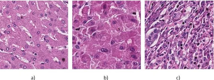
This approach is based on structural changes of the tissue which is associated with CSC presence as can be seen in the examples of Figure 1 that have been graded according to CSC concentration in the corresponding serial section with immuno-staining. In this paper we present an approach based on Convolutional Neural Networks to determine whether digitized samples of liver tissue exhibit characteristics of cancer stem cells or not.
Our paper also aims at investigating the influence of pre-processing, namely color deconvolution (CD) which is supposed to provide a separation of the Hematoxilyn and Eosin components, on the performance of the CNN. Therefore, one classifier is fed with the original RGB images, the other CNN is trained and evaluated on the dataset after color deconvolution. These results are evaluated against texture analysis approach using 1D-SIFT features published by Oguz et al. [9].
Medical Imaging 2017: Digital Pathology, edited by Metin N. Gurcan, John E. Tomaszewski, Proc. of SPIE Vol. 10140, 101400N · © 2017 SPIE · CCC code: 1605-7422/17/$18 · doi: 10.1117/12.2254036
Proc. of SPIE Vol. 10140 101400N-1
Downloaded From: https://www.spiedigitallibrary.org/conference-proceedings-of-spie on 12/22/2018 Terms of Use: https://www.spiedigitallibrary.org/terms-of-use
Figure 1: a) H&E stained liver tissue of healthy patient, b) Grade 1 (well differentiated), c) Grade 2 (poorly differentiated)
2. METHODS
It has been proven that CNNs are a powerful tool for solving different Computer Vision tasks. They expand the concept of classical artificial neural networks (ANN) representing local connectivity, which makes them suitable to deal with pictures as input-layer [3]. These ANNs were originally designed to copy and simulate the information processing of the neural networks in the human brain. Similar to the human brain ANNs are based on so-called cells or nodes. Some cells or nodes are defined as input cells and receive input data being either complete images or characteristic features extrac-ted from images. As long as the output cells do not deliver the expecextrac-ted results, a back-propagation algorithm is used to distribute the error of the result to the cells of the hidden layers and change the weights of the input data of all cells in the artificial neural network. CNNs specifically incorporate the feature extraction and selection process directly in the convolutional lower layers of the CNN, thus selecting the needed low level image features by the extensive variation of convolutional kernels. Janowzcyk et al. [4] present an approach to nuclear segmentation in histology images. Pan et al. [5] apply deep neural networks to cell detection for lung cancer diagnosis. An approach to cell segmentation in skeletal muscle tissue with CNN’s was presented by Sapkota et al. [6]. Chen et al. [7] use so-called deep contour-aware networks to automatically segment glands in colon tissue and won the 2015 MICCAI Gland Segmentation Challenge out of 13 teams. Sharma et al. [8] apply CNN in a very similar context as this paper with the classification of cancerous vs. benign regions in H&E stained tissue of gastric cancer as well as for detection of necrotic regions which performs comparably to conventional texture analysis.
In the field of digital pathology color deconvolution has been around since 2001. This color transformation method assumes that the absorption-properties of all single dyes that are used for a specific histochemical staining method are known. Therefore for each pixel the influence of the different dyes can be determined and staining components can be separated approximately in color space..
2.1 Color Deconvolution (CD)
For the purpose of highlighting nuclei shown in the H&E images we propose the use of Color Deconvolution first introduced by Ruifrok and Johnston [2]. Applying this method leads to three separated channels, the first one representing Hematoxylin, the second one Eosin. The third channel should only contain background, but in case of a chemically saturated region the signal will also appear there. We used a plugin for Fiji to execute the CD with a given stain matrix that is shown in Equ. 1 [10].
Proc. of SPIE Vol. 10140 101400N-2
Downloaded From: https://www.spiedigitallibrary.org/conference-proceedings-of-spie on 12/22/2018 Terms of Use: https://www.spiedigitallibrary.org/terms-of-use
N 11 00 300 30 1 100
'49
4 3 Stride of 3 64 Max Pooling 128 256 Max Max Pooling Pooling 256 4096 409 2 a) 4 ,,.
_tiC .
r."fr'
/y
.,
! J ,i
1 )f
. . 'i
_/`
'
l+á
b) c)(Equ. 1)
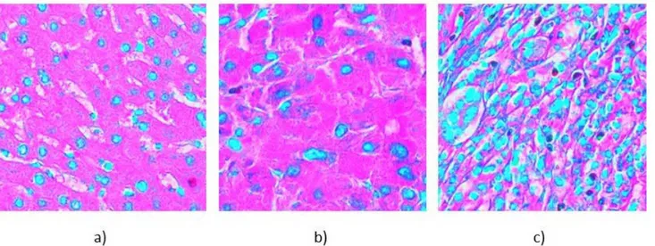
The color separated images are stored in jpg-format where each channel R, G and B represents one of the separated channels. Hence, images after the transformation look quite unnatural which can be seen in Figure 2, where the examples from Figure 1 are shown after the transformation.
Figure 2: Equivalent images as in Figure 1 after transformation
2.2 Convolutional Neural Network (CNN)
We used the so-called AlexNet byKrizhevski et al. [3] as a starting point for our net and changed it slightly to make it fit our needs. We reduced the number of convolutional layers and also changed the kernel sizes to reduce computational complexity. Our resulting net has three convolutional layers, each followed by an activation layer (ReLUs) and a pooling layer. These are followed by two fully connected layers with 4096 neurons each and the two-dimensional output layer (see Figure 3).
To address the problem of overfitting we used two different concepts. Dropout sets the output of a hidden layer to zero with a certain probability, which forces all other neurons to learn features directly related to the problem and prohibits co-adaptions between different neurons. We found 0.2 to be a useful value for the probability.
Figure 3: Schematic view on our CNN
Proc. of SPIE Vol. 10140 101400N-3
Downloaded From: https://www.spiedigitallibrary.org/conference-proceedings-of-spie on 12/22/2018 Terms of Use: https://www.spiedigitallibrary.org/terms-of-use
It is well-known that the number of training data is also crucial for successful training of CNN and the data set we use for evaluation (described in the following section) is comparably small with only 454 image tiles in total. Therefore, with data augmentation the dataset is enlarged by mirroring and rotating the original images. We incorporated three rotations plus horizontal and vertical flipping. In this way we increase the size of our dataset by a factor of 6.
We trained our net on a 64-Bit Windows 7 machine using the Computational Network Toolkit (CNTK) published by Microsoft Research. To speed up computation time we ran the experiments on a system including Nvidia 950GTX graphics hardware.
3. EXPERIMENTS
The dataset we analyzed consists of 454 H&E stained image regions with liver tissue. 270 samples originate from 34 patients diagnosed with cancer, 184 samples originate from 9 healthy patients. The images were acquired using a Leica Aperio CS2 light microscope and 20X objectives. The image size of the extracted tiles was set to 300x300 pixels heuristically balancing the requirements of larger regions for better classification against the possibility to implement a tile-based detection and segmentation of CSC-intense regions within a whole slide. As the number of samples is considered low, we use a Leave-One-Patient-Out approach, meaning we train 43 different models. Therefore, in each case the training set consists of the data, i.e. the image tiles, from 42 different patients, the test set holds the data from the remaining patient.
For evaluation it was intended to compare the results with those by a classical texture approach as published by Oguz et al. [9] who kindly provided their dataset. For this evaluation two different experiments have been performed using the CNN described above. The inputs in the first case are the original RGB-images, in the second case we used the images after color deconvolution as described in section I. For both approaches the maximum number of epochs is set to 20, the learning rate is automatically decreased in case the error has increased in the latest epoch.
4. RESULTS
The results of our experiments for the three different methods, i.e. the CNN on original RGB images, CNN with color deconvolution as pre-processing, and the reference method 1D-Sift texture features are summarized in the confusion matrices in Figure 4. Accuracy, sensitivity and specificity are displayed in corresponding Figure 5.
The accuracy achieved by the new method combining CD and CNN (0.936) is 2.8% higher than the best result using 1D-Sift [9] (0.910). We have as many false negative samples (4) but the number of false positives is reduced from 37 samples to 25. These numbers add up to an increase in specificity from 0.800 to 0.864, while there is no significant change regarding sensitivity. Using only RGB data without any preprocessing showed an accuracy of 0.925, a sensitivity of 0.944 and a specificity of 0.900. The specificity is slightly higher than with CD included, but the sensitivity is lower, which is the number to be increased in the first place. We also noticed a slower decrease of the error during training.
Classification Result 0 1 Ground Truth 0 165 19 1 15 255 (b)
Figure 4: Confusion matrices with classification result versus ground truth for (a) Color Deconvolution and CNN (b) RGB and CNN (c) the 1D-SIFT texture algorithm published by Oguz et al. [9]
Classification Result 0 1 Ground Truth 0 159 25 1 4 266 (a) Classification Result 0 1 Ground Truth 0 147 37 1 4 266 (c)
Proc. of SPIE Vol. 10140 101400N-4
Downloaded From: https://www.spiedigitallibrary.org/conference-proceedings-of-spie on 12/22/2018 Terms of Use: https://www.spiedigitallibrary.org/terms-of-use
1 0,95 0,9 0,85 0,8 0,75 0,7
Accuracy Sensitivity Specificity
L CD + CNN
RGB + CNN
1D -SIFT
Figure 5: Accuracy, sensitivity and specificity are shown for the different approaches
5. CONCLUSION
In this paper we present a new method for the automatic recognition of cancer stem cells that combines color deconvolution and convolutional neural network. The additional color deconvolution leads to better outcomes with regard to the sensitivity. The accuracy reached by CNN is 2.8% higher than the best result presented by Oğuz et al. [9] for CD+CNN and 1.6% higher for RGB+CNN. We show that introducing knowledge about the problem through CD benefits the outcome, as the sensitivity is increased by 4.3%, the accuracy by 1.2%. Automated software systems for the recognition of cancer stem cells could apply such a classifier to shorten examination time and improve objectivity, reliability and reproducibility. More accurate and targeted treatments may become additional benefits in the future in addition to improved quality assurance, documentation and global data availability.
In this paper we presented promising initial results on a new method for automated recognition of cancer stem cells in H&E stained liver tissue using convolutional neural networks. We increased our performance by adding a color deconvolution as preprocessing step. Both approaches, with and without CD, were evaluated on 454 samples from 43 patients. Since the dataset was small, we used a leave-one-patient-out approach. We compared our results to the outcome of an existing method based on 1D-Sift and eigenanalysis on the same dataset. We achieved a better accuracy, mainly because of a significant lower number of false positive samples by adding CD. The same net trained and tested with RGB was not capable of achieving the same results, especially regarding sensitivity. Our higher specificity was not achieved at the expense of sensitivity, as there is no difference. We plan to extend our approach to three classes, by introducing a grading that is based on the density of cancer stem cells.
Proc. of SPIE Vol. 10140 101400N-5
Downloaded From: https://www.spiedigitallibrary.org/conference-proceedings-of-spie on 12/22/2018 Terms of Use: https://www.spiedigitallibrary.org/terms-of-use
6. ACKNOWLEDGEMENT
This project partially as supported by grants of TUBITAK # 213E032 and the German Research Association (Center of Excellence 796).
REFERENCES
[1] Yu, D., Eversole, A., Seltzer, M. L., Yao, K., Huang, Z., Guenter, B., Kuachaiev, O., Zhang, Y., Seide, F., Wang, H., Droppo, J., Zweig, G., Rossbach, C., Currey, J., Gao, J., May, A., Peng, B., Stolcke, A., Slaney, M., "An introduction to computational networks and the computational network toolkit." Technical report, Microsoft Research, (2014).
[2] Ruifrok, Arnout C., and Dennis A. Johnston. "Quantification of histochemical staining by color deconvolution." Analytical and quantitative cytology and histology 23.4 (2001): 291-299.
[3] Krizhevsky, Alex, Ilya Sutskever, and Geoffrey E. Hinton. "Imagenet classification with deep convolutional neural networks." Advances in neural information processing systems. 2012.
[4] Janowczyk A, Dolye S, Gilore H, Madabhushy (2015) A resolution adaptive hierarchical deep learning scheme applied to nuclear segmentation in histology images. In Proc’s 1st Workshop on Deep Learning in Medical Image Analysis (DLMIA 2015); Munich, pp. 73-80
[5] Pan H, Xu Z, Huang J (2015) An Effective Approach for Robust Lung Cancer Cell Detection. In Proc’s 1st Int. Workshop on Patch-based Techniques in Medical Imaging; Munich, pp. 81-88
[6] Sapkota M, Xing F, Liu F, Yan L (2015) Skeletal Muscle Cell Segmentation Using Distributed Convolutional Neural Network. In Proc’s High Performance Computing in Biomedical Image Analysis (HPC-MICCAI); Munich
[7] Chen, Hao, et al. "DCAN: Deep Contour-Aware Networks for Accurate Gland Segmentation." arXiv preprint arXiv:1604.02677 (2016).
[8] Sharma, H., et al. "Deep Convolutional Neural Networks for Histological Image Analysis in Gastric Carcinoma Whole Slide Images." Diagnostic Pathology 1.8 (2016).
[9] Oğuz, O., et al. "Mixture of learners for cancer stem cell detection using cd13 and handle stained images." SPIE Medical Imaging. International Society for Optics and Photonics, 2016.
[10] Landini, G, “Color Deconvolution”, http://www.mecourse.com/landinig/software/cdeconv/cdeconv.html, personal correspondence
Proc. of SPIE Vol. 10140 101400N-6
Downloaded From: https://www.spiedigitallibrary.org/conference-proceedings-of-spie on 12/22/2018 Terms of Use: https://www.spiedigitallibrary.org/terms-of-use