Research Article
Evaluation of Different Restoration Combinations Used in the
Reattachment of Fractured Teeth: A Finite Element Analysis
Nagihan Guven
,
1Ozgur Topuz,
2and
İhsan Yikilgan
31Department of Endodontics, Faculty of Dentistry, Baskent University, Ankara, Turkey
2Department of Endodontics, Gazi University, Ankara, Turkey
3Department of Restorative Dentistry, Gazi University, Ankara, Turkey
Correspondence should be addressed to Nagihan Guven; [email protected] Received 6 December 2017; Accepted 28 January 2018; Published 15 March 2018 Academic Editor: Jan Harm Koolstra
Copyright © 2018 Nagihan Guven et al. This is an open access article distributed under the Creative Commons Attribution License, which permits unrestricted use, distribution, and reproduction in any medium, provided the original work is properly cited.
Objective. The purpose of this study was to test different restoration combinations used for constructing fractured endodontically
treated incisors by reattaching their fractured fragments. Methods. Seven types of 3-D FEM mathematical root canal-filled models
were generated, simulating cases of (OB) reattaching fractured fragments; (CrPL) reattaching fractured fragments + ceramic palatinal laminate; (CmPL) reattaching fractured fragments + composite palatinal laminate; (CM) reattaching fractured
fragments + coronal 1/3 of the root wasfilled using core material; (BP) reattaching fractured fragments + glass fiber post; (CP)
composite resin restoration + glassfiber post; and (OC) composite resin restoration. A 100-N static oblique force was applied to
the simulated teeth with 135°on the node at 2 mm above the cingulum to analyze the stress distribution at the tooth. Results.
For enamel tissue, the highest stress values were observed in model BP, and the lowest stress values were observed in model CmPL. For dentine tissue, the highest stress concentrations were observed around the fracture line for all models. Conclusions. Reattachment of fractured fragments by bonding may be preferred as a restoration option for endodontically treated incisors; also, palatinal laminate decreases the stress values at tooth tissues, especially at the enamel and the fracture line.
1. Introduction
Restoration of endodontically treated teeth is an important issue in dentistry clinical practice. Endodontically treated teeth (ETT) have lower fracture resistance than vital teeth. This situation is caused by loss of substance (because of preexisting decay and endodontic access cavity preparation) and dehydration of teeth [1–3]. Many studies have corrob-orated that the ETT are less disposed to biomechanical fail-ure when less dental hard tissue is removed for endodontic treatment [4]. The prognosis of ETT is also influenced by different parameters. These include the amount of hard tis-sue loss [5], presence of a minimum of 1.5–2.0 mm ferrule height preparation [6], and post and core material use [7]. The anterior teeth, especially the maxillary central inci-sors, are more commonly subjected to injury than the other teeth because of their position in the dental arch. Crown frac-tures represent the majority of dental trauma in permanent
dentition (26–76% of dental injuries) [8–10]. Fractured ante-rior teeth are generally restored using direct composite resin or prosthetic restorations. Prosthetic restorations (especially metal based) may not provide adequate aesthetic harmony with the adjacent teeth. Additional disadvantages are that they require a significant tooth reduction during preparation and there can be inadequate periodontal adaptation.
Because of the excellent retention obtained with advanced bonding systems, the reattachment of tooth frag-ments has become a frequently used option for fractured teeth [8, 9]. The reattachment technique presents some advantages over composite and prosthetic restorations. This technique is generally faster and less complicated; more aes-thetic restoration could be attained by conserving the original translucency and original shape, color, brightness, and con-tours as well as because the restored tooth is more resistant to staining and abrasion compared to resin restorations [11, 12]. Also, the incisal edge will be prevented. Furthermore,
Volume 2018, Article ID 8916928, 8 pages https://doi.org/10.1155/2018/8916928
[15], using a V-shaped enamel notch [16] or a groove with shoulder [17], and placing an internal groove or a superficial contour over the fracture line, while some authors have reported on the use of bonding with no additional prepara-tion [18, 19]. Manju et al. [20] described a case of compli-cated fracture of the maxillary left immature permanent central incisor that was treated endodontically followed by esthetic reattachment of the fractured fragment using a glass fiber post. In addition, some authors have reported that por-celain laminate can be used to reinforce the fractured frag-ments that are bonded to each other. Andreasen et al. [21], in their experimental study, achieved the greatest fracture strength when a laminate veneer alone was used to restore the fractured incisal edge.
Considering the results of many published studies that examined the efficacy of restoration techniques for endodon-tically treated and fractured incisors, it has been revealed that the restoration technique significantly affects such restored teeth. However, to date, there is still no agreement in the lit-erature about which material or technique can optimally restore endodontically treated teeth [22]. Moreover, there are no data about the effect of using the palatinal laminate restoration technique on the biomechanical behavior of restored teeth.
The aim of this study was to evaluate different restoration combinations, especially palatinal laminate, which is a novel approach used for constructing fractured endodontically treated incisors by reattaching their fractured fragments with finite element methods.
2. Materials and Methods
2.1. Preparation of Solid Model and Finite Element Models. This study was conducted using a three-dimensional (3-D) FE method, and a 3-D FEA mathematical model simulating an upper central incisor was created. After image acquisition of microcomputed tomography volume data, the geometric FEM model was performed in Mimics 10.01 (Materialise, Leuven, Belgium) by image thresholding. Then, the FEM model was obtained by importing the solid model into ANSYS 14.5 (ANSYS Inc. Southpoint, 275 Technology Drive, Canonsburg, PA, 15317, USA). Endodontic access cavities were prepared on the tooth models, and root canal fillings were positioned (Figures 1(a)–1(e)). ProTaper F3 gutta percha (Dentsply Maillefer, Switzerland), which is designed for use with the F3 file of the ProTaper rotary instrument for root canal filling, was adapted to the root canal system by modeling to 0.5 mm above the apex. Lamina dura (0.25 mm), periodontal ligament (0.25 mm), and corti-cal and cancellous bone (≥1.5 mm) were designed around the tooth root and matched with the tooth using ANSYS 14.5 (ANSYS Inc. Southpoint, 275 Technology Drive,
based on seven different restoration options, seven different models of the endodontically treated maxillary incisors were developed. In the first clinical scenario, the fractured frag-ment was used for construction; in the second one, it was not used. Three-dimensional (3-D) FEM mathematical models simulated the following: (OB) only the fractured frag-ment was bonded; (CrPL) the fractured fragfrag-ment was bonded, and a ceramic palatinal laminate was designated; (CmPL) the fractured fragment was bonded, and a composite palatinal laminate was designated; (CM) the fractured frag-ment was bonded, and the coronal 1/3 of the root wasfilled using core material; (BP) the fractured fragment was bonded, and a glassfiber post inserted into the root was designated; (CP) composite resin restoration and a glass fiber post inserted into the root were designated without using the frac-tured fragment; (OC) composite resin restoration was desig-nated without using the fractured fragment or adding any other applications (Figure 1).
The palatinal lamina was modeled at a thickness of 2 mm, with limited interproximal contact, incisal edge, and palatinal gingival margin covering only the palatal region of the tooth in models CrPL and CmPL. Palatinal lamina post space was conically prepared and matched with another part, which was exactly the same on models BP and CP. The post cement layer, laminate cement layer, and bonding material on the fractured layer were identified as the surface instead of the thickness. Seven models designed with 7 different restorative approaches were prepared with Mimics Materialise software (MSC. Software, USA), which was transferred to ANSYS 14,5 (ANSYS Inc. Southpoint, 275 Technology Drive, Canonsburg, PA 15317, USA) in the STL format and meshed for analysis.
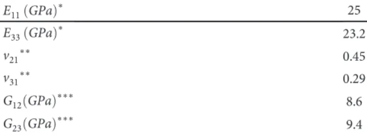
2.3. The Properties of Materials and Anatomical Structures. Young’s modulus and Poisson’s ratio, which describe the physical characteristics of each structure, were loaded into the software to identify the materials from which existing structures on the models prepared with Algor Fempro soft-ware were made (Table 1) [23–28]. Solid features were accepted as linearly resilient, homogenous, and isotropic in the program. It was assumed that the materials and anatom-ical structures were isotropic, linearly elastic, and homoge-neous, except for the glassfiber post and dentine. The glass fiber post was considered as orthotropic so that it showed different mechanical properties along the fiber direction (x direction) and along the other two directions y and z direc-tions (Table 2) [29]. The dentine of each model was assumed as orthotropic (Table 3) [30, 31]. In ANSYS, the postproces-sing function was used to create a stress distribution diagram. We then analyzed the stress distributions and stress concen-trations of the post and remaining dentin of the root for each modeled diameter of the post. For stress analysis, the
required values of mechanical properties of the materials and anatomical structures included Young’s modulus, Poisson’s ratio, and density structures as shown in Tables 1–4. These values must be imported into the software to identify the physical differences of each part of the models.
2.4. Determination of Contact Surfaces on the Models. All interfaces between the modeled materials and anatomical structures were considered completely/tightly adhered.
(a) (b)
(c) (d)
(e) (f)
Figure 1: (a) Model OB, only the fracture fragment was bonded. (b) Model CrPL, the fractured fragment was bonded and a ceramic palatinal laminate was designated, and model CmPL, the fractured fragment was bonded and a composite palatinal laminate was designated. (c) Model
CM, the fractured fragment was bonded and the coronal 1/3 of the root wasfilled using core material. (d) Model BP (bonding and post), the
fractured fragment was bonded and a glassfiber post inserted into the root was designated. (e) Model CP, composite resin restoration and
glassfiber post inserted into the root were designated without using the fractured fragment. (f) Model OC, composite resin restoration
was designated without using the fractured fragment or adding any other applications.
Table 1: Mechanical properties of homogeneous and isotropic default layers.
Material Elasticity modulus
(GPa) Poisson’s ratio (v) Reference Trabecular bone 1.37 0.30 23 Cortical bone 13.7 0.30 23 Enamel 84.1 0.33 24 Periodontal ligament 6.89× 10 −5 0.45 25 Gutta percha 0.14 0.45 26 Composite 16 0.3 27 Ceramic 96 0.22 24 Resin core material 7 0.3 28
Table 2: Mechanical properties of orthotropic glass fiber post [29].
Ex GPa ∗ 37 Ey GPa ∗ 9.5 Ez GPa ∗ 9.5 Gxy∗∗ 0.27 Gxz∗∗ 0.34 Gyz∗∗ 0.27 NUxy∗∗∗ 3.10 NUxz∗∗∗ 3.50 NUyz∗∗∗ 3.10 ∗E
x, Ey, and Ezshow the values of three-dimensional elasticity modules.
∗∗G
xy, Gxz, and Gyz show orthogonal cutting module values in the plane.∗∗∗NUxy, NUxz, and NUyzshow the Poisson’s ratios in the orthogonal plane.
2.5. Border Conditions. Zero motion and rotation were iden-tified at six degrees of freedom from the side and upper sur-faces of dental tissues.
2.6. Loading and Stress Analyses. To simulate the original occlusion, the models were constrained by a force of 100 N that was applied to an area over the cingulum on the palatal surface of the crowns of the models at a 135° angle to the long axis of the tooth (Figure 2). To calculate the stress distribution, the von Mises (equivalent stresses) energetic criterion was preferred [32]. The qualitative stress distribution analyses were recorded in this study using the von Mises criteria.
A view of a midsagittal and oblique-horizontal section from each model was provided to evaluate the stress distribu-tion. Calculated numeric data obtained from each model were transformed into color images. For all structures, the highest von Mises stress values were recorded (Table 4).
3. Results
von Mises stresses at the 3 regions of interest were measured. The von Mises stress distribution in all models and extreme stress values are presented in Figure 2. Table 5 shows the maximum von Mises stress values. At the tooth structure, the highest maximum von Mises stress values were observed in model BP for enamel, which is fractured fragment bonded and glassfiber post inserted into the designated root. How-ever, the lowest stress values were observed in model CmPL for which fractured fragment was bonded and composite
the highest stress values. It was anticipated that model CrPL (fractured fragment was bonded, and ceramic palatinal lam-inate was designated) had the lowest stress values. At dentine, the stress distribution patterns of all models were similar. The highest stress values were observed in model OB, and the lowest stress values were observed in model CM. In root dentine, the highest stress values were observed in model BP (fractured fragment was bonded, and glassfiber post inserted into the root was designated). The lowest stress values were observed in model CrPL (fractured fragment was bonded, and ceramic palatinal laminate was designated).
4. Discussion
In the present study, the FEA method was used to evaluate the pattern of stress distribution in different areas of end-odontically treated central incisor models restored with dif-ferent restorative approaches. The hypothesis of the study, which stated that the restoration approach would not affect the stress distribution of fractured endodontically treated central incisor teeth, was partially rejected.
The lowest values of the von Mises stress equivalents are observed at the connection interface of the models designed as fractured fragment bonded and ceramic or composite palatinal laminate designated. This result indicates that pala-tal laminate can protect the connection interface from stresses caused by occlusal forces. Similarly, in an experimen-tal model using sheep incisors, Andreasen et al. [21] found that the fracture strength was equal to that of intact incisors. This is in contrast to fracture strengths of reattached enamel-dentin tooth fragments without porcelain laminates, which were only 50% of intact incisors. It is suggested that porcelain laminate veneers may be used to supplement fragment bond-ing, enhancing dental esthetics and function. The results of the study support our hypothesis for the beneficial usage of palatinal laminate, which is a novel approach to reinforce the fracture fragments. However, there is no study on the usage of palatinal laminate for any possible clinical scenario. Anindya Bhalla et al. [33], in their recent case report, declared that when the fractured fragments are reattached with post for retention, post provides excellent retention with long-term stability of restored portion as in many previously reported cases [34, 35]. However, in this study, model BP, in which the fractured fragment was bonded and a glass fiber post was inserted into the root, is not superior to the other restoration alternatives. Even in the dental tissues and root surface, the highest stress values were observed in this model. The largest values of the von Mises stress equivalents indicate locations with the highest risk of fracture.
The use of core material (in model CM) has not shown a positive effect on the reduction of stress in the dental tissues, root surface, or connection interface. Moreover, at tooth
G12 GPa ∗∗∗ 8.6
G23 GPa ∗∗∗ 9.4
∗E: Young’s modulus;∗∗v: Poisson’s ratio;∗∗∗G: shear modulus.
Table 4: Density values of layers.
Material Density (10−6kg/mm3) Trabecular bone 1.3 Cortical bone 1.3 Enamel 2.6 Dentine 2.1 Periodontal ligament 1.04 Gutta percha 0.9 Composite 2.1 Ceramic 2.4
Resin core material 2.24
tissues and the root surface in model CM, the von Mises stress value is higher than OB. This result may be from the elastic modulus of the core material being lower than that of the composite resin. Yikilgan and Bala [36] reported that materials with a low elastic modulus cause high stress levels, whereas materials with elastic moduli similar to those of den-tal tissues cause low stress levels.
At the root dentine, the highest stress values were observed in model BP (the fractured fragment was bonded, and a glassfiber post inserted into the root was designated). Although reinforcing the restoration with afiber decreases the stress transmission and thefiber post structure accumu-lated more stress on its own body [37], the present study showed that the use of post material increased the stress values at the root dentine structure. Roscoe et al. [38] evalu-ated the effect of alveolar bone loss, post type, and ferrule presence on the biomechanical behavior of endodontically treated maxillary canines and found that 5.0 mm of bone loss significantly increased the stress concentration and strain on the root dentin. According to the evaluation of the maximum stress values, the maximum values occurred at the root den-tine. Palatinal laminate is thought to be a safe approach for the tooth, which has alveolar bone loss. However, when 2i (direct composite resin restoration and glassfiber post) and
OC (direct composite resin restoration) were compared at the connection interface and root surface in model 2i, the von Mises stress value was lower than CP. It was observed that the use of afiber post reduces the stress buildup when fractured parts cannot be used.
These results demonstrated that palatinal laminate is an acceptable restorative approach for decreasing the stress con-centration at the connection interfaces when autologous reat-tachment is preferred as a restorative option. When compared to laminate groups at enamel, the maximum von Mises stress value in model CrPL (in which the fractured fragment was bonded and a ceramic palatinal laminate was designated) is higher than that in model CmPL (in which the fractured fragment was bonded and a composite palatinal laminate was designated). The high elastic modulus of ceramic materials most likely causes this difference. Compos-ite materials have mechanical properties that are similar to those of dentin. Consequently, they behave like monobloc units and withstand chewing forces [39].
The biomechanical behavior of endodontically treated teeth can be investigated with fracture strength tests and stress analysis methods. Fracture strength tests illustrate the maximum strength that the tooth endures until it becomes fractured. However, stress analysis methods can show
long-(a) (b) (c) (d)
(e) (f) (g) (h)
(i) (j)
Figure 2: (a) At the tooth structure, the highest maximum von Mises stress values were observed in model BP for enamel. (b) The lowest stress values were observed in model. (c) For dentine tissue, the highest stress values were observed in model CrPL. (d) The lowest stress values were found in model CP. (e) On connection interfaces, at enamel, model CP had the highest stress values. (f) Model CrPL had the lowest stress values. (g) At dentine, the highest stress values were observed in model OB. (h) The lowest stress values were observed in model CM. (i) In root dentine, the highest stress values were observed in model BP. (j) The lowest stress values were observed in model CrPL.
term deformations that can occur in the dental tissues and restorative materials by masticator forces. In the stress anal-ysis test,finite element analysis is considered ideal in terms of enabling reproducible situations and repeatable results. Because of these advantages, we used thefinite element anal-ysis in our study.
One of the limitations of FEA studies is that when the models are created, dentine has been modelled as a homoge-neous/isotropic material, the structure of which is generally assumed to be homogeneous and isotropic [27, 40, 41]. In this study, the layer represented as dentine structure was con-sidered orthotropic material. Therefore, the effects of den-tinal tubules, intrapulpal hydrostatic pressure, and the elastic modulus gradient on the mechanical properties of dentine were not ignored [42].
Endodontically treated teeth are susceptible to biome-chanical failure. The main factors of the tendency of such teeth to fracture are the loss of tooth tissue, altered physical properties of dentine, and altered proprioception/nocicep-tion, which cumulatively interact to influence the tooth load-ing and distribution of stresses [43, 44]. Therefore, the choice of the esthetic restorative treatment of fractured anterior teeth poses dilemmas to clinicians [45]. Direct composite resin restoration, fragment reattachment, and ceramic resto-rations (full crowns, laminate veneers, or ceramic fragments) are treatment options that may be preferred according to clinical situations or based on clinical decision. Two impor-tant criteria, such as the aesthetics and function of the diseased tooth, should be provided to perform an ideal treat-ment for a complicated crown fracture, which is dictated by various factors. With the significant development of adhesive
concluded the following:
(i) The model restored with only composite resin material had high stress values at tooth tissues. Hence, fragment reattachment is superior to com-posite restorations, although it may not match the intact tooth.
(ii) The viability of tooth fragment reattachment along with post insertion as a restorative technique is unclear.
(iii) Tooth fragment reattachment along with palatinal laminate is a viable technique.
(iv) Palatinal laminate can safely be used as a novel restor-ative approach for teeth with alveolar bone loss. In any possible clinical scenario, reattachment of the fractured tooth segment should be attempted as a priority. Reattachment of fractured fragments can be considered a good alternative treatment option when the fractured fragment is available. However, additional applications are required to prevent biomechanical failure of the tooth.
Conflicts of Interest
The authors declare that they have no conflicts of interest.
References
[1] W. M. Fennis, R. H. Kuijs, C. M. Kreulen, F. J. Roeters, N. H.
Creugers, and R. C. Burgersdijk,“A survey of cusp fractures
in a population of general dental practices,” The International
Journal of Prosthodontics, vol. 15, no. 6, pp. 559–563, 2002.
[2] D. Arola and R. K. Reprogel,“Effects of aging on the
mechan-ical behavior of human dentin,” Biomaterials, vol. 26, no. 18,
pp. 4051–4061, 2005.
[3] C. J. Soares, F. R. Santana, N. R. Silva, J. C. Preira, and C. A.
Pereira,“Influence of the endodontic treatment on mechanical
properties of root dentin,” Journal of Endodontics, vol. 33,
no. 5, pp. 603–606, 2007.
[4] W. Karzoun, A. Abdulkarim, A. Samran, and M. Kern,
“Frac-ture strength of endodontically treated maxillary premolars
supported by a horizontal glassfiber post: an in vitro study,”
Journal of Endodontics, vol. 41, no. 6, pp. 907–912, 2015.
[5] K. Bitter, J. Noetzel, O. Stamm et al.,“Randomized clinical trial
comparing the effects of post placement on failure rate of
post-endodontic restorations: preliminary results of a mean period of 32 months,” Journal of Endodontics, vol. 35, no. 11,
pp. 1477–1482, 2009.
[6] P. L. B. Tan, S. A. Aquilino, D. G. Gratton et al.,“In vitro
frac-ture resistance of endodontically treated central incisors with
varying ferrule heights and configurations,” The Journal of
Prosthetic Dentistry, vol. 93, no. 4, pp. 331–336, 2005. (1) Groups that used fractured fragment for construction
OB Enamel 17.555 Enamel 43.528 30.432 Dentine 5.5604 Dentine 6.5187 CrPL Enamel 10.82 Enamel 14.999 29.456 Dentine 9,0007 Dentine 5,1146 CmPL Enamel 10.347 Enamel 18.442 31.256 Dentine 7.3247 Dentine 6.0642 CM Enamel 20.929 Enamel 39.193 40.146 Dentine 6.4163 Dentine 4.1036 BP Enamel 22.816 Enamel 38.619 47.999 Dentine 7.0146 Dentine 5.5755
(2) Groups that did not use fractured fragment for construction
CP Enamel 17.263 Enamel 33.651 40.127
Dentine 6.5185 Dentine 5.096
OC Enamel 16.399 Enamel 45.521 42.04
[7] S. F. Chuang, P. Yaman, A. Herrero, J. B. Dennison, and C. H.
Chang,“Influence of post material and length on
endodonti-cally treated incisors: an in vitro and finite element study,”
The Journal of Prosthetic Dentistry, vol. 104, no. 6, pp. 379–
388, 2010.
[8] G. V. Macedo, P. I. Diaz, C. A. D. O. Fernandes, and A. V.
Ritter,“Reattachment of anterior teeth fragments: a
conserva-tive approach,” Journal of Esthetic and Restorative Dentistry,
vol. 20, no. 1, pp. 5–18, 2008.
[9] J. O. Andreasen and F. M. Andreasen, Textbook and Color Atlas of Traumatic Injuries to the Teeth, Mosby- Year Book, Inc, St. Louis, 3rd ed edition, 1994.
[10] A. Caprioglio, V. Conti, C. Caprioglio, and D. Caprioglio,“A
long-term retrospective clinical study on MTA pulpotomies in immature permanent incisors with complicated crown
frac-tures,” European Journal of Paediatric Dentistry, vol. 15, no. 1,
pp. 29–34, 2014.
[11] F. C. S. Chu, T. M. Yim, and S. H. Y. Wei,“Clinical
consider-ations for reattachment of tooth fragments,” Quintessence
International, vol. 31, no. 6, pp. 385–391, 2000.
[12] M. D. Turgut, N. Gönül, and N. Altay,“Multiple complicated
crown–root fracture of a permanent incisor,” Dental
Trauma-tology, vol. 20, no. 5, pp. 288–292, 2004.
[13] R. J. Simonsen,“Traumatic fracture restoration: an alternative
use of the acid etch technique,” Quintessence International,
Dental Digest, vol. 10, pp. 15–22, 1979.
[14] E. Amir, B. Bar-Gil, and H. Sarnat,“Restoration of fractured
immature maxillary central incisors using the crown frag-ments,” Pediatric Dentistry, vol. 8, pp. 285–288, 1996. [15] F. M. Andreasen, J. G. Norén, J. O. Andreasen, S. Engelhardtsen,
and U. Lindh-Strömberg, “Long-term survival of fragment
bonding in the treatment of fractured crowns: a multicenter
clinical study,” Quintessence International, vol. 26, no. 10,
pp. 669–681, 1995.
[16] A. J. Diangelis and M. Jungbluth,“Reattaching fractured tooth
segments: an esthetic alternative,” The Journal of the American
Dental Association, vol. 123, no. 8, pp. 58–63, 1992.
[17] R. VamsiKrishna, K. Madhusudhana, A. Swaroopkumarreddy,
A. Lavanya, C. Suneelkumar, and G. Kiranmayi,“Shear bond
strength evaluation of adhesive and tooth preparation combi-nations used in reattachment of fractured teeth: an ex-vivo
study,” Journal of Indian Society of Pedodontics and Preventive
Dentistry, vol. 33, no. 1, pp. 40–43, 2015.
[18] L. C. Martens, H. M. Beyls, L. G. de Craene, and R. F.
D'Hauwers,“Reattachment of the original fragment after
ver-tical crown fracture of a permanent central incisor,” The
Jour-nal of Pedodontics, vol. 13, no. 1, pp. 53–62, 1988.
[19] A. Reis, C. Francci, A. D. Loguercio, M. R. Carrilho, and
L. E. Rodriques Filho,“Re-attachment of anterior fractured
teeth: fracture strength using different techniques,” Operative
Dentistry, vol. 26, no. 3, pp. 287–294, 2001.
[20] M. Manju, S. L. Shanthraj, K. C. Savitha, and N. Sethi,“Esthetic
and biologic mode of reattaching incisor fracture fragment
uti-lizing glassfiber post,” Journal of Natural Science, Biology and
Medicine, vol. 6, no. 2, pp. 446–448, 2015.
[21] F. M. Andreasen, E. Flugge, J. Daugaard-Jensen, and E. C.
Munksgaard, “Treatment of crown fractured incisors with
laminate veneer restorations. An experimental study,” Dental
Traumatology, vol. 8, no. 1, pp. 30–35, 1992.
[22] V. L. Ortega, L. F. Pegoraro, P. C. R. Conti, A. L. Valle, and
G. Bonfante, “Evaluation of fracture resistance of
endodontically treated maxillary premolars, restored with
ceromer or heat-pressed ceramic inlays and fixed with
dual-resin cements,” Journal of Oral Rehabilitation, vol. 31, no. 4,
pp. 393–397, 2004.
[23] C.-C. Ko, C.-S. Chu, K.-H. Chung, and M.-C. Lee,“Effects of
posts on dentin stress distribution in pulpless teeth,” The
Journal of Prosthetic Dentistry, vol. 68, no. 3, pp. 421–427, 1992.
[24] F. Zarone, R. Sorrentino, D. Apicella et al.,“Evaluation of the
biomechanical behavior of maxillary central incisors restored by means of endocrowns compared to a natural tooth: a 3D
static linear finite elements analysis,” Dental Materials,
vol. 22, no. 11, pp. 1035–1044, 2006.
[25] J. S. Rees, P. H. Jacobsen, and J. Hickman,“The elastic
modu-lus of dentine determined by static and dynamic methods,”
Clinical Materials, vol. 17, no. 1, pp. 11–15, 1994.
[26] C. Veríssimo, P. C. Sinamoto Júnior, C. J. Soares, P. Y. Noritomi,
and P. C. F. Santos-Filho, “Effect of the crown, post, and
remaining coronal dentin on the biomechanical behavior of endodontically treated maxillary central incisors,” The Journal
of Prosthetic Dentistry, vol. 111, no. 3, pp. 234–246, 2014.
[27] G. Eskitascioglu, S. Belli, and M. Kalkan,“Evaluation of two
post core systems using two different methods (fracture
strength test and a finite elemental stress analysis),” Journal
of Endodontics, vol. 28, no. 9, pp. 629–633, 2002.
[28] H. Kermanshah, A. Geramy, S. F. Ebrahimi, and T. Bitaraf, “IPS-Empress II inlay-retained fixed partial denture reinforced
with zirconia bar: three-dimensional finite element and
in-vitro studies,” Acta Odontologica Scandinavica, vol. 70, no. 6,
pp. 569–576, 2012.
[29] A. Lanza, R. Aversa, S. Rengo, D. Apicella, and A. Apicella, “3D FEA of cemented steel, glass and carbon posts in a
maxil-lary incisor,” Dental Materials, vol. 21, no. 8, pp. 709–715,
2005.
[30] J. H. Kinney, S. J. Marshall, and G. W. Marshall,“The
mechan-ical properties of human dentin: a critmechan-ical review and re-evaluation of the dental literature,” Critical Reviews in Oral
Biology & Medicine, vol. 14, no. 1, pp. 13–29, 2003.
[31] M. Ferrari, R. Sorrentino, F. Zarone, D. Apicella, R. Aversa,
and A. Apicella,“Non-linear viscoelastic finite element
analy-sis of the effect of the length of glass fiber posts on the
biome-chanical behaviour of directly restored incisors and
surrounding alveolar bone,” Dental Materials Journal,
vol. 27, no. 4, pp. 485–498, 2008.
[32] L. J. Ricks-Williamson, P. G. Fotos, V. K. Goel, J. D. Spivey,
E. M. Rivera, and S. C. Khera, “A three-dimensional
finite-element stress analysis of an endodontically prepared maxil-lary central incisor,” Journal of Endodontics, vol. 21, no. 7,
pp. 362–367, 1995.
[33] A. Bhalla, R. K. Khatri, A. Choudhary, and R. Garg, “Tooth
fragment reattachment: an esthetic, biological restoration,”
Journal of Natural Science, Biology and Medicine, vol. 6, no. 1, pp. 205–207, 2015.
[34] G. J. Christensen,“When to use fillers, build-ups or posts and
cores,” The Journal of the American Dental Association, vol. 127, no. 9, pp. 1397-1398, 1996.
[35] M. Hayashi, Y. Takahashi, S. Imazato, and S. Ebisu,“Fracture
resistance of pulpless teeth restored with post-cores and
crowns,” Dental Materials, vol. 22, no. 5, pp. 477–485, 2006.
[36] İ. Yıkılgan and O. Bala, “How can stress be controlled in
“Influence of alveolar bone loss, post type, and ferrule presence on the biomechanical behavior of endodontically treated
max-illary canines: strain measurement and stress distribution,”
The Journal of Prosthetic Dentistry, vol. 110, no. 2, pp. 116–
126, 2013.
[39] F. R. Tay and D. H. Pashley, “Monoblocks in root canals:
a hypothetical or a tangible goal,” Journal of Endodontics,
vol. 33, no. 4, pp. 391–398, 2007.
[40] S. Belli, O. Eraslan, O. Eraslan, M. Eskitascioglu, and
G. Eskitascioglu,“Effects of NaOCl, EDTA and MTAD when
applied to dentine on stress distribution in post-restored roots
withflared canals,” International Endodontic Journal, vol. 47,
no. 12, pp. 1123–1132, 2014.
[41] J. Borcic, I. Anic, I. Smojver, A. Catic, I. Miletic, and S. P.
Ribaric,“3D finite element model and cervical lesion
forma-tion in normal occlusion and in malocclusion,” Journal of
Oral Rehabilitation, vol. 32, no. 7, pp. 504–510, 2005.
[42] A. Kishen and S. Vedantam,“Hydromechanics in dentine: Role
of dentinal tubules and hydrostatic pressure on mechanical
stress–strain distribution,” Dental Materials, vol. 23, no. 10,
pp. 1296–1306, 2007.
[43] K. Gulabivala,“Restoration of the root-filled tooth,” in Color
Atlas and Text of Endodontics, K. Gulabivala, R. T. Walker, J. R. Goodman, and C. J. R. Stock, Eds., pp. 241–272, Times Mir-ror International Publishers, Mosby-Wolfe, London, 2nd edi-tion, 1995.
[44] S. Belli, Ö. Eraslan, O. Eraslan, and G. Eskitaşcıoğlu, “Effect of
restoration technique on stress distribution in roots withflared
canals: an FEA study,” The Journal of Adhesive Dentistry,
vol. 16, no. 2, pp. 185–191, 2014.
[45] K. P. Bharath, R. U. Patil, H. V. Kambalimath, and
A. Alexander, “Autologous reattachment of complicated
crown fractures using intra canal anchorage: report of two cases,” Journal of Indian Society of Pedodontics and Preventive
International Journal of
Aerospace
Engineering
Hindawi www.hindawi.com Volume 2018Robotics
Journal of Hindawi www.hindawi.com Volume 2018 Hindawi www.hindawi.com Volume 2018Active and Passive Electronic Components VLSI Design Hindawi www.hindawi.com Volume 2018 Hindawi www.hindawi.com Volume 2018
Shock and Vibration
Hindawi
www.hindawi.com Volume 2018
Civil Engineering
Advances inAcoustics and VibrationAdvances in
Hindawi
www.hindawi.com Volume 2018
Hindawi
www.hindawi.com Volume 2018
Electrical and Computer Engineering Journal of Advances in OptoElectronics Hindawi www.hindawi.com Volume 2018
Hindawi Publishing Corporation
http://www.hindawi.com Volume 2013 Hindawi www.hindawi.com
The Scientific
World Journal
Volume 2018 Control Science and Engineering Journal of Hindawi www.hindawi.com Volume 2018 Hindawi www.hindawi.com Journal ofEngineering
Volume 2018Sensors
Journal of Hindawi www.hindawi.com Volume 2018 Machinery Hindawi www.hindawi.com Volume 2018 Modelling & Simulation in Engineering Hindawi www.hindawi.com Volume 2018 Hindawi www.hindawi.com Volume 2018 Chemical EngineeringInternational Journal of Antennas and
Propagation International Journal of Hindawi www.hindawi.com Volume 2018 Hindawi www.hindawi.com Volume 2018 Navigation and Observation International Journal of Hindawi www.hindawi.com Volume 2018