63
Therapeutic plasma exchange in neurologic diseases: An experience
with 91 patients in Seven Years
Nörolojik Hastalıklarda Terapötik Plazma Değişimi: 91 Hasta ile Yedi Yıllık Deneyim Sonuçları
Cor res pon den ce Ad dress/Ya z›fl ma Ad re si
Sibel Karaca MD, Başkent University Faculty of Medicine, Adana Research and Implementation Center, Department of Neurology, Adana, Turkey Gsm: +90 505 675 74 47 E-mail: [email protected] received/ge liş ta ri hi: 09.10.2012 Ac cep ted/Ka bul ta ri hi: 04.12.2012
© Arc hi ves of Neu ropsy chi atry, pub lis hed by Ga le nos Pub lis hing. / © Nö rop si ki yat ri Ar şi vi Der gi si, Ga le nos Ya yı ne vi ta ra f›n dan ba s›l m›fl t›r.
Sibel KArAcA1, İlknur KozAnoĞlu2, Başak KArAKuruM gÖKSel1, Mehmet KArATAş1, Meliha TAn1, V. deniz Yerdelen1, Semih gİrAY1, zülfikar Arlıer1,
1Başkent University Faculty of Medicine, Adana Research and Implementation Center, Department of Neurology, Adana, Turkey 2Başkent University Faculty of Medicine, Adana Research and Implementation Center, Department of Hematology, Adana, Turkey
ÖZET
Amaç: Bu çalışmada, terapötik plazma değişimi (TPD) ile tedavi ettiğimiz nöro-immünolojik olgulara ait 7 yıllık deneyimimizin sonuçları rapor edilmiştir.
Yön tem : TPD uyguladığımız 91 olgunun (53 erkek, 38 kadın) medikal kayıtları geriye dönük olarak incelenmiştir.
Bul gu lar: Tanısal olarak sınıflandığında, bu olguların 60’ının Guillain-Barre sendromu (GBS), 23’ünün Miyastenia Gravis (MG), 4’ünün ise kronik inflammatuar demiyelinizan polinöropati (KİDP) tanısıyla TPD aldığı görülmüştür. Birer olguya ise, polimiyozit, septik ensefalopati, akut dissemine ensefalomiyelit (ADEM) ve Opsoklonus-Myoklonus sendromu (OMS) tanılarıyla TPD uygulanmıştır.
GBS hastalarımızın %26,7’sinde tam düzelme, %61,7’sinde kısmi düzelme izlenmiş olup, disabilitesi yüksek %11,7 hasta solunum yetmezliği nedeniyle kaybedilmiştir. MG’li hastaların %13,4’ünde tedaviye rağmen ölüm, %78’inde tam klinik düzelme gözlenmiştir. KIDP’li 4 hastamızın üçünde total, birinde kısmi düzelme gözlenmiş, ADEM’li olgumuz TPD ile önce kısmen düzelmiş ancak tedaviden 2 ay sonra aspirasyon pnömonisine bağlı solunum yetmezliği nedeniyle kaybedilmiş, polimiyozitli olgumuzda kısmi, septik ensefalopati ve OMS’li hastalarımızda tam düzelme gözlenmiştir. TPE uygulamasında karşılaştığımız yan etkiler hipotansiyon, hipokalsemi ve anemi gibi hafif ve yönetilebilir düzeydedir.
So nuç: Çalışmamızın sonuçları otoimmün kökenli nörolojik hastalıklarda TPE tedavisinin etkili ve güvenilir bir yöntem olduğunu göstermektedir (Nö rop si ki yat ri Ar fli vi 2014; 51: 63-68)
Anah tar ke li me ler: Terapötik Plazma Değişimi, Guillain-Barre sendromu, Kronik İnflammatuar Demiyelinizan Polinöropati, Miyastenia Gravis
çıkar çatışması: Yazarlar bu makale ile ilgili olarak herhangi bir çıkar çatışması bildirmemişlerdir.
ABS TRACT
ıntroduction: In this study, we report the results of our experience of therapeutic plasma exchange (TPE) for neuroimmunologic disorders performed at our hospital over a seven-year period.
Met hods: We retrospectively reviewed the medical records of 91 patients (53 male, 38 female) who had been treated at our center with TPE.
re sults: 60 patients with Guillain-Barrè syndrome (GBS), 23 with myasthenia gravis (MG), 4 with chronic inflammatory demyelinating polyneuropathy (CIDP) and 1 patient each with polymyositis, septic encephalopathy, acute disseminated encephalomyelitis (ADEM) and Opsoclonus-Myoclonus syndrome (OMS) received TPE. 26.7% of GBS patient’s made complete recovery, 61.7% had partial recovery and 11.7% patients died due to respiratory failure. Despite our best efforts and effective TPE treatments, 13.4% of MG patients deceased, however, 78% had full recovery. Three patients with CIDP were discharged with full and 1 patient with partial recovery. The patient with ADEM had partial recovery with TPE at first, but deceased 2 months later due to pneumonia-related respiratory insufficiency. While, patient with polymyositis had slight-partial recovery, we obtained full recovery with TPE in septic encephalopathy and OMS patients. The side effects and complications of treatments with TPE, which included hypotension, hypocalcaemia and anemia, were mild and manageable.
conc lu si on: The improvement rates were encouraging and we concluded that significant benefit can be achieved with TPE for the treatment of neuroimmunological disorders. (Arc hi ves of Neu ropsy chi atry 2014; 51: 63-68) Key words: Therapeutic Plasma Exchange, Guillain-Barre syndrome, Chronic Inflammatory Demyelinating Polyneuropathy, Myasthenia Gravis
conflict of interest: The authors reported no conflict of interest related to this article.
Arc hi ves of Neu ropsy chi atry 2014; 51: 63-68 Nö rop si ki yat ri Ar fli vi 2014; 51: 63-68
Introduction
Therapeutic plasma exchange (TPE) has been used to remove immunoglobulins and other immunologically active substances, such as complements or cytokines, from the blood for the treatment of neurologic diseases in which autoimmunity plays a major role (1). The number of diseases treated with TPE increases with further understanding of the etiopathogenesis of neurologic diseases and improved techniques. It is a standard treatment regimen for some neurologic diseases, such as GBS, MC and CIDP (2,3,4,5,6,7,8). In a recent report of the Therapeutics and Technology and Assessment Subcommittee of the American Academy of Neurology, TPE was established as an effective course of treatment for many diseases; it is offered in cases of severe acute inflammatory demyelinating polyneuropathy (AIDP)/GBS, in the short-term management of CIDP (Class I, Level A), and is probably effective and should be considered for mild AIDP/GBS (9). There have been some other case reports and small studies in which it was claimed that TPE might be effective for some other neurologic diseases such as multiple sclerosis (10), neuromyelitis optica (11), acute disseminated encephalomyelitis (ADEM) (12), Stiff-man syndrome (13), Bickerstaff’s encephalitis (14) and hemorrhagic leucoencephalitis (15). Additionally, it has been suggested that TPE may also be successful in treating complications of the central nervous system resulting from systemic hematologic diseases such as thrombocytopenic purpura (16). An alternative treatment option for TPE is intravenous immunoglobulin (IVIG), however, IVIG is very expensive and in many countries is not covered by insurance (17). Plasma exchange typically requires central venous access that can lead to severe complications such as thrombosis, septic infections or pneumothorax. However, TPE is a safe procedure for the treatment of appropriate neurological illnesses in a specialized unit with a high patient volume (9).
Neurologic disorders constitute the largest group of indications for TPE and, the number is increasing due to growing knowledge of pathogenic relevance of auto-antibodies. Although some traditional indications are supported by properly designed randomized trials, others are not. In order to determine the utility of plasmapheresis in different diseases, especially in rarely encountered diseases, we need more results. Therefore, we aimed to make contribution to the literature by reporting our results, considering other publications on this subject.
Methods
We reviewed the medical records of 91 neurologic patients who had been consecutively treated with TPE therapy between January 2004 and June 2011, in the Department of Neurology at Başkent University Faculty of Medicine, Adana Research and Practice Center. During this period, 457 procedures of TPE were applied to 91 patients with neurologic disorders.
The medical records were analyzed for the patients’ demographic details (sex and age), types of neuroimmunologic
diseases, treatment modalities, prognoses, and any complications of the treatment.
Neurological indications included GBS, MG, CIDP, polymyositis, septic encephalopathy, OMS and ADEM. There were 38 female and 53 male patients. The mean age was 51.1±16.8 years with a range of 17-80 years (Table 1).
All TPE procedures were done via central venous catheter. The device used in all procedures was COBE Spectra (Lakewood, Colorado, USA) which works with continuous flow. Total blood and plasma volume were calculated by using standard formulation. Albumin-saline, ISOHES, ringer lactate and fresh frozen plasma were selected as replacement fluids according to the patient’s clinical and laboratory parameters. The total protein, albumin and calcium levels of all patients were measured before and after each TPE therapy. TPE was given every other day for most of the patients and a total of 1.51 plasma volumes were exchanged per cycle. The mean TPE session number was 5.06±1.73 and the mean plasma volume was 50 ml/kg for each cycle.
For patients with GBS and CIDP, we considered a mean improvement in the disability grade, measured 4 weeks after the end of the TPE therapy and based on the 7-point Hughes Disability Grade Scale (18) and a mean improvement in the Osserman scale (19) for myasthenic patients, as the primary outcome measure. We have already been using the Hughes Disability Grading Scale for the monitoring of efficacy of TPE for the inflammatory neuropathy patients, the scores were taken from the patients’ files. In addition, for the resolution of crises in myasthenic cases, we considered the duration of ventilation, time needed for independent walking, residual disabilities, and death, 4 weeks after treatment as secondary outcome measures. We also recorded the complications due to TPE. The approvals for off-label treatments were used.
Table 1. Diagnostic and demographic details of the patients treated with plasmapheresis
diagnosis number of the patients Mean age±Sd Sex (Female/ Male) GBS 60 52.5±15.86 27/33 CIDP 4 49.3±17 2/2 MG (prethymectomy) 9 41.1±18.8 5/4 MC 14 51.6±15.6 3/11 ADEM 1 28 0/1 Septic Encephalopathy 1 18 1/0 Polymyositis 1 51 0/1 OMS 1 49 0/1
GBS: Guillain Barré Syndrome, CIDP: Chronic Inflammatory Demyelinating Polyneuropathy, MG: Myasthenia Gravis, MC: Myasthenic Crisis, ADEM: Acute Disseminated Encephalomyelitis, OMS: Opsoclonus Myoclonus Syndrome
Results
GBS
The clinical diagnoses, the disability levels accordingly (18), and demographic features of the patients are summarized in Table 1 and 2. Twenty (33.3%) patients with GBS were not able to walk without assistance (Hughes 3), 25 patients (41.7%) were confined to a wheelchair or bed (Hughes 4), and 15 patients (25%) were in need of mechanical ventilation (Hughes 5). In all cases, TPE therapy was started within 10 days after the onset of the disease. Table 3 summarizes the prognoses of these patients. In a four-week therapy period, during which all patients also received physical therapy and rehabilitation, 16 patients (26.7%) experienced complete recovery (≥4 degree improvement in grades), 37 patients (61.7%) showed partial improvement (2-3 degree improvement in grades) and, when they were discharged, all of them were able to walk unaided or with little support. Seven GBS patients died (11.7%) with higher initial Hughes scores and respiratory failure. Among deceased patients from GBS, only 4 completed all the TPE sessions, 2 received only 1
session, and 1 received 2 sessions. Two of deceased patients suffered from acute motor sensory axonal neuropathy (AMSAN), and 5 of them had AIDP. Fifteen of 60 GBS patients were classified as acute motor axonal neuropathy (AMAN), among which there were no deaths.
CIDP
All of the CIDP patients required assistance with walking when first admitted to hospital and they either had not benefitted from at least 3-month corticosteroid treatment, or it had caused marked blood glucose elevation. However, after TPE therapy, all of them improved significantly. One of the patients was already on a cyclic TPE treatment since 2 years; he had not benefitted from steroid treatment which he had received at another center for 1 year, but we were able to achieve significant improvement with 5 cycles of TPE in one of his exacerbation periods. Another male patient, who was initially treated with IVIG successfully, had to be treated with cyclic TPE due to insurance problems and marked improvement was recorded. One female patient was treated with methylprednisolone for 3 months with no improvement, but prominent recovery was achieved with 6 cycles of TPE. A second female patient was treated for 3 exacerbations of CIDP with TPE due to steroid-related severe blood glucose elevation and unresponsiveness to IVIG in the 6th months of her diagnosis. Additionally, all the patients with CIDP were on azathioprine treatment (2 mg/kg/day).
MG
Twenty tree patients with MG were treated with TPE therapy; 9 (F/M: 4/5) received TPE therapy during the preparation stages of thymectomy, and 14 (F/M: 3/11) were treated for either MC’s (6) or severe bulbar signs and myasthenia-related respiratory insufficiency (8). The patients with bulbar signs were treated with TPE to quickly stabilize the clinical outlook and to prevent deterioration of their clinical status because of newly started 1 mg/kg prednisone daily. Their Osserman gradings are shown on Table 4. Three patients died in myasthenic group; 1 due to infection in the early post-thymectomy period (day 4); 2 due to MC. Last 2 died in their second crises and, they had needed 10 sessions of TPE for the resolution of their first crises: One of them had congenital lung anomaly, and critical illness neuropathy was observed in the second. In addition, second crises of these patients were more severe and they died in the initial 1-2 sessions of TPE.
Table 2. Demographics, electrophysiological subtypes (for the cases with GBS) and Hughes grading’s of the cases with demyelinating polyneuropathy treated with TPE number of the patients Aıdp n=35 AMAnn=15 AMSAnn=10 cıdpn=4 F/M 16/19 8/7 3/7 2/2 Mean Age 52.5±13.2 52.6±21.1 58.1±19.2 49.3±17 Grade 0 0 0 0 0 Grade 1 0 0 0 0 Grade 2 0 0 0 1 Grade 3 13 4 3 3 Grade 4 14 7 5 0 Grade 5 8 4 2 0 Grade 6 0 0 0 0
GBS: Guillain Barre Syndrome, TPE: Therapeutic Plasma Exchange, AIDP: Acute Inflammatory Demyelinating Polyneuropathy, AMAN: Acute Motor Axonal Neuropathy, AMSAN, Acute Motor Sensory Axonal Neuropathy, CIDP: Chronic Inflammatory Demyelinating Polyneuropathy
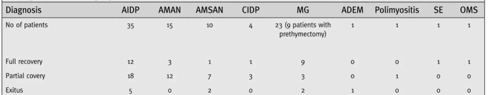
Table 3. Details of the prognoses of cases after the treatment with TPE
diagnosis Aıdp AMAn AMSAn cıdp Mg AdeM polimyositis Se oMS
No of patients 35 15 10 4 23 (9 patients with
prethymectomy) 1 1 1 1 Full recovery Partial covery Exitus 12 18 5 3 12 0 1 7 2 1 3 0 9 3 2 0 0 1 0 1 0 1 0 0 1 0 0
AIDP: Acute Inflammatory Demyelinating Polyneuropathy, AMAN: Acute Motor Axonal Neuropathy, AMSAN: Acute Motor Sensory Axonal Neuropathy, CIDP: Chronic Inflammatory Demyelinating Polyneuropathy, MG: Myasthenia Gravis, ADEM: Acute Demyelinating Encephalomyelitis, SE: Septic Encephalopathy, OMS: Opsoclonus Myoclonus Syndrome
The targeted neurological deficits of the remaining 12 patients were improved by TPE therapy. Nine had a complete recovery, while the bulbar signs were resolved in 3 patients with MC. None of our myasthenic patients received IVIG treatment.
In addition, 7 patients from the prethymectomy group and 7 patients from MC group were found to be positive for AchR antibodies, and 1 patient in the MC group was positive for anti-titin. The mean duration of the disease was estimated to be 12 months in the MC patients and 18 months in the prethymectomy group.
Polymyositis
A 51-year-old man has complained severe quadriparesis since 6 months. Elevation of creatine kinase and pseudomyotonic discharges on electromyography were shown. Muscle biopsy revealed necrotizing myopathy. We performed 7 sessions of TPE therapy on alternate days, but the patient achieved only slight improvement in a 4-week period. IVIG therapy also proved ineffective. However, the patient made a complete recovery after a 3-month azathioprine therapy with 2.5 mg /kg/day.
Septic Encephalopathy
An 18-year-old girl showing a confused state of mind was examined in the intensive care unit. She had facial cellulitis and it had progressed into septic state. She made a full recovery with 3 cycles of TPE.
OMS
A 49-year-old man with rapid, dysrhythmic and uncoordinated involuntary eye movements and ataxia by 4-day was hospitalized. It is known that most adults with OMS have neoplastic, infectious, metabolic, or idiopathic etiologies. We excluded the possibility of a cancer with appropriate investigations. After the treatment failed with steroids (1 g/day for 5 days), we got full recovery by 5 courses of TPE on alternate day.
ADEM
A 28-year-old man was referred with 2-month duration of severe quadriparesis, he was unresponsive to 7-day high-dose methylprednisolone pulses and also, IVIG (35 gram/day) for 5 days had been failed. He received 7 sessions of TPE on alternate days, and underwent respiratory physiotherapy. Marked improvement was achieved in breathing problems, and the patient was removed from mechanical ventilation. He
was discharged as quadriplegic but, 2 months after he was hospitalized with pneumonia due to aspiration, and he died.
Results
Thirty-one of our 91 patients made a complete recovery, and 49 showed partial improvement. Despite our best efforts, 11 of our patients died.
During the procedures, no patient mortality was related to the TPE therapy. A total of 7 patients; 3 GBS patients, 2 MG patients and 2 CIDP patients experienced adverse events. In our patients, only one of them had tetany due to hypocalcaemia during apheresis, and the procedure was stopped until the calcium levels were restored. Mild and manageable complications such as hypotension and hypocalcaemia were also observed in these 7 patients.
Discussion
It is believed that cross reaction between the antibodies against a microbial agent and the neuronal tissues cause GBS (20). GBS is a major cause of acute neuromuscular paralysis with an annual incidence of 1.3-2 per 100 000 throughout the world (21), and is treated both medically and supportively. Medical treatment methods include IVIG and plasmapheresis, although TPE was introduced as a possible alternative treatment in 1978 (22), and was shown to offer significant benefit by a randomized trial published in 1985 (23). In a study comparing the effects of TPE, IVIG, and a combined regimen of TPE followed by IVIG, no difference was found in terms of clinical outcome between the three groups (24). The Cochrane systematic meta-analysis reported that TPE was the only treatment for GBS found to be superior to supportive treatment (25). Furthermore, TPE was more beneficial when applied within the first 7 days of the disease, although it was still effective when performed within the first month (25). Sixteen (26.7%) of our GBS patients made a complete recovery, 37 (61.7%) had partial recovery improvement with 2-3 degrees on the Hughes scale after TPE at the end of first month, and 7 (11.7%) died. However, no other treatment modalities have been adequately tested in these deceased GBS patients. Yucesan et al. reported that their all 6 patients with GBS improved with TPE treatment and their recovery rate was 100% (7). In our experience, the recovery rate of GBS patients was 88.3%, but our study population was 10 times larger with 60 GBS patients. In the report of Van Doorn et al. related to GBS patients treated with TPE, 3-10% of patients died and 20% were unable to walk after 6 months (26). In our study group, 11.7% of GBS patients deceased; these proportions are similar with the literature.
Overall, we may say that TPE is effective for GBS and improved therapeutic strategies are urgently needed for the unresponsive cases. When we followed TPE with IVIG therapy, significant improvement was seen in the GBS patients, and in particular in those with AMAN. However, a study by the Plasma Exchange/Sandoglobulin Guillain-Barré Syndrome Table 4. Osserman grading’s of the patients with
Myasthenia Gravis
numbers of patients n=23
F/M 7/16
Mean age (year) 46.1±17.1
Grade 1 5
Grade 2A 2
Grade 2B 1
Grade 3 9
Trial Group showed that IVIG and TPE were equally effective in the treatment of GBS, although sequential application did not yield any additional benefit (24). However, in the Miller-Fischer variant of GBS, neither IVIG nor TPE is recommended; owing to the benign nature of the disease, the regimens are not found to be superior to supportive therapy alone (27). We considered scores of 1-3 in the Hughes Scale as showing good prognosis, and higher scores were accepted as poor prognostic factors.
In CIDP, efficacy of TPE was confirmed in two small controlled trials (4,5). The use of TPE is recommended in any patient with CIDP who cannot walk unaided or has a more severe deficit (Class I, Level A) (9), who has compromised respiratory function, whose disease cannot be controlled adequately with two months of corticosteroid therapy with a dosage of 1-1.5 mg/kg every other day (8). Recently, due to a change in policy implemented by the Ministry of Health in Turkey, social security did not cover the IVIG treatment for CIDP patients and thus, those patients who did not respond to corticosteroids had to be treated by regular TPE’s for a while. We achieved significant improvement with our 4 CIDP patients using TPE therapy. Although there were not enough patients to determine how beneficial TPE is in the treatment of patients with CIDP, we suggest that it might be an effective treatment choice for CIDP patients who do not respond well to corticosteroids and have unpredictable blood glucose elevation.
MG is a relatively rare disease with a variable prevalence of 5 to 15/100.000 (28,29,30). About 15% to 20% of patients with MG experience MC at some point in the course of their disease. Current statistics still report a mortality rate of 3% to 8% related to MC’s, despite newer treatment and intense medical care (30). Infections, including lower and upper respiratory tract infections, are responsible for at least 70% of MCs (31). MC is traditionally defined as an acute respiratory failure due to worsening MG, requiring admission to an intensive care unit (32,33). The basis of the action of plasmapheresis in MC is that it rapidly eliminates the pathological antibodies via mechanical separation or more recently, by immunoadsorption or double filtration techniques (34,35,36). A standard course in myasthenia exacerbation entails 5 to 6 exchanges on alternating days utilizing 2 to 4 L per exchange (34,35). Although there is inadequate data to evaluate the use of plasmapheresis in the treatment of MC or the prethymectomy period in MG, in the last report of the American Academy of Neurology, TPE appears to be just as beneficial as IVIG therapy in patients with MG (9). In addition, a Cochrane review compared the efficacy of IVIG with plasma exchange, other treatments and placebo; it was concluded that IVIG and TPE are equally effective on MC (36). The efficacy of TPE in MG varies from 55 to 100% in various reports (37,38,39). In addition, although it was shown that TPE was optimal when it was performed 4 sessions in one course, the number of sessions can be changed according to the patients’ clinical response. For advanced MG, a daily schedule was reported to be more effective than an alternate-day regimen. Although plasma
exchange procedures are mostly done via plasmapheresis, in clinical practice, immunoadsorption can also be used, and may in fact be a superior method in MC cases (40). We treated 18 MCs of 14 patients with TPE, and 16 of these crises were resolved, 2 died in the initial TPE sessions of their second crises. In the report of Yucesan et al., all of the 30 MG patients treated with TPE improved. It is important to note that, one of our deceased myasthenics had congenital lung problems and critical illness neuropathy was observed in the second and, during their first crises, 10 sessions of TPE was needed for considerable improvement. The efficacy of TPE in MG varies from 55% to 100% in various reports and our results were similar to that in the literature. Co-morbid diseases may increase the mortality.
The side effects of plasmapheresis include pneumothorax, hypotension, hypocalcaemia and septicemia, pulmonary embolism, bleeding from the catheter region or obstruction, anemia, thrombocytopenia, and impairment of coagulation parameters (2). We recorded hypotension, hypocalcemia and anemia in our patients. These results were similar to those of the study by Yucesan et al., which was also carried out in Turkey. They also reported a case with septicemia due to venous catheter used for TPE. From these points of view, it can be said that TPE is a safe procedure when performed in experienced units, and may be preferable for controlling symptoms in the early stages of neuroimmunological disorders in which autoimmunity plays a major role in the pathogenesis. Despite all clinical observations, further prospective research are necessary to establish the role of plasmapheresis in mild AIDP/GBS, long-term management of CIDP, treatment of neuropathy associated with IgM gammopathy, MCs and the prethymectomy period of MG, and in fulminant central nervous system demyelinating diseases.
References
1. Hughes RAC, Swan AV, Raphael JC, Annane D, van Koningsveld R, van Doorn P. Immunotherapy for Guillain-Barré syndrome: a systematic review. Brain 2007; 130:2245-2257.
2. Koç AF, Kılıç NB, Yerdelen D, Bozdemir H. Guillain-Barré syndrome; Etiology, Clinical Findings and Plasma Exchange. Journal of Neurological Sciences (Turkish) 2005; 22:267-273.
3. Kuwabara S. Guillain-Barré Syndrome. Curr Neurol Neurosci Rep 2007; 7:57-62.
4. Hahn AF, Bolton CF, Pillay N, Chalk C, Benstead T, Bril V, Shumak K, Vandervoort MK, Feasby TE. Plasma-exchange therapy in chronic inflammatory demyelinating polyneuropathy: a double blind, sham-controlled, crossover study. Brain 1996; 119:1055-1066.
5. Dyck PJ, Daube J, O’Brien P, Pineda A, Low PA, Windebank AJ, Swanson C. Plasma exchange in chronic inflammatory demyelinating polyradiculoneuropathy. N Eng J Med 1986; 314:461-465.
6. Ubogu Eroboghene E, Zaidat Osama O, Suarez José I. Acute Motor-Sensory Axonal Neuropathy Associated with Active Systemic Lupus Erythematosus and Anticardiolipin Antibodies. Journal of Clinical Rheumatology 2001; 7:326-331.
7. Yücesan C, Arslan O, Arat M, Yücemen N, Ayyildiz E, Ilhan O, Mutluer N. Therapeutic plasma exchange in the treatment of neuroimmunologic disorders: Review of 50 cases. Transfusion and Apheresis Science 2007; 36:103-107.
8. Tindall RSA, Rollins JA. Assessment of therapeutic plasmapheresis in demyelinating neurologic disorders. Ther Plasmapher 1986; 79:991-997.
Evidence-based guideline update: Plasmapheresis in neurologic disorders. Neurology 2011; 76:294-300.
10. Schilling S, Linker RA, König FB, Koziolek M, Bähr M, Müller GA, Paulus W, Gärtner J, Brück W, Chan A, Gold R. Plasma exchange therapy for steroid unresponsive multiple sclerosis relapses: Clinical experience with 16 patients. Nevenarzt 2006; 77 :430-438.
11. Watanabe S, Nakashima I, Misu T, Miyazawa I, Shiga Y, Fujihara K, Itoyama Y. Therapeutic efficacy of plasma exchange in NMO-IgG positive patients with neuromyelitis optica. Mult Scler 2007; 13:128-132. 12. Rodriguez M, Karnes WE, Bartleson JD, Pineda AA. Plasmapheresis
in acute episodes of fulminant CNS inflammatory demyelination. Neurology 1993; 43:1100-1104.
13. Shariatmadar S, Noto TA. Plasma exchange in stiff-man syndrome. The Apher 2001; 5:64-67.
14. Odaka M, Yuki N, Yamada M, and et al. Bickerstaff’s brainstem encephalitis: clinical features of 62 cases and a subgroup associated with Guillain-Barre syndrome. Brain 2003; 126:2279-2290.
15. Ryan LJ, Bowman R, Zantek ND, Sherr G, Maxwell R, Clark HB, Mair DC. Use of therapeutic plasma exchange in the management of acute hemorrhagic leukoencephalitis: a case report and review of the literature. Transfusion 2007; 47:981-986.
16. Basic-Jukic N, Kes P, Bubic-Filipi L, Brunetta B. Treatment of thrombotic microangiopathies with plasma exchange. Hematology 2007; 12:63-67. 17. Bayry J, Kazatchkine MD, Kaveri SV. Shortage of human intravenous
immunoglobulin: Reasons and possible solutions. Nat Clin Ract Neurol 2007; 3:120-121.
18. Hughes RAC, Newsom-Davis JM, Perkin GD, Pierce JM. Controlled trial of prednisolone in acute polyneuropathy. Lancet 1978; 2:750-753. 19. Osserman KE, Genkins G. Studies in myasthenia gravis: Review of
twenty year experience in over 1200 patients. Mount Sinai J Med 1971; 38:497-537.
20. Hughes RA, Rees JH. Clinical and epidemiologic features of Guillain-Barre syndrome. J. Infect Dis 1997; 176:92-98.
21. Van Koningsveld R, Van Doorn PA, Schmitz PI, Ang CW, Van der Meché FG. Mild forms of Guillain-Barré syndrome in an epidemiologic survey in The Netherlands.Neurology 2000; 8;54:620-625.
22. Brettle RP, Gross M, Legg NJ, Lockwood M, Pallis C. Treatment of acute polyneuropathy by plasma exchange. Lancet. 1978; 18:1100. 23. The Guillain-Barré syndrome Study Group. Plasmapheresis and acute
Guillain-Barré syndrome. Neurology 1985; 35:1096-1104.
24. Plasma Exchange / Sandoglobulin Guillain Barre Syndrome Trial Group. Randomized Trial of Plasma Exchange, Intravenous Immunoglobulin, and Combined Treatments in Guillain-Barre syndrome. Lancet 1997; 349:225-230.
for Guillain Barre’ syndrome. Cochrane Database Syst Rev 2002: CD001798.
26. Van Doorn PA, Ruts L, Jacobs BC. Clinical features, pathogenesis, and treatment of Guillain-Barre syndrome. Lancet Neurol 2008; 7:939-950.
27. Mori M, Kuwabara S, Fukutake T, Hattori T. Intravenous immunoglobulin therapy for Miller-Fischer syndrome. Neurology 2007; 68:1144-1146.
28. Gajdos P, Chevret S, Toyka K. Plasma exchange for myasthenia gravis. Cochrane Database Sys Rev 2002; 68:CD002275.
29. Tzartos SJ, Barkas T, Cung MT, Mamalaki A, Marraud M, Orlewski P, Papanastasiou D, Sakarellos C, Sakarellos-Daitsiotis M, Tsantili P, Tsikaris V. Anatomy of the antigenic structure of a large membrane autoantigen, the muscle-type nicotinic acetylcholine receptor. Immunol Rev 1998; 163:89-120.
30. Philips LH. The epidemiology of myasthenia gravis. Semin Neurol 2004; 24:17-20.
31. Elechi CA, Shah A, Lisah RP. Infections occurring in hospitalized myasthenia gravis patients. Ann NY Acad Sci 1993; 681:561-62. 32. Keesey JC. Clinical evaluation and management of myasthenia gravis.
Muscle Nerve 2004; 29:484-505.
33. Keesey JC. “Crisis” in myasthenia gravis: an historical perspective. Muscle Nerve 2002; 26:1-3.
34. Natarajan N, Weinstein R. Therapeutic apheresis in neurology critical care. J Intensive Care Med 2005; 20:212-225.
35. Weinstein R. Therapeutic apheresis in neurological disorders. J Clin Apher 2000; 15:74-128.
36. Gajdos P, Chevret S, Toyka K. Intravenous immunoglobulin for myasthenia gravis. Cochrane Database Syst Rev 2006; 19:CD002277.
37. Yeh JH; Chen WH, Chiu HC. Double filtration plasmapheresis in the treatment of myasthenic crisis-analysis of prognostic factors and efficacy. Acta Neurol Scand 2001; 104:78-82.
38. Yeh JC, Chiu HC. Comparison between double-filtration plasmapheresis and immunoadsorption plasmapheresis in the treatment of patients with myasthenia gravis. J Neurol 2000; 247:51-53.
39. Yeh S-H, Chiu HC. Plasmapheresis in myasthenia gravis. Acta Neurol Scand 1999; 99:147-151.
40. Flachenecker P, Taleghani BM, Gold R, Grossmann R, Wiebecke D, Toyka KV. Treatment of severe myasthenia gravis with protein A immunoadsorbtion and cyclophosphamide. Transfus Sci 1998; 19(Suppl):43-46.