Rhinocerebral Mucormycosis
Rinoserebral Mukormikozis
Mine Hayriye Sorgun
1, Buket Tuğan Yıldız
1, Canan Yücesan
11 Ankara Üniversitesi Tıp Fakültesi Nöroloji Anabilim Dalı
Rinoserebral mukormikozis nadir görülen, ilerleyici ve ölümcül olabilen oportunistik fungal bir infeksiyondur. Yüksek mortalite ve morbiditeye sahiptir. Erken tanı, medikal ve cerrahi tedavi prognozun düzelmesi için önemlidir. Biz, predispozan faktörü aplastik anemi ve akut myelob-lastik lösemi olan iki olgu sunduk. Mukormikozis, olgularımızın etkilenen gözünde oküler hare-ketlerde bozulmaya, propitozise, kemozise ve hiperemiye neden olmuștur. Ayrıca ilk olgu-muzda mukormikozis nedeni ile trigeminal sinirin birinci ve ikinci dalıda etkilenmiștir. Her iki olgumuzda da mukormikozis infeksiyonu histopatolojik olarak doğrulanmıștır. İlk olgumuzun manyetik rezonans görüntülemesinde sol taraftaki maksiller, sfenoid ve etmoid sinuste yumușak doku artıșı ve etkilenen bölge çevresindeki dura ve ekstraokuler kaslarda kontrast tutulumu mevcuttur. İkinci olgunun orbitofrontal bilgisayarlı tomografisinde sol frontal, mak-siller, etmoid ve sfenoid sinuste radio-opasite ve sol etmoid sinusun kemik duvarında delikli destruksiyon vardır. Her iki olgumuzda antifungal tedavi verilmesine rağmen ölmüștür. Mu-kormikozisin bașarılı tedavisi için erken tanı, altta yatan predispozan risk faktörün düzelebilir olması, cerrahi debridman ve erken antifugal tedavi gerekmektedir.
Anahtar Sözcükler: Rinoserebral mukormikozis, oküler palsi, presdispozan faktör, erken tanı, antifungal tedavi
Rhinocerebral mucormycosis is a rare, progressive and fatal opportunistic fungal infection. It has high morbidity and mortality. Early diagnosis, medical and surgical management is neces-sary for improving prognosis. We reported two cases. Aplastic anemia and acute myeloblastic leukemia were predisposing factors in our cases. Mucormycosis caused ocular movement disorders, amaurosis, proptosis, chemosis and hyperemia on the affected eye in our cases. Furthermore, the first case had trigeminal palsy involving first and second divisions of the nerve due to mucormycosis. We confirmed mucormycosis infection with histopathological examination in both cases. In our first case, magnetic resonans imaging revealed increase of soft tissues in the maxillary, sphenoid and ethmoid sinuses and contrast-enhancement of dura around the affected area and of extraocular muscles on the left side. Orbitoparanasal com-puted tomography of the second case revealed radiopacity involving the left frontal, maxiller, ethmoid, sphenoid sinuses and spotty destruction of bony walls of the left ethmoid sinus. Although we gave prompt antifungal therapy in both cases, they died. Successful treatment of mucormycosis requires early diagnosis, reversal of underlying predisposing risk factors, sur-gical debridement and prompt antifungal therapy.
Key Words: Rhinocerebral mucormycosis, ocular palsy, predisposing factor, early diagnosis, antifungal therapy
Rhinocerebral mucormycosis is a rare, progressive and fatal opportunistic fungal infection (1). Mucormycosis was first described by Paultauf in 1885 (1, 2). The majority of cases have diagnosed in patients with immunologic and metabolic dis-orders such as diabetes mellitus, lymphoma, leukemia, myelodys-plastic syndromes, prolonged cor-ticosteroid therapies, acute renal failure, and severe burns (1-11).
Rhinocerebral mucormycosis has high morbidity and mortality. Early di-agnosis, medical and surgical man-agement is necessary for improving prognosis (7, 8).
Case Report 1
A 50-year-old male who had aplastic anemia was referred to our neurol-ogy clinic because of amaurosis and pain on the right eye. He had proptosis, chemosis and mydriatic pupil with no response to light and
Ankara Üniversitesi Tıp Fakültesi Mecmuası 2012, 65 (3) DOI: 10.1501/Tıpfak_000000828
DAHİLİ TIP BİLİMLERi/MEDICAL SCIENCES
Olgu Sunumu / Case Reports
Received: 25.04.2012 Accepted: 12.02.2014 Correspondig author
Uz.Dr. Mine Hayriye SORGUN GSM: 0543 890 09 34
E-mail : [email protected]
Ankara Üniversitesi Tıp Fakültesi İbni Sina Hastanesi Nöroloji Anabilim Dalı 11.Kat, Samanpazarı / ANK.
Ankara Üniversitesi Tıp Fakültesi Mecmuası 2012, 65 (3)
Rhinocerebral Mucormycosis 160
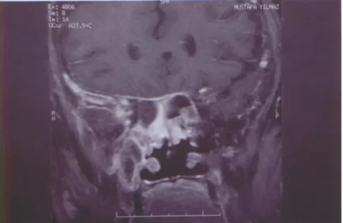
complete visual loss with no light perception on the right eye. Hori-zontal movements were impaired on the right eye. He had hypoes-thesia within the first and second division of the trigeminal nerve. Magnetic resonance imaging (MRI) of brain and orbita revealed soft tissue in the maxillary, sphenoid and ethmoid sinuses and contrast-enhancement of dura and extraocu-lar muscles (Figure 1). We thought he had mucormycosis and started amphotericin B 3mg/kg daily. The biopsy of maxillary sinuses con-firmed our diagnosis of mucormy-cosis. Because of pancytopenia the patient could not be operated and died in one week.
Case Report 2
A 46-year-old female referred to our clinic from haematology with the chief complaint of headache, pain over the left side of his face and left eye, nasal obstruction and left eye swelling. She had acute myeloblas-tic leukemia. On physical examina-tion, there was edema around the left eye, her left eye was hyperemic and swollen. There was no ocular palsy at once. Orbitoparanasal CT scan revealed radiopacity involving the left frontal, maxiller, ethmoid, sphenoid sinuses, spotty destruc-tion of bony walls of the ethmoid sinus (Figure 2). Surgical debride-ment of paranasal inflamed tissue was done; mucormycosis was diag-nosed by histopathological exami-nation and amphotericin B treat-ment was started. Despite of early diagnosis and treatment, the pa-tient got worse. Total ophtalmop-legia developed on the left eye and the patient died one week later.
Discussion
Zygomycetes (mucormycosis) is a malignant opportunistic fungus that cause central nervous system infection in the setting of obvious
Figure 1: Soft tissue increased of maxillary, sphenoid and ethmoid sinus and
contrasten-hancement of dura and extraocular muscles in MRI.
Figure 2: Orbitoparanasal CT scan revealed radiopacity involving left frontal, maxiller,
Journal of Ankara University Faculty of Medicine 2012, 65 (3)
Mine Hayriye Sorgun, Buket Tuğan Yıldız,Canan Yücesan 161
immune dysfunction or anatomic abnormalities (5, 12, 13). It occurs as a rare complication in patients with diabetic acidosis, in drug ad-diction, and in patients with leu-kemia and lymphoma, particularly those treated with corticosteroids and cytotoxic agents, severe burns, cirrhosis, severe dehydration, asthma, nephrotic syndrome, ne-crotizing gingivitis (1-13). Some of the cases have no predisposing fac-tors (14, 15). Aplastic anemia and acute myeloblastic leukemia were the predisposing factors in our cas-es.
Zygomycetes can invade cerebral blood vessels, causing an arteritis that can trombose or rarely, rup-ture. Zygomycetes infection often extends from an initial sinus or nasal infection and spreads along infected vessels to the retro-orbital tissues and then to the adjacent brain, producing a localized mass with adjacent cerebral infarctions due to thrombosis from hyphal in-vasion of arteriole vessel walls (5, 12, 13). Numerous hyphae are present within trombi and vessel wall, often invading the surround-ing parenchyma. Sometimes, it may cause hemorrhagic infarction (5).
Zygomycetes location involves the cavernous sinus with the presenta-tion of facial pain, cranial nerve palsies (cranial nerves III, IV, V and VI), proptosis and edema of the lids and retina (1, 3-15). Palatal ulcer was reported with rhinocere-bral mucormycosis in literature (9).
Mucormycosis caused ocular movement palsies, amaurosis, proptosis, chemosis and hyperemia on the affected eye in our cases. Furthermore, the first case had tri-geminal palsy involving first and second divisions of the nerve due to mucormicosis.
Fungi are difficult to isolate or identi-fy from cerebrospinal fuid (CSF). Culturing the fungus from CSF occurs in less than 5% for Zygo-mycetes (12, 13). Histopathological examination shows aseptate and ir-regular fungal hyphae branching with neutrophilic infiltrate invad-ing the smaller blood vessels and causing necrosis (1, 6, 9-11, 14). We confirmed mucormycosis infection with histopathological examina-tion in both cases.
Radiological studies such as CT or MRI show contrast enhancing masses within sinuses, cavernous sinus thrombosis, intracranial masses, brain abscesses and ischae-mic infarcts in cases with mucor-mycosis (1, 6, 9-11, 14). The our first case, MRI revealed increase of soft tissues in the maxillary, sphe-noid and ethmoid sinuses and con-trast-enhancement of dura around the affected area and of extraocular muscles on the left side. Orbitopa-ranasal CT of the second case re-vealed radiopacity involving the left frontal, maxillary, ethmoidal, and sphenoidal sinuses and spotty destruction of bony walls of the ethmoid sinus.
The cerebral form of mucormycosis is usually fatal in short order. Rapid
correction of hyperglycemia and acidosis, and treatment with am-photericine B have resulted in re-covery in some patients (5). Am-photericine B is a antifungal that has been proven to be efficacious. The daily dosage is slowly in-creased from 0.5 to 1 mg/kg/day or higher for treatment of Zygo-mycetes infections. Amphotericine B is frequently nephrotoxic. Pa-tients should be followed with fre-quent serum creatinine and urine analysis; red or white blood cells can be seen in urine analysis. Lipo-somal amphotericine B should be preferred because of least nephro-toxic effect (13). Surgical manage-ment is necessary for improving prognosis (7, 8). Although we started antifungal therapy as early as possible in both cases, they died. Successful treatment of mucormycosis requires early diagnosis, reversal of underlying predisposing risk fac-tors, surgical debridement and prompt antifungal therapy. How-ever, mucormycosis is frequently a fatal disease despite early diagnosis and prompt treatment such as in our cases. ophtalmology, otorhino-laryngology, neurology, hematolo-gy and internal medicine specialists should be aware of the disease and begin amphotericine B treatment as soon as possible when the first manifestations of the disease occur. The treatment should be begun even before histopathological con-firmation of the disease for not be-ing late for effective treatment.
Ankara Üniversitesi Tıp Fakültesi Mecmuası 2012, 65 (3)
Rhinocerebral Mucormycosis 162
REFERENCES
1. O’Neill BM, Alessi AS, George EB, et
al. Disseminated rhinocerebral mu-cormycosis: a case report and review of literature. J Oral Maxillofac Surg 2006; 64:326-333.
2. Blitzer A, Lawson W, Meyers BR, et
al. Patient survival factors in paranasal sinus mucormycosis. Laryngoscope 1980;90:635-648.
3. Sugar AM. Mucormycosis. Clin Infect
Dis 1992;14:126-129.
4. Naussbaum ES, Holl WA.
Rhinocerebral mucormycosis: changing patterns of disease. Surg Neurol 1994;41:152-156.
5. Victor M, Ropper AH. Infections of
the nervous system and sarcoidosis. In: Principles of neurology. 8th ed. New York: McGraw-Hill; 2005. p. 1110-1177.
6. Mohinra S, Mohinra S, Gupta R, et al.
Rhinocerebral mucormycosis: the dis-ease spectrum in 27 patients. Mycosis 2007;50:290-296.
7. Yohai RA, Bullock JD, Aziz AA, et al.
Survival factors in rhino- orbital- cere-bral mucormycosis. Surv Ophthalmol 1994;39:3-22.
8. Ladurner R, Brandacher G, Steurer W,
et al. Lessons to be learned from a complicated case of rhinocerebral mu-cormycosis in a renal allograft reci-pient. Transpl Int 2003;16:885-889.
9. Garg R, Grupta VV, Ashok L.
Rhi-nomaxillary mucormycosis: A palatal ulcer. Contemp Clin Dent. 2011;2:119-123.
10. Thomas S, Singh VD, Vaithilimgam Y,
et al. Rhinocerebral mucormycosis: a case report. Oral Maxillofac Surg. 2012; 16(2):233-236.
11. Viterbo S, Fasolis M, Garzino-Demo
P, et al. Management and outcomes of three cases of rhinocerebral mucormy-cosis. Oral Surg Oral Med Oral Pathol Oral Radiol Endod 2011; 112(6): e69-74
12. Cortez KJ, Walsh TJ. Space-occupying
fungal lesions. In Scheld WM, Whitley RJ, Marra CM, editors: Infections of the central nervous system, ed 3, Philadephia, Lippincott Willams & Wilkins; 2004. p. 713-733.
13. Johnson RT, Griffin JW, McArthur
JC. Fungal Infections. In: Current Therapy in Neurologic Disease. 7th ed. Philadephia: Mosby Inc; 2006. p. 161-169.
14. Roden MM, Zaoutis TE, Buchanan
WL, et al. Epidemiology and outcome of zygomycosis: a review of 929 re-ported cases. Clin Infect Dis 2005;41:634-653.
15. Elinav H, Zimhony O, Cohen MJ, et
al. Rhinocerebral mucormycosis in pa-tients without predisposing medical conditions: a review of the literature. Clin Microbiol Infect 2009;15:693-697.