The light microscopic investigation of the effects of in-ovo
administered bisphenol A (BPA) on the development of testes
Banu KANDİL
1, Emrah SUR
21Siirt University, Faculty of Veterinary Medicine, Department of Histology and Embryology, Siirt; 2Selçuk University, Faculty of Veterinary Medicine, Department of Histology and Embryology, Konya, Turkey.
Summary: The aim of this study was to determine the effects of BPA on testes development in chicken embryos. For this purpose, 310 fertile eggs of Isa Brown laying parent stock were divided into five groups as control, vehicle-control, 50, 100, and 250 µg/egg BPA. Test solutions were injected into the yolk before incubation. At the 13th, 18th and 21st days of incubation, the eggs were opened until six living male embryos were obtained from each group. Tissue samples were fixed in 10% buffered formalin (pH 7.4). After routine histological processes, tissue samples were embedded in paraffin. Six µm thickness sections were stained with the Crossmon’s trichrome method. All histological evaluation and histometrical measurements were performed on the left testes. On the 13th, 18th and 21st days of the incubation, the groups that were treated with BPA showed growth retardation in testicular tissues, fewer cell cords and poorly cellular organization. At 21 days of the incubation, there were a significant decrease in the mean diameter of the seminiferous tubule in all experimental groups compared to control groups (p<0.05). The increase in mean cortical thickness was observed in the BPA treated groups compared to the control groups (p<0.05). The mean testes surface area was higher at 50 µg/egg and 100µg/egg BPA treated groups compared to the control groups and at 250 µg/egg BPA administered group (p<0.05). In 50 and 100 µg/egg BPA treated chicken embryos, BPA triggered ovo-testis formation by characterizing thickened cortex containing oocyte-like cell clusters whereas BPA had toxic effects at 250 μg/egg. It was concluded that BPA can induce both estrogen-oocyte-like and toxic effects in the developing testes of chicken embryos in a dose-dependent manner.
Keywords: BPA, chicken embryo, testes.
Yumurtaya verilen bisfenol A (BPA)' nın testislerin gelişimi üzerindeki etkilerinin ışık mikroskobik
seviyede belirlenmesi
Özet: Bu çalışmanın amacı Bisfenol A (BPA)ʹnın tavuk embriyolarında testis gelişimi üzerindeki etkisini belirlemektir. Bu amaçla Isa Brown ırkı yumurtacı tavuklara ait 310 adet döllü yumurta kontrol, taşıyıcı kontrol, 50, 100, 250 µg/yumurta BPA olmak üzere 5 gruba ayrıldı. Test solüsyonları inkübasyondan önce yumurta sarısına enjekte edildi. İnkübasyonun 13. 18. ve 21. günlerinde her gruptan 6 adet canlı erkek embriyo elde edilene kadar yumurtalar açıldı. Alınan testis dokuları %10'luk tamponlu formalin (pH 7.4) solüsyonunda tespit edildi. Dokular rutin histolojik metotlarla takip edilerek parafinde bloklandı. Bloklardan alınan 6 μm kalınlığındaki kesitlere ise Crossmon'un üçlü boyama yöntemi uygulandı. Tüm histolojik ve histometrik değerlendirmeler sol testis’ler üzerinde yapıldı. İnkübasyonun 13. 18. ve 21. günlerinde BPA uygulanan gruplarda testis dokularında gelişme geriliği, az sayıda hücre kordonu ve zayıf hücresel organizasyon gözlendi. İnkübasyonun 21. gününde deney gruplarının ortalama seminifer tubul çaplarında kontrol gruplarına kıyasla belirgin düşüşler tespit edildi (p<0.05). Kontrol gruplarına kıyasla deney gruplarında ortalama korteks kalınlığında artış gözlendi (p<0.05). Elli ve 100 µg/yumurta dozunda BPA uygulanan gruplar kontrol grupları ve 250 µg/yumurta dozunda BPA uygulanan grupla karşılaştırıldığında ortalama testis yüzey alanında önemli bir artış tespit edildi (p<0.05). Elli ve 100 µg/yumurta dozunda BPA uygulanan tavuk embriyolarında BPA'nın oosit benzeri hücre toplulukları içeren kalınlaşmış korteksle karakterize ovo-testis gelişimini tetiklediği, buna karşın 250 µg/yumurta dozunda uygulanan BPA'nın ise toksik etkiye sahip olduğu görüldü. BPA'nın tavuk embriyolarında gelişmekte olan testisler üzerinde doza bağlı olarak hem toksik hem de östrojen benzeri etkilere neden olabileceği sonucuna varıldı.
Anahtar sözcükler: BPA, tavuk embriyosu, testis.
Introduction
Endocrine disrupter compounds (EDCs) are the chemical substances that may impede the reproductive, neurological, and immunological process related to endocrine system at different dose levels because many
systems controlled by hormones (14, 33). EDCs mimic naturally hormones binding to the receptors and alter the hormonal functions. These disruptive effects can result in range from male and female infertility to diabetes, obesity, and hormone-sensitive cancers. One of the important
concern about estrogenic compounds is their adverse effect on embryonic development of the gonads. Although there are many EDCs having estrogenic effects such as diethylstillbestrol and ethynylestradiol, the most widely used of the EDC is Bisphenol A (BPA) which is a monomer of polycarbonate plastic (3, 4, 20).
BPA [2.2-bis (4-hydroxyphenyl) propane] is a widely used in the cover coating on food and drink packages, dental sealants, thermal papers, toys, optical lenses and as an additive in other plastics. BPA mimics the actions of endogenous estrogen and also acts as a xenoestrogen on the reproductive system of many species. Exposure to BPA, especially during embryonic development may cause functional or morphological disorders on various systems (7).
Although the undifferentiated sex organs are bilateral in the early embryonic period, the female gonads develop asymmetrically in many avian species. The gonads contain two main histologic structure called medulla and germinal epithelium. During differentiation process, the germinal epithelium of the left gonad surrounding the medulla thickens to form an ovary in female whereas the sex cords containing the germ cells develop to form the seminiferous tubules located in the medulla in both gonads of male. Consequently, in most avian species, only the left gonad develops into an ovary in female whereas both gonads develop into testes in male (4, 15).
In poultry, the estrogen hormone plays a critical role in the development and differentiation of gonads in both sexes by binding to estrogen receptor. If the testosterone concentration is higher than the estrogen level, both gonads develop into testicular tissue whereas when the estrogen concentration is higher than the testosterone level, the only left gonad develops into ovarian tissue because estrogen receptors are asymmetrically expressed in avian embryonic gonads during sexual differentiation (25). Thus, differences in the concentration of estrogen during the embryonic period may cause the formation of ovo-testis identified by thickened cortex containing oocyte-like cells in male embryos. So, the chicken embryos are generally preferred in studies investigating the estrogenic effects of EDCs such as BPA because sexual differentiation in avian depends on the estrogen hormone (3).
In this study, we aimed to investigate the effects of BPA on the development of testes in chicken embryos at the light microscopic level.
Material and Methods
Ethical approval: All experimental procedures used in this study were approved by the Ethical Committee of Selçuk University, Veterinary Faculty, Experimental
Animal Production and Research Center dated 29.01.2016 and numbered 2016/11.
Experimental design: 310 fertile eggs of Isa Brown
parent stock were obtained from a local breeder. Fertile eggs weighing 50-55 g were randomly selected and divided into five groups; Control (n=45 egg), vehicle-control (n=50 egg), 50 μg/egg BPA-treated (n=60 egg), 100 μg/egg BPA-treated (n=70 egg), and 250 μg/egg BPA-treated (n=85 egg). Fertile eggs were disinfected for 15 minutes with steam obtained with a mixture of 21 g
potassium permanganate and 42 mL formaldehyde/m3 in
a closed container.
Preparation of vehicle solution: 1 g lecithin, is
known as emulsifier and is used for homogenous dispersion of BPA in yolk, was dissolved in 1.5 mL dichloromethane and 10 mL peanut oil was added. The solution was incubated overnight at 60 ˚C to dissolve dichloromethylene (3).
Preparation of test solutions: BPA required for each
group was weighed and dissolved in ethanol. Then the vehicle solution was added (3). Before the incubation, the test solutions were injected into the yolk via a small hole in the shell, punched with a needle. Holes were sealed with melted paraffin wax. Eggs were incubated under optimal
conditions [Incubation temperature 37.7 oC, RH (Relative
Humidity) 55%] in the incubator at the Selçuk University, Faculty of Science, Department of Biology Laboratory.
Histological investigations: At days 13, 18 and 21 of
incubation, the eggs from each group were opened until six living male embryos were obtained from each group. The left testicular tissue of each embryo was carefully removed and fixed in a 10% buffered formalin (pH 7.4) solution for 24 h. After routine histological methods, tissue samples were embedded in paraffin. The serial sections (6 μm thickness) from paraffin blocks were stained with Crossmon’s trichrome method (8). Histological examination was performed using a light microscope (Leica DM-2500 model with DFC-320 camera attachment giving digital images). Images were digitally recorded.
Evaluations of specimens: Histometric evaluations
were performed on 21st day of incubation. The sections cut
most centrally through the left testes were evaluated.
Evaluation of testes surface area: The measurements of testes surface area were performed on seven transverse serial sections taken from the midline.
Thickness of cortex: The mean thickness of cortex was found by measuring 30 different regions from the specimens used for testes surface area evaluations.
Diameter of the seminiferous tubules: The mean diameter of seminiferous tubules was found by evaluating the clear cross-sections of 30 different tubules from the specimens used for testes surface area evaluations.
Statistical analysis: The data obtained from this
study were analyzed by using one-way analysis of variance and followed by post hoc Duncan multiple comparisons test using the Statistical Package for Social Sciences (SPSS version 10.0; SPSS Inc. Corp., USA). Results were considered at significant at p<0.05.
Results
Day 13 of incubation: Histological examinations of
the sections from the left testes tissues of the control groups revealed a medulla surrounded by a cortex layer with a few cell layers thick, called tunica albuginea. Primary sex cords were seen in the medullar region
composed of mesenchymal connective tissue.
Spermatogonia with large-rounded nuclei and Sertoli cells characterized by triangular-shaped nuclei located on the basal membrane were seen in these cords, which called later seminiferous tubules. Blood vessels were seen in the mesenchymal tissue between the cords (Figure 1a and c). Although the histological structure of testes tissues in vehicle-control (Figure 1b) and 50 µg/egg BPA-treated groups (Figure 1c) was similar to control group (Figure 1a), especially in 100 (Figure 1d and e), and 250 µg/egg (Figure 1f) BPA-treated groups showed impaired cellular organization and retarded development of the primary sex cords, and the mesenchimal tissue occupied a relatively large space (Figure 1d and e). Thickening caused by oocyte-like cell clusters were observed in the cortex layer (Figure 1f). Ovarian-like extension was also observed in the sections of the testicular tissue showing ovo-testis abnormality (Figure 1e).
Day 18 of incubation: The primary sex cords
developed and the number of these cell cords increased in the control groups (Figure 2a and b) compared with BPA-treated groups (Figure 2c, d, e, and f). It was observed that the retarded development of the cell cords and poorly organization of spermatogonia and Sertoli cell was distinct in the 50, 100 and 250 µg/egg BPA-treated groups (Figure 2c, 2d, 2e, and 2f).
Day 21 of incubation: It was observed that the tubule
development became more prominent in control groups but the tubule lumen was not yet fully formed in any groups. Sertoli cells and spermatogonia were found to be more distinct (Figure 3a and b). It was found that the development of primary sex cords was retarded, and that the organization of spermatogonia and Sertoli cells was also impaired in all experimental groups (Figure 3c, d and Figure 4b). Cortical thickening was distinct in BPA-treated groups (Figure 4b) compared to control group (Figure 4a) and oocyte-like cell clusters were observed in these regions (Figure 4b).
Histometric Results
Testicular surface area: The highest value of the
mean testis surface area was obtained from low (50 μg/egg BPA) and mid-level (100 µg/egg BPA) dose BPA-treated groups. It was showed that these values were significantly different from the control groups and 250 μg/egg BPA-treated group (p<0.05). The difference between the control groups and 250 µg/egg BPA-treated group was no statistically important, but it was noted that the values obtained from the high (250µg/egg BPA) dose BPA group were lower than the control groups (Table 1).
Seminiferous tubule diameter: There was a
statistically significant difference in the diameter of the seminiferous tubule between experimental groups and control groups. The BPA-treated groups showed a decrease in the diameter of their seminiferous tubules (p<0.05, Table 1).
Cortical thickness: It was determined that the results
of cortical thickness measurement obtained from the BPA-administered groups were higher than the control groups (p<0.05, Table 1). Especially in low (50 μg/egg BPA-treated) and mid-level (100 μg/egg BPA) doses groups, the increase in cortical thickness was distinct. It was noted that the difference between the low/mid-level dose groups and high (250μg/egg BPA) dose group was statistically significant (p<0.05, Table 1).
Table 1. The histometric measurements performed on the left testes on the 21st day of incubation. Tablo 1. İnkübasyonun yirmi birinci gününde sol testisler üzerinde yapılan histometrik ölçümler.
Groups (n=6)
Mean testicular surface area (µm2)
Mean±SE
Mean diameter of seminiferous tubule (µm)
Mean±SE
Mean cortical thickness (µm) Mean±SE Control 697106.56±38257.85b 38.81±040a 14.72±0.23c Vehicle-control 664742.36±30269.02b 38.41±0.43a 15.78±0.27c 50 µg/egg BPA-treated 840774.62±37355.58a 29.07±0.45b 20.40±0.33a 100 µg/egg BPA-treated 857630.05±22990.35a 28.75±0.47b 20.68±0.36a 250 µg/egg BPA-treated 586920.07±15509.13b 28.06±0.46b 18.22±0.38b
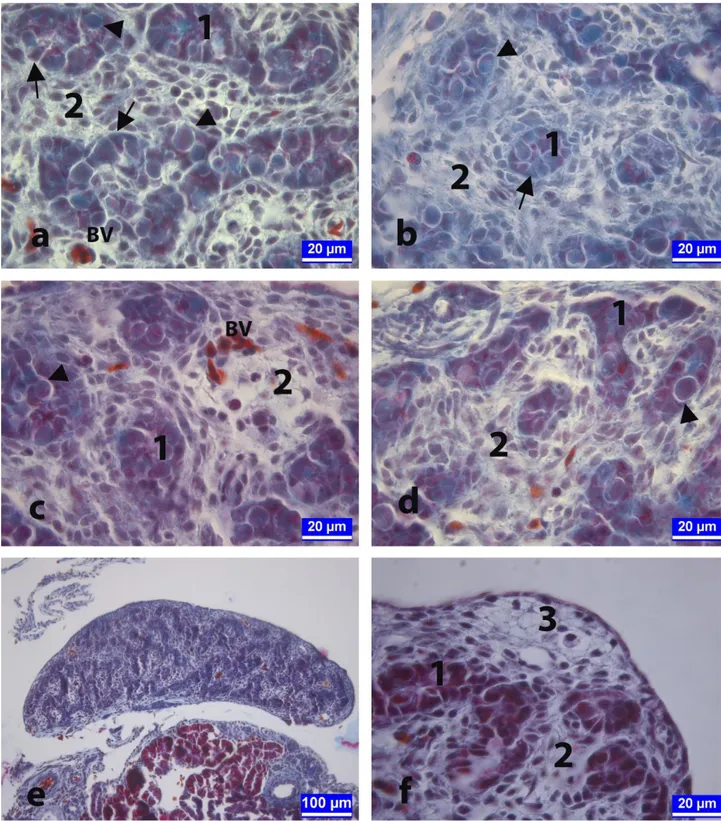
Figure 1. On the 13th day of the incubation. The sections from testes of embryos in control (a), vehicle-control (b), 50 μg/egg BPA-treated (c), 100 μg/egg BPA-BPA-treated (d, e), and 250 μg/egg BPA-BPA-treated (f) groups. Testicular tissue of an embryo showing ovo-testis abnormality and extending like ovarium development in 100 μg/egg BPA-treated group (e). Arrows: Sertoli cells, Arrow heads: Spermatogonia, BV: Blood vessels, 1: Primary sex cords, 2: Mesenchymal tissue, 3: Oocyte-like cell clusters. Crossmon's trichrome staining. Şekil 1. İnkübasyonun on üçüncü günü. Kontrol (a), taşıyıcı-kontrol (b), 50 μg/yumurta (c), 100 μg/yumurta (d, e) ve 250 μg/yumurta (f) dozunda BPA uygulanan gruplara ait embriyolardan alınan testis dokusu kesitleri. Yüz μg/yumurta dozunda BPA uygulanan gruptan bir embriyoya ait ovo-testis anomalisi gösteren ve ovaryum gibi uzamış testis dokusu (e). Oklar: Sertoli hücreleri, Ok başları: Spermatogonyumlar, BV: Kan damarları, 1: Primer seks kordonları, 2: Mezenşimal doku, 3: Oosit benzeri hücre kümeleri. Crossmon'un üçlü boyaması.
Figure 2. The sections of developing testes at day 18 of the incubation from the control (a), vehicle-control (b), 50 μg/egg BPA-treated (c), 100 μg/egg BPA-treated (d, e), and 250 μg/egg BPA-treated (f) groups. Arrows: Sertoli cells, Arrow heads: Spermatogonia, 1: Cross-sections of primary sex cords that will form seminiferous tubules, 2: Mesenchymal tissue. Crossmon's trichrome staining. Şekil 2. İnkübasyonun on sekizinci gününde kontrol (a), taşıyıcı-kontrol (b), 50 μg/yumurta (c), 100 μg/yumurta (d, e) ve 250 μg/yumurta dozunda (f) BPA uygulanan gruplara ait embriyolardan alınan testis dokusu kesitleri. Oklar: Sertoli hücreleri, Ok başları: Spermatogonyumlar, 1: Seminifer tubülleri oluşturacak olan primer seks kordonlarının enine kesiti, 2: Mezenşimal doku. Crossmon'un üçlü boyaması.
Figure 3. On day 21 of incubation. The sections from developing testes of chickens in the control (a), vehicle-control (b), 50 μg/egg BPA-treated (c), and 250 μg/egg BPA-treated (d) groups. Arrows: Sertoli cells, Arrow heads: Spermatogonia, 1: Seminifer tubule, 2: Connective tissue. Crossmon's trichrome staining.
Şekil 3. İnkübasyonun yirmi birinci günü. Kontrol (a), taşıyıcı-kontrol (b), 50 μg/yumurta (c) ve 250 μg/yumurta (d) dozunda BPA uygulanan gruplara ait embriyolardan alınan testis dokusu kesitleri. Oklar: Sertoli hücreleri, Ok başları: Spermatogonyumlar, 1: Seminifer tubüller, 2: Bağ doku. Crossmon'un üçlü boyaması.
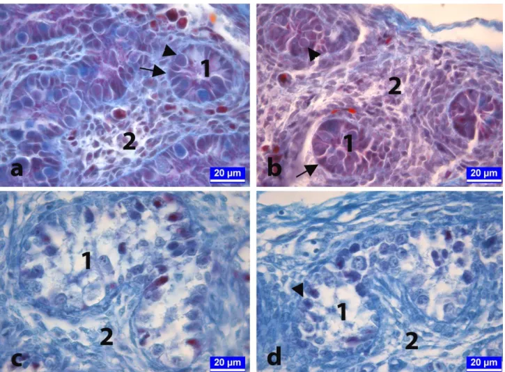
Figure 4. The sections of developing testes at day 21 of the incubation from the control (a), and 100 μg/egg BPA-treated (b) groups. Cortical thickening was distinct in BPA-treated group. 1: Seminifer tubules, 2: Cortex, 3: Oocyte-like cell clusters. Crossmon's trichrome staining.
Şekil 4. İnkübasyonun yirmi birinci gününde kontrol (a) ve 100 μg/yumurta (b) dozunda BPA uygulanan gruplara ait civcivlerden alınan testis dokusu kesitleri. Kortikal kalınlaşmanın BPA uygulanan grupta belirgin olduğu görülmekte. 1: Seminifer tubüller, 2: Korteks, 3: Oosit benzeri hücre kümesi. Crossmon'un üçlü boyaması.
Discussion and Conclusion
Endocrine systems regulate of developmental,
metabolic, and reproductive processes including
embryonic development, gonadal formation, sex
differentiation, growth, and digestion. Endocrine-disrupting compounds (EDCs) may affect these processes by either binding to or blocking hormone receptors, thereby triggering or preventing hormonal response. BPA is the most commonly known EDC and has estrogenic potency (7, 12).
BPA, which has an estrogenic effect, is a major problem for public health because almost all humans can be exposed to BPA and the placental transmission can occur. BPA is also environmental pollutant to threat to the wild life and poultry industry because the egg yolk is relevant route of BPA (2, 7, 12, 17). Studies have reported that BPA is detected in blood samples taken from pregnant women, cord blood, ovarian follicular fluid and amniotic fluid specimens. Researchers are concerned that in recent years there has been an increase in genital anomalies in boys and early puberty in girls, and that one of the possible causes of female breast cancer may be BPA and similar EDCs to which we are exposed in daily life (17).
There are many studies investigating the estrogenic effects of BPA on the embryonic development of the gonads and reproductive functions in poultry. In these studies, different amounts of BPA-containing emulsions were injected into fertilized chicken or quail eggs (3, 10, 16, 26). Berg et al. (3) injected BPA into fertilized quail and chicken eggs at 67 and 134 μg/egg doses in 20 and 100 μl emulsion, respectively. In that study, Müllerian duct anomaly in female quail embryos and ovo-testis anomaly expressing ovary-like development in the left testis in male chicken embryos were observed (3).
The chicken and quail embryos have been preferred as materials to determine the embryotoxic and teratogenic effects of certain drugs, various chemical substances and some EDCs (4, 27). Jelinek (18) has developed a method called the Chicken Embryotoxicity Screening Test (CHEST), and the results obtained from the test could have been adapted to pregnant mammals. It is suggested that the predictive dose calculated by multiplying the concentration of dilutions determined by the CHEST
method as embryotoxic dose ranges by 10-2 is considered
as the toxic limits per kg for the body weight of the pregnant mother (18). This technique can be performed easily in a short time even at the mediocre laboratory conditions and, it is also reproducible, reliable, and cost-effective. One of the most important advantages of this technique is to reduce the number of experiments and the number of experimental animals to be used in toxicological studies in mammals. In this respect, the pain and suffering caused to a living organism is minimized
and ethical rules, legal restrictions and the Animal Rights are not violated (19, 21, 31).
In embryotoxicity studies using fertilized avian eggs, different approaches are put forward about the injection routes of the test materials. It is reported that the aim of the study and the chemical structures of the test substances and vehicle solutions must be taken into consideration (21, 28). Egg yolk is known to be the route of elimination of lipophilic substances (4). It is also reported that the most suitable method for testing the distribution and effects of some hydrophilic and hydrophobic substances is egg yolk injection technique (9). In this study, it was thought that the egg yolk was the most suitable region where the lipophilic and hydrophilic substances used in this study can be most homogeneously dispersed and absorbed the most efficiently as described in material and method section. (4).
At early stages of avian embryonic growth, the testicular and ovarian differentiation is not fully developed yet. Morphological differentiation starts approximately at day 6.5 of the incubation. At this stage, the cell cords develop to form seminiferous tubules in the medullar region of the gonads bilaterally in male embryos whereas the cortex is thickened increasing somatic and germ cells unilaterally in female embryos. In later periods, both gonads develop to form testes in male embryos while the right gonad regresses and only the left gonad continues to develop an active ovary in female embryos (6, 22, 27). This gonadal asymmetry observed in female is similar in male in that the left testis is larger (5, 11, 30). The cause of this asymmetry has been suggested to be estrogen receptors that express in different amounts in the right and left gonads at early stages of the embryonic process (25). For these reasons, the left testicular tissue of avian embryos is particularly preferred in many studies evaluated the effects of estrogenic endocrine disruptors on reproductive system in avian species (3, 16, 23).
In this study, BPA dissolved at a dose of 50, 100 and 250 μg/egg in a 100 μl volume of vehicle solution was injected into the egg yolk at the beginning of the incubation of fertile hen's eggs. On the 13th and 18th days
of incubation, histological examinations were carried out on the left testes tissues of male embryos. On the first day of hatching (day 21 of incubation), the left testes were also evaluated histometrically. Histological examinations have shown that BPA has an adverse effect on the development of testicular tissue. BPA-treated groups had poorly developed cell cords, mis-shaped tubules having few spermatogenetic cells whereas the well-developed testicular architecture consists of uniform tubules containing well-organized germ cells were observed in control and vehicle-control animals (Figures and Table 1). In addition to these findings, increased cortical thickness
and mean testis surface area showed the estrogenic effect of BPA on testicular tissue of newly hatched male chicks in the low and mid-level dose groups (Table 1). On the 21st
day of incubation, it was also observed that although the mean diameter of the seminiferous tubule was significantly reduced, the mean cortical thickness increased in all BPA groups compared to the control groups (Table 1). It is notable that this increase was more pronounced in the low (50μg/egg) and mid-level dose (100μg/egg) BPA groups (Table 1). In the high-dose BPA group, the cortical thickness was higher than the control groups but lower than the low and mid-level dose BPA groups (p<0.05). It was found that the significant increase in the mean testicular surface area in the low and mid-level dose BPA groups compared to the other groups (p<0.05) but no statistically significant difference was seen between the control groups and the high dose BPA group. Besides, the results of the mean testis surface area observed in the high-dose BPA group is lower than the control group. These findings suggested that BPA might have been toxic effect at high doses compared to the low and mid-level doses. Although it appears as a contradictory situation, the studies have showed that BPA is a biphasic effective endocrine disruptor (13, 24, 32). It was known that BPA has many adverse effects such as cancer, obesity, diabetes, and infertility etc. It is thought that altering the hormone synthesis, and impairing the epigenetic regulation by binding to estrogen receptors are responsible for the possible mechanisms underlying the multiple effects of BPA. Some investigators have reported that the exposure to BPA results in unusual dose-response relationship instead of classical positive dose-response effect. This confusing situation is called as "non-monotonic dose response" and it is shown "non-linear dose curve" expressed as "U" or "Ո". Unfortunately, these paradoxal results make difficult the risk assessment of BPA (1, 29).
In conclusion, in ovo administrated BPA caused the poorly development of the testicular tissue and retarded formation of the seminiferous tubules in the left testes. Especially low (50μg/egg) and mid-level (100μg/egg) doses BPA exposure result in thickened cortex containing oocyte-like germ cells classified as ovo-testis formation indicated feminization in male embryos.
Acknowledgements
This study, is part of the Master of Science Thesis of Banu KANDİL, for which we thank the “Selcuk University Academic Staff Training Program (ÖYP) Coordination Unit”, Project no: 2015-ÖYP-038, for financial support and it was presented in 2. International Conference on Science Ecology and Technology (ICONSETE).
References
1. Acconcia F, Pallottini V, Marino M (2015): Molecular mechanisms of action of BPA. Int J, 13, 1-9.
2. Berg C, Blomqvist A, Holm L, et al. (2004): Embryonic exposure to oestrogen causes eggshell thinning and altered shell gland carbonic anhydrase expression in the domestic hen. Reproduction, 128, 455-461.
3. Berg C, Halldin K, Brunström B (2001): Effects of bisphenol A and tetrabromobisphenol A on sex organ development in quail and chicken embryos. Environ Toxicol Chem, 20, 2836-40.
4. Berg C, Halldin K, Fridolfsson AK, et al. (1999): The avian egg as a test system for endocrine disrupters: effects of diethylstilbestrol and ethynylestradiol on sex organ develop. Sci Total Environ, 233, 57-66.
5. Birkhead T, Fletcher F, Pellatt E (1998): Testes asymmetry, condition and sexual selection in birds: an experimental test. P Roy Soc Lon B Bio, 265, 1185-89. 6. Chang G, Chen R, Qin Y, et al. (2012): The development
of primordial germ cells (PGCs) and testis in the quail embryo. Pakistan Vet J, 32, 88-92.
7. Crain DA, Eriksen M, Iguchi T, et al. (2007): An ecological assessment of bisphenol-A: evidence from comparative biology. Reprod Toxicol, 24, 225-239. 8. Crossmon G (1937): A modification of Mollory’s
connective tissue stain with a discussion of the principles involved. Anat Rec, 69, 33-38.
9. Drake VJ, Koprowski SL, Lough JW, et al. (2006): Gastrulating chick embryo as a model for evaluating teratogenicity: a comparison of three approaches. Birth Defects Res A Clin Mol Teratol, 76, 66-71.
10. El Gawish R, Ghanem M, Maeda T (2013): Effects of bisphenol A and DDT on mRNA expression of vitellogenin II in liver of quail embryos. Iran J Vet Res, 14, 237-240. 11. Elbajory SIA, El Tingari MD, Abdalla PA (2013):
Morphological study of the testis of adult Sudanese duck (Anas platyrhynchos). Int J Anim Vet Adv, 5, 103-7. 12. Flint S, Markle T, Thompson S, et al. (2012): Bisphenol
A exposure, effects, and policy: a wildlife perspective. J Environ Manage, 104, 19-34.
13. Ge LC, Chen ZJ, Liu H, et al. (2014): Signaling related with biphasic effects of bisphenol A (BPA) on Sertoli cell proliferation: a comparative proteomic analysis. Biochim Biophys Acta, 1840, 2663-73.
14. Goldman JM, Laws SC, Balchak SK, et al. (2000): Endocrine-disrupting chemicals: prepubertal exposures and effects on sexual maturation and thyroid activity in the female rat. Crc Cr Rev Toxicol, 30, 135-96.
15. Halldin K (2005): Impact of endocrine disrupting chemicals on reproduction in Japanese quail. Domest Anim Endocrin, 29, 420-429.
16. Halldin K, Berg C, Bergman A, et al. (2001): Distribution of bisphenol A and tetrabromobisphenol A in quail eggs, embryos and laying birds and studies on reproduction variables in adults following in ovo exposure. Arch Toxicol, 75, 597-603.
17. Ikezuki Y, Tsutsumi O, Takai Y, et al. (2002): Determination of bisphenol A concentrations in human
biological fluids reveals significant early prenatal exposure. Hum Reprod, 17, 2839-41.
18. Jelinek R (1977): The Chick Embryotoxicity Screening Test (CHEST). 381-386. In: D Neubert, H Merker, T Kwasigrooh (Eds), Methods in prenatal toxicology. Georg Thieme, Stutgart.
19. Jelinek R, Peterka M, Rychter Z (1985): Chick embryotoxicity screening test-130 substances tested. Indian J Exp Biol, 23, 588-595.
20. Kabir ER, Rahman MS, Rahman I (2015): A review on endocrine disruptors and their possible impacts on human health. Environ Toxicol Pharm, 40, 241-258.
21. Kemper F, Luepke N (1986): Toxicity testing by the hen's egg test (HET). Food Chem Toxicol, 24, 647-648. 22. Lambeth LS, Raymond CS, Roeszler KN, et al. (2014):
Over-expression of DMRT1 induces the male pathway in embryonic chicken gonads. Dev Biol, 389, 160-172. 23. Mattsson A, Mura E, Brunström B, et al. (2008):
Selective activation of estrogen receptor alpha in Japanese quail embryos affects reproductive organ differentiation but not the male sexual behavior or the parvocellular vasotocin system. Gen Comp Endoc, 159, 150-157.
24. Miyawaki J, Sakayama K, Kato H, et al. (2007): Perinatal and postnatal exposure to bisphenol a increases adipose tissue mass and serum cholesterol level in mice. J Atheroscler Thromb, 14, 245-252.
25. Nakabayashi O, Kikuchi H, Kikuchi T, et al. (1998): Differential expression of genes for aromatase and estrogen receptor during the gonadal development in chicken embryos. J Mol Endocrinol, 20, 193-202.
26. Oshima A, Yamashita R, Nakamura K, et al. (2012): In ovo exposure to nonylphenol and bisphenol A resulted in dose‐independent feminization of male gonads in Japanese quail (Coturnix japonica) embryos. Environ Toxicol Chem, 31, 1091-97.
27. Smith CA, Sinclair AH (2004): Sex determination: insights from the chicken. Bioessays, 26, 120-132.
28. Stoloff L, Verrett MJ, Dantzman J, et al. (1972): Toxicological study of aflatoxin P1 using the fertile chicken egg. Toxicol Appl Pharm, 23, 528-531.
29. Vandenberg LN, Ehrlich S, Belcher SM, et al. (2013): Low dose effects of bisphenol A: An integrated review of in vitro, laboratory animal, and epidemiology studies. Endocr Disruptors, 1, e26490.
30. Vatsalya V, Arora KL (2012): Allometric growth of testes in relation to age, body weight and selected blood parameters in male Japanese quail (Coturnix japonica). Int J Poult Sci, 11, 251-258.
31. Veselý D, Vesela D (1991): The use of chick embryo for prediction of some embryotoxic effects of mycotoxins in mammals. Vet Med Praha, 36, 175-181.
32. Wetherill YB, Petre CE, Monk KR, et al. (2002): The xenoestrogen bisphenol A induces inappropriate androgen receptor activation and mitogenesis in prostatic adenocarcinoma cells 1. Mol Cancer Ther, 1, 515-524. 33. Yiğit F, Aktaş A, Dağlıoğlu S (2013): Effects of bisphenol
A and diethylstilbestrol on the involution of bursa of Fabricius in the hens. İstanbul Üniv Vet Fak Derg, 39, 168-174.
Geliş tarihi: 16.05.2017 / Kabul tarihi: 22.09.2017 Address for correspondence:
Dr.Emrah SUR
Selçuk University, Faculty of Veterinary Medicine, Department of Histology and Embryology, Konya, Turkey. e-mail: [email protected]