EXPERIMENTAL STUDY
Neuronal regeneration in injured rat spinal cord after human
dental pulp derived neural crest stem cell transplantation
Kabatas S
1, Demir CS
2, Civelek E
1,
Yilmaz I
2, Kircelli A
4, Yilmaz C
4, Akyuva Y
1, Karaoz E
2,3University of Health Sciences, Gaziosmanpaşa Taksim Training and Research Hospital, Department of Neurosurgery, Karayollari Mahallesi, Gaziosmanpasa, Istanbul, Turkey. [email protected]
ABSTRACT
OBJECTIVE: This study aimed to analyze the effect of human Dental Pulp-Neural Crest Stem Cells (hDP-NCSCs) delivery on lesion site after spinal cord injury (SCI), and to observe the functional recovery after transplantation. METHODS: Neural Crest Stem Cells (NCSCs) were isolated from human Dental Pulp (hDP). The experimen-tal rat population was divided into four groups (n = 6/24). Their behavioral motility was scored regularly. After 4-weeks, rats were sacrifi ced, and their spinal cords were examined for Green Fluorescent Protein (GFP) la-beled hDP-NCSCs by immunofl uorescence (IF) staining.
RESULTS: In early post-injury (p.i) period, the ultrastructure of spinal cord tissue was preserved in Group 4. The majority of cells forming the ependymal region around the central canal were found to be hDP-NCSCs. While the grey-and-white-matter around the ependymal region was composed of e.g. GFP cells, with astrocytic-like appearance. The scores showed signifi cant motor recovery in hind limb functions in Group 4. However, no ob-vious change was observed in other groups.
CONCLUSION: Cells e.g., mesenchymal (Vimentin+) which express GFP+ cells in the gray-and-white-matter around the ependymal region could indicate the potential to self-renewal and plasticity. Thus, transplantation of hDP-NCSCs might be an effective strategy to improve functional recovery following spinal cord trauma (Fig. 10, Ref. 32). Text in PDF www.elis.sk.
KEY WORDS: functional recovery, human dental pulp, neural crest stem cells, spinal cord injury.
1University of Health Sciences, Gaziosmanpasa Taksim Training and Research Hospital, Department of Neurosurgery, Istanbul, Turkey, 2Liv Hospital, Center for Regenerative Medicine and Stem Cell Research&Manufacturing (LivMedCell), Istanbul, Turkey, 3University of Istinye, Faculty of Medicine, Department of Histology&Embryology, Istanbul, Turkey, and 4Baskent University School of Medicine, Department of Neurosurgery, Ankara,Turkey
Address for correspondence: S. Kabatas, MD, University of Health Sciences,
Gaziosmanpasa Taksim Training and Research Hospital, Department of Neurosurgery, Karayollari Mahallesi, Osmanbey Caddesi 616, Sokak No: 10, 34255 Gaziosmanpasa, Istanbul, Turkey.
Phone: +90.212.9453000-3628, Fax: +90.212.9453180
Abbreviations: B BB-scoring – Basso, Beattie and Bresnahan
scoring, BDNF – Brain derived neurotrophic factor, BFGF – Basic fi broblast growth factor, CNS – Central nervous system, CNPase – 2’,3’-cyclic-nucleotide 3’-phosphodiesterase, CNTF – Cili-ary neurotrophic factor, DAPI – 4’,6-diamidino-2-phenylindole, DMEM – Dulbecco’s modifi ed Eagle’s medium, DPSCs – Dental pulp stem cells, EGF – Epidermal growth factor, EP3 – Prosto-glandin E2 receptor, FBS – Fetal bovine serum, GDNF – Glial cell-derived neurotrophic factor, GFAP – Glial fi brillary acidic pro-tein, GFP – Green fl uorescent propro-tein, hDP – human Dental pulp, hDPSCs – human Dental pulp stem cells, hDP-NCSCs – human Dental pulp-neural crest stem cells, HE – Hematoxylin&Eosin, IF – Immunofl uorescence, L+T – Laminectomy and trauma, MHC-1 – Major histocompatibility complex 1, ms – Milliseconds,
MSC – Mesenchymal stem cell, NCSCs – Neural crest stem cells, NF – Neural factor, NGF – Nerve growth factor, PBS – Phosphate-buffered saline, PFA – Paraformaldehyde, pGFP – plasmid Green fl uorescent protein, p.i. – Postinjury, RT – Room temperature, SCI – Spinal cord injury, V – Volts, VEGF – Vascular endothelial growth factor
Introduction
Spinal cord injury (SCI) has many distinct factorial aspects including primary mechanical damage, secondary cell apoptosis, reactive gliosis, and axons inability to regenerate (1–3). Due to the non-responsive environment of the injured spinal cord, axon regeneration does not occur (4). Besides, the loss of function af-ter SCI might be from both the primary mechanical insult and the subsequent, multifaceted secondary degenerative response. However, some experimental studies in last decades proved that the injured spinal cord could be restored (5–9). Nowadays, stem cell based therapy is promising some valuable strategies for func-tional recovery of the injured spinal cord (10). In this content, human dental pulp-neural crest stem cells (hDP-NCSCs) were used in addition to neural progenitor stem cells for functional re-covery in SCI (11). hDPSCs are neural crest-derived stem cells residing within the perivascular niche of the dental pulp. hDPSCs appear to be an excellent source of stem cells because they can be obtained without adverse health effects and can be obtained
non- invasively from extracted teeth discarded as medical waste, avoiding many ethical issues. hDPSCs show self-renewal abil-ity, immunomodulatory capacabil-ity, and proliferation in vitro, and they can differentiate into muscle, cartilage, bone, and other cell types in vitro and in vivo (12, 13, 14). In this study, it was aimed to analyze the healing effect of hDP -NCSCs on lesion site after SCI and to observe the degree of functional recovery in motility after transplantation.
Materials and methods
Animals
The SCI study included about 2 months old 24 female, non-pregnant Wistar albino rats with a weight of 200–300 g. Rats were divided into four groups (n = 6/24): only laminectomy (Group 1); laminectomy+trauma (L+T) (Group 2); L+T+ phosphate-buffered saline (PBS) (Group 3); L+T+hDP-NCSCs (Group 4), respectively. Rats were sacrifi ced 4 weeks after transplantation. The experimen-tal design and all procedures were approved by the Ethics Com-mittee of Kocaeli University.
Isolation and culture of hDP-NCSCs
To isolate hDP-NCSCs, normal human third molars (n = 5) were obtained from adults (17- to 25-yr-old) at the Dental Hos-pital of Kocaeli University according to the guidelines provided by ethics committee. Isolation and culture of hDP-NCSCs were performed according to protocols described elsewhere (15, 16). Briefl y, the pulp tissue of each sample was gently separated from the crown and root, digested with collagenase type I (Sigma Al-drich, St. Louis MO) to generate single cell suspensions. To cul-ture the isolated NCSCs derived from dental pulp; MEM-Earle (Biochrom-FG0325) medium was used including 10 % fetal bovine serum (FBS; Invitrogen/ GIBCO, Grand Island, NY, USA) and 100 IU/mL penicillin–100 μg/mL streptomycin (P/S; Invitrogen/ GIBCO). Cells were cultured in tissue culture fl asks and incubated at 37 °C in a humidifi ed atmosphere containing 5 % CO2. The ad-herent cells were grown to passage three (P3). At passage three, cells were used for characterization and differentiation studies.
Characterization of hDP-NCSCs by fl ow cytometry
Undifferentiated hDP-NCSCs at P3 were subjected to fl ow cytometry analysis [FACSCalibur (BD Biosciences, San Jose, CA)]. Immunophenotyping analysis was performed for these an-tigens: CD45/34/11b/ MHC Class-II/19, CD44, CD73, CD90 and CD105 (BD Bioscience).
In vitro differentiation of hDP-NCSCs
To induce adipogenic differentiation, cells were seeded onto 6-well plates (P3; 3000 cells/cm2) and cultured with Mesenchymal
stem cell (MSC) Basal Medium (StemCell Technologies Inc., Van-couver, BC, Canada) supplemented with 10 % adipogenic supple-ment and 1 % P/S for 3 weeks. The medium was refreshed every 3–4 days. The differentiated cells were stained with 0.5 % oil red O (Sigma-Aldrich) to confi rm the formation of intracellular lipid droplets, which indicates adipogenic differentiation.
For osteogenic differentiation, cells (P3; 3000 cells/cm2)
were seeded on collagen precoated cover slips in 6-well plates. The differentiation medium (LDMEM supplemented with 0.1 μM dexamethasone (Sigma-Aldrich), 0.05 μM ascorbate-2-phosphate (Wako Chemicals, Richmond, VA, USA), 10 mM β-glycerophosphate (Sigma-Aldrich), 2 % primocin (Invivogen) and 10 % FBS) was replaced twice a week. After 4 weeks, os-teogenic differentiation was estimated by alkaline phosphatase activity. Cells were fi xed for 5 min in ice-cold 70 % ethanol. The cells were stained with alizarin red S (2 %, pH 4.2). Stained cells were dehydrated in pure acetone, fi xed in acetone-xylene (1:1) solution, cleared with xylene.
To induce neurogenic differentiation, cells were cultured for 3–5 days in differentiation medium (DMEM supplemented with 0.5 mM isobutylmethylxanthine, 10 ng/ml brain-derived neuro-trophic factor (BDNF), 10 ng/ml epidermal growth factor (EGF), 10 ng/ml basic fi broblast growth factor (BFGF), 20 % neural stem cell proliferation supplement (StemCell Technologies Inc.) and 1 % penicillin-streptomycin. The cells stained antibodies which are associated with neural like cells (GFAP/c-fos, NF/c-fos, Nestin/c-fos and CNTF/BDNF).
Green fl uorescent protein (GFP) labeling of hDP-NCSCs
The pGFP plasmid was obtained from Clontech (Palo Alto, CA), amplifi ed in XL-1 strain of E. coli and purifi ed using an endotoxin-free plasmid isolation kit (Qiagen). The plasmid was transfected into the target cells using a Neon Transfection System (Invitrogen, Carlsbad, CA). In total, 2×10⁵ target cells were mixed
with 1 μg plasmid DNA in 10 μl transfer buffer that was supplied with the transfection system kit (Invitrogen). The transfection pa-rameters were adjusted to 1200 volts (V), 40 milliseconds (ms), and a single pulse. The transiently transformed cells were trans-ferred into a tube containing 1 ml DMEM supplemented with 10 % FBS. Following 48 h of incubation, the cells were selected with G418 (Gibco, 200 μg/ml in media) for 6 weeks.
Surgical procedure and cell transplantation
For skin preparation of T10–11 spinal cord surgery, dorsal laminectomy of tracer injection, dermal surface of the related regions was cleared by hair razor, and the skin was washed by antibacterial soap followed with betadine and 70 % ethanol ap-plication (17). After an overnight fast with unrestricted access to water, all 24 rats were anesthetized with intramuscular ketamine (50 mg/kg) and xylazine (5 mg/kg) prior to surgery. Under dissec-tion stereo microscope; 3 mm long laminectomy, encompassing the caudal end of T10 vertebra and the rostral end of T11 vertebra, was performed. For SCI groups (total: n = 18), a severe T10–T11 contusive injury was introduced by dropping the impounder rod (1 g) from a height of 50 mm. GFP labeled hDP-NCSCs (3x10⁵
cells/5μL) were transplanted into the injured spinal cord via Ham-ilton syringe (HamHam-ilton company, Reno, NV) connected to a sy-ringe pump (KD Scientifi c Inc., Holliston, MA, USA) for 5 min, respectively. PBS group received 5 μL of PBS at the injured spinal cord with the same technique. The needle was removed 10 min after subcutaneous transplantation, and muscle & skin layers were
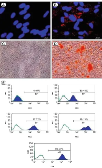
Fig. 1. Characterization of hDP-NCSCs. The cells differentiated into the adipogenic lineage after 15 days of incubation and osteogenic dif-ferentiation (day 8) after osteogenic induction. Adipogenic differen-tiation was identifi ed by neutral lipid vacuole formation (stained with Oil Red O, A – Control, B – Differentiated hDP-NCSCs). Mineral nodules were stained positive with Alizarin Red S stain (C – Control, D – Differentiated hDP-NCSCs). Immunophenotypic properties of hDP-NCSCs evaluated with fl ow cytometry (E). Predefi ned markers that specifi ed MSCs were used to defi ne the characteristics of cultured cells. The hDP-NCSCs expressed all MSC markers including; CD44, CD90 and CD105 but not CD45, CD34 or MHC class II (Scale bars: A,B–20 μm; C,D–100μm).
closed in layers. The urinary bladders of SCI rats were evacuate twice daily during the entire study.
Functional tests
Functional tests were performed using the Basso, Beattie and Bresnahan (BBB) locomotor rating scale at pre-surgery, at days 1, 7, 14, 21, and 28 postinjury (p.i.). Two independent, blinded exam-iners observed each animal for 4 min. Hind limb movements were recorded by video camera and locomotor functions were assessed (18). The BBB scores were presented as mean± standard error.
Tissue harvesting and immunohistochemical examination
To detect GFP+ hDP-NCSCs, an immunofl uorescence double staining protocol was performed on the paraffi n-embedded tissues (19). Slides were deparaffi nized with two changes of xylene for 5 min each and then rehydrated in a series of graded alcohol solu-tions of 100, 90, and 80 %. Antigen retrieval was performed on the slides using a steamer-citrate buffer antigen retrieval method. Endogenous peroxidase activity was blocked by incubating the slides in fresh 3 % H2O2 in PBS buffer. Nonspecifi c staining was blocked with a mixture of two different sera at 1.5 % in PBS for 30 min at room temperature (RT). The sections were incubated for 1 h at RT with the following primary antibodies: anti-GFP antibody (sc-9996; Santa Cruz, Heidelberg, Germany), Vimentin (sc-7557; Santa Cruz), GFAP (MS-280-P, Thermo Scientifi c, Rockford, IL), BDNF (sc-20981; Santa Cruz), Nestin (SC-33677; Santa Cruz) and NF (SC-12980; Santa Cruz). Also the sections stained for pro-infl ammatory cytokines such as; MPO (SC-34161; Santa Cruz), IL-1β (SC-23460; Santa Cruz), IL-6 (SC-1265; Santa Cruz), MIP-2 (SC-1388; Santa Cruz) and anti-infl ammatory cytokines such as IL-1ra (SC-25444; Santa Cruz) and EP3 (SC-16019; Santa Cruz). The sections were incubated in a mixture of two fl uorescence-conjugated secondary antibodies, which included goat anti-mouse FITC (SC-2010; Santa Cruz), donkey anti-goat TR (SC-2783; Santa Cruz), and goat anti-mouse TR (SC-2781; Santa Cruz) at a dilution of 1:50 in PBS for 30 min at RT. The sections were mounted with mounting medium containing DAPI (Santa Cruz Biotechnology). Then, the mounted cells were examined under a fl uorescence microscope (Leica DMI 4000B, Wetzlar, Germany).
Statistical analyses
Statistical analysis was performed with SPSS software for Windows (SPSS for Windows, Version 21, SPSS Inc, Armonk, NY, USA). Descriptive analyses were presented using means and standard deviation for normally distributed variables. Univariate parametric variance analysis was used for groups which did not fulfi ll the requirements of parametric test (ANOVA). Nonpara-metric data were analyzed with Kruskall – Wallis test. In order to investigate differences between the groups, Tukey test was used for parametric and Dunn test was used for nonparametric data. Statistical signifi cance was defi ned by p < 0.05.
Results
Culture and characterization of hDP-NCSCs
hDP-NCSCs attached to the culture fl asks sparsely and non-adherent small cells presented in the primary culture were gradu-ally depleted in further passages. Following 3–4 days of incuba-tion, proliferation started and the cells gradually grew into small colonies. A pure population of fi broblast-like, spindle-shaped cells appeared after three passages. The expression of hDP-NC-SCs markers was analyzed by fl ow cytometry, revealing cells that were positive for CD44, CD73, CD90 and CD105 and nega-tive for CD45/34/11b/ major histocompatibility complex (MHC) Class-II/19 (Fig. 1 A–E). The multipotency of the isolated cells was determined by adipogenic, neurogenic and osteogenic
dif-A B
C D
ferentiation. After three weeks of culture, hDP-NCSCs in the ad-ipogenic differentiation cocktail developed enlarged lipid droplets that invaded the entire cytoplasm of each cell. Their differentiation into adipocytes was confi rmed by Oil Red O staining (Fig. 1 B).
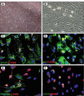
Moreover, the cells cultured in the osteogenic differentiation me-dium formed cellular aggregates that were characterized by the presence of amorphous material deposits. The amorphous deposits were observed under a microscope. Calcium deposits were stained by Alizarin Red S (Fig. 1 D). The neurogenic differentiation of iso-lated cells was observed in 5-day-old culture media that included a chemical cocktail. Neuron-like cells displayed distinct morpholo-gies that ranged from extensively simple, bipolar cells to large, branched and multipolar cells. These cells positively stained for some neuron or glial cell-specifi c markers, including GFAP/c-fos, NF/c-fos, Nestin/c-fos and CNTF/BDNF (Fig. 2 A–F).
Survival and migration of hDP-NCSCs
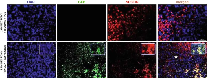
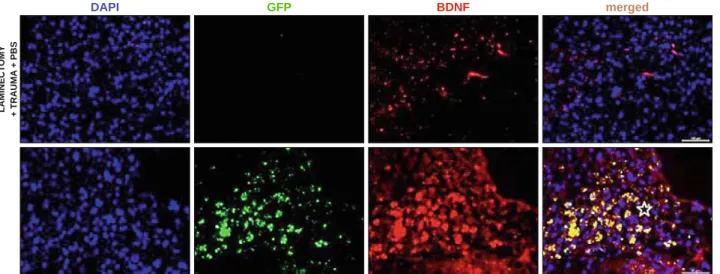
After 4 weeks, the immunofl uorescence microscopic analyses of longitudinal and transversal sections of rat spinal cords from experimental groups were performed with double staining of GFP together with Nestin, GFAP, BDNF, Vimentin, NF, S100. In all sections of 1st, 2nd and 3rd groups, they showed negative staining for GFP. Noticeably, the center of the lesion was severely cavitated in the 2nd and 3rd groups (Figs 3, 4, 5, 6 and 7). However, GFP+ cells were observed in the vicinity of the damage site of Group 4 at the end of 4 weeks, and most of the surviving cells were located at the periphery of the lesion site next to healthier tissue (Figs 3, 4, 5, 6 and 7). GFP+ hDP-NCSCs migrated into cavitated area from the injection sites, and the majority of these still survived and expressed some stem cell and neural markers such as such as GFAP (Fig. 3), nestin (Fig. 4), NF (Fig. 5), BDNF (Fig. 6) and vimentin (Fig. 7). Interestingly, the sections of low scored animals of Group 4 in tests showed considerably lower number of GFP+ cells migrated to the damage site than the sections of higher scored animals. Additionally, the increase of GFAP expression in this re-gion is remarkable (Fig. 3).
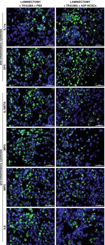
Tissue sections were also stained for pro-infl ammatory (MPO, IL-1β, IL-6 and MIP-2) and anti-infl ammatory cytokines (IL-1ra
Fig. 2. Differentiation of hDP-NCSCs into neuron-glial like cells
in--vitro and expression of neural markers. Phase contrast image after 5
days; neuron-like cells displayed distinct morphologies (A – Control, B – Morphology of neural differentiated cells). Differentiated cells represented intensifi ed staining of C – GFAP (green) and c-fos (red); D – Neurofi lament (green) and c-fos (red); E – Nestin (green) and c-fos (red); F – CNTF (green) and BDNF (red) (Scale bars: 50 μm).
A B C D E F LAMINECT OMY + TRAUMA + PBS LAMINECT OMY + TRAUMA + hDP-NCSCs
DAPI GFP GFAP merged
Fig. 3. The immunostaining of longitudinal sections of rat spinal cords for GFAP expression. GFAP is expressed by numerous cell types of the CNS; its different expression levels in the tissue sections may indicate neuronal regeneration. In the 3rd group; GFAP expression was weak. By
contrast, the 4th group demonstrated strong staining patterns in the lesion area (stars), indicating that the hDP-NCSCs might be directly
and EP3) (Fig. 8). After 4 weeks of stem cell injection, staining of the tissue sections with pro-infl ammatory and anti-infl ammatory cytokines showed different statistical results. In Group 4, the ex-pression of pro-infl ammatory cytokines decreased compared to the other groups (p ˂ 0.05), and the expression of anti-infl ammatory cytokines signifi cantly increased (p ˂ 0.05) (Fig. 9).
Functional recovery
To confi rm the traumatic impact of the standardized severe weight-drop contusion injury to the T10–T11 spinal cord, the hind limb locomotion of the SCI rats was evaluated. At each assess-ment time point, consistent functional defi cits were noted among SCI rats with the BBB locomotion scores showing profound loss initially, which was then gradually improved and approaching
a plateau level of spontaneous recovery typical for this type of injury by 4 weeks p.i. According to BBB locomotor activity test the performances of the Group 4 were statistically different than of the Group 2 and 3 (p < 0.05). The injured rats (Group 2 and 3) showed marked lower activity score than MSC injected group (Group 4) in the BBB locomotor rating score. All experimental groups (Group 2–4) showed BBB locomotion score increment to some extent, but the highest scores were observed in the MSC injected group (Group 4) (Fig.10).
Discussion
MSC shows its therapeutic effect not only by transdifferenti-ating into necessary tissue cells in the damaged region, but also
DAPI GFP NESTIN merged
LAMINECT OMY + TRAUMA + PBS LAMINECT OMY + TRAUMA + hDP-NCSCs
Fig. 4. Detection of Nestin positive cells in the SCI lesion site (star). The expression of Nestin - a protein marker for neural stem cells – was induced in the tissue by the signals derived p.i. The expression level of Nestin was limited in the 3rd group. By contrast, the expression of Nestin by
GFP-positive hDP-NCSCs near the lesion epicenter increased signifi cantly. The nucleus of cells was stained with DAPI (blue) (Scale bars: 100 μm).
DAPI GFP NF merged LAMINECT OMY + TRAUMA + PBS LAMINECT OMY + TRAUMA + hDP-NCSCs
Fig. 5. The IF staining of Neurofi lament in the SCI lesion site (star). Neurofi lament is a major component of the neuronal cytoskeleton, and believed to function primarily to provide structural support for the axon and to regulate axon diameter. In the 4th group, expression of
by increasing the growth factor and cytokine secretions, which have different paracrine and autocrine activities in the damaged region. However, it also has immunomodulatory and anti-infl am-matory effects (13, 20). Cytokines are an important regulator of infl ammation following acute SCI (21). Regarding the IF stain-ing results in our study, anti-infl ammatory cytokines (IL-1ra and EP3) expressions were higher in the 4th group than other groups. Increase in anti-infl ammatory cytokines indicated that the damaged area was repaired. The reduction in the infl ammatory cytokines (MPO, IL-1β, IL-6 and MIP-2) also supports this result. IL-1β has been implicated as a pro-infl ammatory cytokine, whose inhi-bition may prove to be benefi cial in SCI (22). The levels of IL-1β
in the 4th group signifi cantly decreased. IL-6 remains a cytokine with dual functions. On the one hand, it has been demonstrated to modulate the immune response by increasing infl ammation after SCI. High levels of IL-6 can shift NSC differentiation toward as-trocytes rather than neurons, thus promoting glial scar formation (23). Additionally, suppression of IL-6 levels reduces secondary injury by reducing infl ammatory cell infi ltration and may result in improved recovery after SCI (24, 25). Moreover, a medium level of IL-6 is important for preserving neurogenesis and neuroprotec-tion following central nervous system (CNS) injuries (26). In the present study, hDP-NCSCs partially decreased IL-6 levels, while high levels of IL-6 were observed in the other groups.
LAMINECT
OMY
+ TRAUMA
+ PBS
DAPI GFP BDNF merged
Fig. 6. Detection of BDNF in the SCI lesion site (star). The expression of this NF was induced in the tissue by the signals derived post injury. The expression level of BDNF was limited in the 3rd group. In contrast, the expression of BDNF by GFP-positive hDP-NCSCs near the lesion epicenter
increased signifi cantly, which might support axonal outgrowth. The nucleus of cells was stained with DAPI (blue) (Scale bars: 100 μm, 50 μm).
DAPI GFP VIMENTIN merged
LAMINECT OMY + TRAUMA + PBS LAMINECT OMY + TRAUMA + hDP-NCSCs
Fig. 7. The infi ltration of hDP-NCSCs into the lesion site. The stem cells transplanted into the spinal cord were labeled previously with GFP. Vimentin was used as an MSC marker and was used along with GFP to track the hDP-NCSCs in the tissue. The GFP/vimentin-positive cells collected around the lesion site (star) in the hDP-NCSCs -transplanted tissues at 28 days p.i. The nucleus of cells was stained with DAPI (blue) (Scale bars: 100 μm, 50 μm).
Previous studies reported that hDPSCs display an increased level of immunosuppressive activities when compared with Bone Marrow-Mesenchymal Stem Cells (BM-MSCs). Huang et al. showed that the implantation of DPSCs derived from rhesus mon-keys into the hippocampus of mice did not cause any immune rejection. Similarly, higher expressions of human MHC-1, which indicates a stronger degree of immunosuppression, were seen in mouse livers, which were incorporated with hDPSCs (14). In the present study, an acute host immune rejection, such as major infi l-tration of lymphocytes, directed toward the transplanted cells was not observed (27). The IF results at 4th week showed that parts of implanted MSCs have transformed into nerve cells and can survive for long time. They basically adapt to the body’s environ-ment and continue to promote the recovery of the injured spinal cord function. The Hematoxylin&Eosin (HE) staining results of 1st week and 4th week showed that immune rejection of MSCs transplantation was not signifi cantly different from the group of non-implanted cells.
In vitro studies have shown that hDPSCs can differentiate
to-ward functionally active neurons under appropriate culture condi-tions (14). In this study, we determined that hDP-NCSCs could be isolated and amplifi ed effectively in vitro. It could grow well in the special medium for MSCs. Redundant MSCs can be obtained through passage, which can provide experimental foundation for future studies and clinical applications.
Meanwhile, our results showed that hDP-NCSCs transplan-tation for the treatment of rat SCI can signifi cantly improve the neurological function of the damaged spinal cord at 2 weeks after treatment. And at 4 weeks after the treatment, the improvement was much more signifi cant, which was consistent with the results of previous researches on treatment of SCI by transplantation of MSCs (28). This indicated that the damaged spinal cord could be recovered and regenerated by the implantation of hDP-NCSCs. The mechanism still remains unclear on how the transplanted MSCs survive, concentrate and migrate to the damaged spinal cord. Our fi ndings suggested that at 2 weeks after hDP–NCSCs transplan-tation into the damage spinal cord, MSCs begin to adapt to the microenvironment of the spinal cord and spontaneously secrete or induce other cells to produce nerve repair factors to promote regional nerve repair and stimulate the surviving nerve axons to stretch out lateral branches to the damaged axons. Then, spinal cord function was improved (11, 29, 30). It also may be associated with the integration of transplanted cells to the neural circuits (31, 32).
Conclusion
Our study demonstrated that the MSCs can be isolated from the dental pulp and cultured and passaged in vitro. After trans-plantation of the passaged MSCs into rats with SCI, we found that the isolated MSCs can survive in rat bodies without any im-mune rejection. The implanted MSCs can differentiate into nerve cells and are involved in the recovery of the damaged spinal cord. Thus, it improves the scores of motion behavior and promotes the recovery of motor function after SCI. These results provide a theoretical and experimental basis for hDP-NCSCs transplantation
LAMINECTOMY + TRAUMA + PBS LAMINECTOMY + TRAUMA + hDP-NCSCs IL1ra EP3 IL1BET A MPO MIP2 IL6 anti-in fl ammatory cytokines pro-in fl ammatory cytokines
Fig. 8. The IF staining of paraffi n-embedded spinal cord tissue sec-tions for IL1ra, EP3, IL1β, MPO, MIP2 and IL6 after transplantation in Group 3 and 4. The density of IL1β, MPO, MIP2 and IL6 positive cells signifi cantly decreased and IL1ra, EP3 expression was increased after hDP-NCSCs injection. The nucleus of cells was stained with DAPI (blue) (Scale bars: 50 μm).
applied in the treatment of SCI. Compared with other stem cells, hDP-NCSCs have advantages such as good primitiveness, strong amplifi cation ability, simple acquisition, and weak in vivo rejec-tion, without any damage to donor. hDP-NCSCs can provide new cell source and new method for treatment of SCI. As the study is further improved and deepened, it’s possible to apply the hDP-NCSCs transplantation to clinical treatment of SCI.
References
1. Teng YD, Kabatas S, Li J, Wakeman DR, Snyder EY, Sidman RL.
Functional Multipotency of Neural Stem Cells and Its Therapeutic Impli-cations. In: Ulrich H, editor. Perspectives of Stem Cells: From tools for studying mechanisms of neuronal differentiation towards therapy, 1 st ed. Dordrecht, Heidelberg, London, New York: Springer Science+Business Media B.V.; 2010: 255–270.
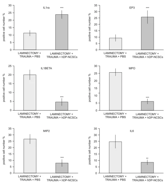
Fig. 9. Quantifi cation of cells expressing proteins associated with infl ammation. Stem cell treatment greatly suppressed the expression of the infl ammatory markers (IL1ra, EP3, IL-1β, IL6, MIP-2 and MPO) in the tissue. After stem cell treatment, cells expressing pro-infl ammatory proteins (IL-1β, IL6, MIP-2 and MPO) decreased and IL1ra and EP3 positive cells were increased in the tissue (*p; 0.01 < p < 0.05, **p; 0.001 < p < 0.01, ***p; 0.001 < p).
Fig. 10. Effect of T10-11 SCI on general hind limb function over time after SCI. Defi cits are expressed as a BBB locomotion score.
2. Teng YD, Yu D, Ropper AE et al. Functional multipotency of stem
cells: a conceptual review of neurotrophic factor-based evidence and its role in translational research. Curr Neuropharmacol 2011; 9 (4): 574–585.
3. Fernández-Martos CM, González-Fernández C, González P, Maque-da A, Arenas E, Rodríguez FJ. Differential expression of Wnts after
spi-nal cord contusion injury in adult rats. PLoS One 2011; 6 (11): e27000.
4. Xie F, Zheng B. White matter inhibitors in CNS axon regeneration
failure. Exp Neurol 2008; 209: 302–312.
5. Joosten EA, Veldhuis WB, Hamers FP. Collagen containing neonatal
astrocytes stimulates regrowth of injured fibers and promotes modest loco-motor recovery after spinal cord injury. J Neurosci Res 2004; 77: 127–142.
6. Karaoz E, Kabatas S, Duruksu G et al. Reduction of lesion in
in-jured rat spinal cord and partial functional recovery of motility after bone marrow derived mesenchymal stem cell transplantation. Turk Neurosurg 2012; 22 (2): 207–217.
7. Sonmez E, Kabatas S, Ozen O et al. Minocycline Treatment Inhibits
Lipid Peroxidation, Preserves Spinal Cord Ultrastructure and Improves Functional Outcome after Traumatic Spinal Cord Injury in the Rat. Spine (Phila Pa 1976) 2013; 38 (15): 1253–1259.
8. Aras Y, Sabanci PA, Kabatas S et al. The Effects of Adipose
Tissue-Derived Mesenchymal Stem Cell Transplantation During the Acute and Subacute Phases Following Spinal Cord Injury. Turk Neurosurg 2016; 26 (1): 127–139.
9. Cömert S, Altinörs N, Kabataş S et al. Human Mesenchymal Stem
Cell Therapy in Thoracic Spinal Cord Injury and Evaluation of the Results-an experimental study in rats. J Turk Spinal Surg 2017; 28 (2): 83–90.
10. Kabatas S, Teng YD. Potential roles of the neural stem cell in the
res-toration of the injured spinal cord: review of the literature. Review. Turk Neurosurg 2010; 20 (2): 103–110.
11. Yamamoto A, Matsubara K, Kano F, Sakai K. Analysis of the
neu-roregenerative activities of mesenchymal stem cells in functional recov-ery after rat spinal cord injury. Methods Mol Biol 2014; 1213: 321–328.
12. Karaöz E, Demircan PC, Sağlam O, Aksoy A, Kaymaz F, Duruksu G. Human dental pulp stem cells demonstrate better neural and epithelial
stem cell properties than bone marrow-derived mesenchymal stem cells. Histochem Cell Biol 2011; 136 (4): 455–473.
13. Özdemir AT, Özgül Özdemir RB, Kirmaz C et al. The paracrine
immunomodulatory interactions between the human dental pulp derived mesenchymal stem cells and CD4 T cell subsets. Cell Immunol 2016; 310: 108–115.
14. Song M, Lee JH, Bae J, Bu Y, Kim EC. Human Dental Pulp Stem
Cells Are More Effective Than Human Bone Marrow-Derived Mesen-chymal Stem Cells in Cerebral Ischemic Injury. Cell Transplant 2017; 26 (6): 1001–1016.
15. Karaöz E, Doğan BN, Aksoy A et al. Isolation and in vitro
charac-terisation of dental pulp stem cells from natal teeth. Histochem Cell Biol 2010; 133 (1): 95–112.
16. Akpinar G, Kasap M, Aksoy A, Duruksu G, Gacar G, Karaoz E.
Phenotypic and proteomic characteristics of human dental pulp derived mesenchymal stem cells from a natal, an exfoliated deciduous, and an impacted third molar tooth. Stem Cells Int 2014; 457059.
17. Choi H, Liao WL, Newton KM et al. Respiratory abnormalities
result-ing from midcervical spinal cord injury and their reversal by serotonin 1A agonists in conscious rats. J Neurosci 2005; 25 (18): 4550–4559.
18. Basso DM, Beattie MS, Bresnahan JC. A sensitive and reliable
lo-comotor rating scale for open fi eld testing in rats. J Neurotrauma 1995; 12: 1–21.
19. Adas G, Arikan S, Karatepe O et al. Mesenchymal stem cells
im-prove the healing of ischemic colonic anastomoses (experimental study. Langenbecks Arch Surg 2011; 396: 115–126.
20. Yuksel S, Guleç MA, Gultekin MZ et al. Comparison of the early
pe-riod effects of bone marrow-derived mesenchymal stem cells and platelet-rich plasma on the Achilles tendon ruptures in rats. Connect Tissue Res 2016; 57 (5): 360–373.
21. Urdzíková LM, Růžička J, LaBagnara M et al. Human
mesenchy-mal stem cells modulate infl ammatory cytokines after spinal cord injury in rat. Int J Mol Sci 2014; 15 (7): 11275–11293.
22. Ruzicka J, Machova-Urdzikova L, Gillick J et al. A Comparative
Study of Three Different Types of Stem Cells for Treatment of Rat Spinal Cord Injury. Cell Transplant 2017; 26 (4): 585–603.
23. Mukaino M, Nakamura M, Okada S, Toyama Y, Liu M, Okano H.
Role of IL-6 in regulation of infl ammation and stem cell differentiation in CNS trauma. Nihon Rinsho Meneki Gakkai Kaishi 2008; 31 (2): 93–98.
24. Nakamura M, Okada S, Toyama Y, Okano H. Role of IL-6 in
spi-nal cord injury in a mouse model. Clin Rev Allergy Immunol 2005; 28 (3): 197–204.
25. Arima H, Hanada M, Hayasaka T et al. Blockade of IL-6 signaling
by MR16-1 inhibits reduction of docosahexaenoic acidcontaining phos-phatidylcholine levels in a mouse model of spinal cord injury. Neurosci-ence 2014; 269: 1–10.
26. Herrmann O, Tarabin V, Suzuki S et al. Regulation of body
tempera-ture and neuroprotection by endogenous interleukin-6 in cerebral ischemia. J Cereb Blood Flow Metab 2003; 23 (4): 406–415.
27. Demircan PC, Sariboyaci AE, Unal ZS, Gacar G, Subasi C, Karaoz E. Immunoregulatory effects of human dental pulp-derived stem cells on
T cells: comparison of transwell co-culture and mixed lymphocyte reac-tion systems. Cytotherapy 2011; 13 (10): 1205–1220.
28. Nosrat IV, Widenfalk J, Olson L, Nosrat CA. Dental pulp cells
pro-duce neurotrophic factors, interact with trigeminal neurons in vitro, and rescue motoneurons after spinal cord injury. Dev Biol 2001; 238: 120–132.
29. Yamamoto A, Sakai K, Matsubara K, Kano F, Ueda M. Review.
Multifaceted neuro-regenerative activities of human dental pulp stem cells for functional recovery after spinal cord injury. Neurosci Res 2014; 78: 16–20.
30. Sakai K, Yamamoto A, Matsubara K et al. Human dental
pulp-de-rived stem cells promote locomotor recovery after complete transection of the rat spinal cord by multiple neuro-regenerative mechanisms. J Clin Invest. 2012; 122 (1): 80–90.
31. Leong WK, Henshall TL, Arthur A et al. Human adult dental pulp
stem cells enhance poststroke functional recovery through non-neural re-placement mechanisms. Stem Cells Transl Med 2012; 1: 177–187.
32. Crigler L, Robey RC, Asawachaicharn A, Gaupp D, Phinney DG.
Human mesenchymal stem cell subpopulations express a variety of neuro-regulatory molecules and promote neuronal cell survival and neuritogen-esis. Exp Neurol 2006; 198: 54–64.
Received November 8, 2017. Accepted December 20, 2017.