Full Terms & Conditions of access and use can be found at
https://www.tandfonline.com/action/journalInformation?journalCode=ibih20
Biotechnic & Histochemistry
ISSN: 1052-0295 (Print) 1473-7760 (Online) Journal homepage: https://www.tandfonline.com/loi/ibih20
Effects of ciprofloxacin on fetal rat liver during
pregnancy and protective effects of quercetin
Z Dogan, H Elbe, E Taslidere, H Soysal, A Cetin & S Demirtas
To cite this article: Z Dogan, H Elbe, E Taslidere, H Soysal, A Cetin & S Demirtas (2017) Effects of ciprofloxacin on fetal rat liver during pregnancy and protective effects of quercetin, Biotechnic & Histochemistry, 92:7, 481-486, DOI: 10.1080/10520295.2017.1356469
To link to this article: https://doi.org/10.1080/10520295.2017.1356469
Published online: 24 Aug 2017.
Submit your article to this journal
Article views: 187
View related articles
View Crossmark data
Ll
Taylor & Francis~ Tllylorf.J,;i11caCr11u1,
~'
[?1.111
tl1
[?®
[? CrossMdrkEffects of cipro
floxacin on fetal rat liver during
pregnancy and protective effects of quercetin
Z Dogan
1, H Elbe
2, E Taslidere
3, H Soysal
4, A Cetin
5, S Demirtas
61
Department of Anatomy, Faculty of Medicine, Adiyaman University, Adiyaman,2Department of Histology and Embryology, Faculty of Medicine, Mugla Sıtkı Kocman University, Mugla,3Department of Histology and Embryology, Faculty of Medicine, Bezmialem Vakif University, Istanbul,4Department of Anatomy, Faculty of Medicine, Baskent University, Ankara,
5
Departments of Anatomy, and6Biochemistry, Inonu University, Faculty of Medicine, Malatya, Turkey Accepted July 13, 2017
Abstract
Urinary tract infections are common in pregnant women and ciprofloxacin frequently is used as a broad spectrum antibiotic. It has been suggested that ciprofloxacin causes liver damage in fetuses. Quercetin is a flavonoid with antioxidant properties. We investigated the efficacy of quercetin treatment for preventing fetal liver damage caused by ciprofloxacin. Pregnant rats were divided into four groups: untreated control group (C), 20 mg/kg quercetin for 21 days group (Q), 20 mg/kg twice/day cipro-floxacin for 10 days group (CP), and 20 mg/kg, ciprocipro-floxacin + quercetin for 21 days group (CP + Q). Fetal livers were removed on day 21 of gestation to measure antioxidants and for histological observa-tion. Malondialdehyde (MDA) and glutathione (GSH) levels, and superoxide dismutase (SOD), cata-lase (CAT) and glutathione peroxidase (GSH-Px) activities were measured in tissue samples. GSH-Px, SOD and CAT activities were significantly lower in the CP group compared to group C. A significant increase in MDA was observed in the CP group compared to group C. There was no significant difference in GSH levels in any group. MDA levels were lower and CAT, SOD and GSH-Px enzyme activities were higher in the CP + Q group compared to group CP. Liver samples of the CP group exhibited central vein dilation, portal vein congestion, pyknotic nuclei and cytoplasmic vacuolization in some hepatocytes. Histological changes were less prominent in the rats treated with quercetin. Use of ciprofloxacin during pregnancy caused oxidative damage in fetal liver tissue. Oxidative stress was ameliorated by quercetin. Quercetin supports the antioxidant defense mechanism and it is beneficial for treating fetal liver damage caused by ciprofloxacin.
Key words: ciprofloxacin, fetus, liver, pregnancy, quercetin, rat
Recurrent urinary tract infection is a common problem, particularly for pregnant and older women (Dwyer PL 2002). Bacterial urinary tract infections during pregnancy increase perinatal and maternal morbidity and mortality (Onoh et al.2013). The fetus is at risk for prematurity, low birth weight,
intrauterine growth retardation and fetal death (Abdul and Onile2001, Ezechi et al.2003).
Fluoroquinolones are recommended for women infected with resistant microorganisms (Dwyer PL 2002); they are potent antibiotics with a wide spec-trum of activity. The antibiotics interact with DNA gyrase and topoisomerase IV to block DNA repli-cation, which causes bacterial cell death (Robicsek et al.2006). Ciprofloxacin is a synthetic quinolone that exhibits wide spectrum antibiotic activity (Channa et al.2012).
Some toxic effects of ciprofloxacin have been reported in humans and animals including hepato-toxicity (Orman et al.2011, Moreno et al.2015). Correspondence: Zumrut Dogan, Department of Anatomy, Faculty
of Medicine, Adiyaman University, 02100 Adıyaman, Turkey.
Phone: 0 416 2233800 (1485), e-mail: [email protected],
Color versions of one or more of thefigures in the article can be
found online atwww.tandfonline.com/ibih
© 2017 The Biological Stain Commission
Biotechnic & Histochemistry 2017, 92(7): 481–486
~ Taylor & Francis
~ Taylor&FrancisGroup
Channa and Janjua (2003) reported that cipro-floxacin administration during gestation caused hepatocyte damage in Wistar albino rats. Some anti-biotics, including ciprofloxacin, increase the num-ber of free radicals and cause oxidative damage to tissues (Elbe et al.2015, Taslıdere et al.2015).
Malondialdehyde (MDA) levels are increased by oxidative damage (Kletkiewicz et al. 2016). Glutathione (GSH) is an antioxidant that prevents damage to cellular components caused by reactive oxygen species (ROS) (Pal et al. 2015). Oxidative stress may reduce GSH levels (Martin et al. 2000, Kletkiewicz et al. 2016). Superoxide dismutase (SOD), catalase (CAT) and glutathione peroxidase (GSH-Px) enzymes participate in defense against oxidative damage (Ismail et al.2016).
Flavonoids are naturally occurring secondary metabolites that are widely distributed as in the plant kingdom (Kanter et al. 2007, Coskun et al. 2015). Quercetin is among the most widely distributed flavonoids in plants and has been reported to be beneficial to human health (Liu et al. 2010). Quercetin scavenges free radicals directly, inhibits biomolecule oxidation, prevents lipid damage and stimulates antioxidant defense path-ways in vivo and in vitro (Sanders et al. 2001, Coldiron et al.2002).
We investigated the antioxidant and protective effects of quercetin on ciprofloxacin-induced fetal liver damage in Wistar albino rats. We analyzed histopathological findings and performed bio-chemical tests to determine tissue MDA and GSH levels, and SOD, CAT and GSH-Px activities.
Material and methods
Animals and experimental design
We used twenty-eight 6-month-old 250 g female Wistar albino rats. All experimental procedures were reviewed and approved (2012/A-23) by the local ethics committee of the Medical Sciences Experimental Search and Application Center at the Inonu University. Two females and one male were housed together in a cage from 5 pm until 8 am the following day; the males then were separated from the females. Vaginal smears were examined under the microscope. Spermatozoa were found in the vaginal smears of 22 rats; this was considered 0.5 day gestation.
The 22 pregnant rats were divided randomly into four groups. For controls (C), corn oil was adminis-tered by gavage for 21 days to six pregnant rats and
intraperitoneal (i.p.) saline was administered twice/ day, between days 7 and 17 of gestation. For quercetin group (Q), 20 mg/kg quercetin was administered daily by gavage for 21 days tofive rats throughout gestation (Ciftci and Ozdemir 2011). For the ciproflox-acin group (CP),five rats were injected i.p. with 20 mg/kg CP for 10 days twice/day from day 7 through day 17 of gestation (Channa and Janjua2003). For the ciprofloxacin + quercetin group (CP + Q), six rats were injected i.p. with 20 mg/kg ciprofloxacin twice/day for 10 days and 20 mg/kg quercetin was administered by gavage for 21 days.
Fetuses were delivered by C section on day 20 of pregnancy. A pool was prepared by choosing ran-domly one or two pups from each mother. The his-tology and biochemistry of 28 pups, seven for each group, were examined at 20 days of gestation.
Histology
At the end of the study, the rats were sacrificed by ketamine and xylazine anesthesia. The right lobe of the fetal liver was removed following laparotomy and divided into two equal parts. Tissue samples for his-tology werefixed immediately in 10% formalin solu-tion (100 ml formalin, 8.5 g sodium chloride in 900 ml distilled water) (Drury and Wallington 1967). The specimens werefixed for 24 h. After fixation the speci-men was washed thoroughly with water and passed through ascending grades of ethyl alcohol, and cleared with xylene, then impregnated with molten wax. After trimming, the paraffin blocks were cut at 4 μm, mounted on slides and stained with hematoxylin and eosin (H & E) (Rekha et al.2013).
All sections were examined using a Leica DFC280 light microscope and a Leica Q Win and Image Analysis system (Leica Micros Imaging Solutions Ltd., Cambridge, UK).
Biochemistry
The second portion of the tissue sample was stored at −80° C for use in measuring levels of MDA and GSH, and SOD, CAT and GSH-Px activities. Samples were homogenized in ice-cold 0.1 M Tris–HCl buffer, pH 7.5, containing 1 mM phenylmethylsulfonylfluoride, a protease inhibitor, with a homogenizer (T25 Ultra Turrax; IKA, Staufen, Germany) at 16,000 rpm for 2 min at 4–8° C. The homogenates were used to mea-sure the levels of MDA and GSH, and the super-natants were used to measure the activities of SOD, CAT and GSH-Px.
MDA
Lipid oxidation was determined as described pre-viously (Uchiyama and Mihara 1978). The reaction mixture contained 2 ml freshly prepared reaction solution [15% (w/v) trichloroacetic acid, 0.375% (w/v) thiobarbituric acid (TBA)] and 250 μl tissue homogenate; the mixture was boiled for 15 min. The mixture was cooled to room temperature and centrifuged at 10,000 × g for 10 min, then the super-natant was collected. Absorbance of the supersuper-natant was recorded at 535–520 nm. The MDA levels in nmol were calculated from the plotted standard curve prepared from 1,1,3,3-tetramethoxypropane. MDA results were expressed as nmol/g wet tissue in the homogenate.
GSH
GSH concentrations in the homogenates were mea-sured using a spectrophotometric method (Elman 1959). This method is based on the reduction of GSSG to GSH in the presence of glutathione reduc-tase and NADPH, and formation of the colored product which is formed by the reaction of GSH with DTNB [5,5′-dithio-bis(2-nitrobenzoic acid)], fol-lowed by measuring its absorbance at 410 nm. Results are recorded as nmol/g wet tissue.
SOD
SOD activity was measured by determining the reduction of nitroblue tetrazolium by measuring the superoxide anion produced by xanthine and xanthine oxidase (Sun et al.1988). One unit of SOD was defined as the amount of protein that inhibited the rate of NBT reduction by 50%, and the results were recorded as U/mg protein. Protein in the liver tissue was determined using the method of Lowry et al. (1951).
CAT
CAT activity was measured by determination of the rate constant of H2O2 at 240 nm using a spectrophotometer (Aebi 1974). H2O2 solution [0.036% (w/v)] was prepared in 50 mM potassium phosphate buffer, pH 7.0 at 25° C. The reaction mixture was prepared by adding 50μl of superna-tant to 950μl freshly prepared H2O2solution. The decreasing absorbance was recorded at 240 nm for 60 sec. The molar extinction coefficient of H2O2was used to determine CAT activity as 40/M/cm. One unit of CAT activity is defined as the amount of enzyme that catalyzes the degradation of 1μmol of H2O2/min at 37° C. Specific activity of CAT was expressed as U/mg protein.
Statistical analysis
Statistical analysis was performed using SPSS for Windows, version 13.0 (SPSS Inc., Chicago, IL) statistical package program. Data are presented as means ± SE. Normality for continuous variables was determined using the Shapiro Wilk test. The variables did not show normal distributions; there-fore, the Kruskal-Wallis and Mann Whitney U tests were used to compare the variables among the study groups. Values for p≤ 0.05 were considered significant.
Results
Histology
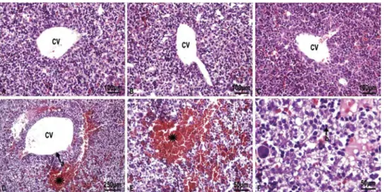
The histological appearance was normal for sec-tions of both C and Q groups (Fig. 1A and B). Some histopathological changes including dilation of central veins, congestion in portal veins and sinusoids, pyknotic hepatic nuclei and vacuoliza-tion were evident in the liver samples of the rats in group CP. Lymphocytic infiltration and degenerat-ing cells with eosinophilic cytoplasm were signi fi-cantly more frequent in the CP group compared to C and Q groups (Fig. 1D−F). Histopathological changes were reduced in the CP + Q group com-pared to the CP group (Fig. 1C).
Biochemistry
The mean tissue MDA levels were significantly higher in the CP group than in the C and Q groups (p = 0.002 and 0.013, respectively). Also, the mean MDA levels were significantly lower for the CP + Q group than for the CP group (p = 0.0035). The mean GSH level was significantly lower for the CP group than for the quercetin group (p = 0.047). The mean GSH level for the CP group was lower than for the C group, but the difference was not statistically significant. The mean tissue SOD and CAT activities of the CP group were significantly lower than for the C group (p = 0.002 and 0.035, respectively). On the other hand, we found significantly higher SOD and CAT activ-ities for the CP + Q group than for CP group (p = 0.035 and 0.002, respectively). Although GSH-Px levels for the CP group were significantly lower than for the C group (p = 0.035), there was no significant difference between the CP and CP + Q groups. The biochemical findings for all groups are summarized in Table 1.
Discussion
Fluoroquinolones often are used for genitourin-ary infections, especially for treatment of hospi-tal-acquired infections such as urinary catheter-related infection (Rawi et al.2011). Ciprofloxacin is a well tolerated fluoroquinolone (Petri 2001). Fluoroquinolone antibiotics, including cipro flox-acin, have been reported to cause fetal liver and epiphyseal cartilage damage, and increased risk of skeletal malformations during the prenatal period (Channa and Janjua 2003, 2006, 2008). We found that ciprofloxacin administered dur-ing gestation caused morphological changes in the liver parenchyma of the offspring due to oxidative damage. Adikwu and Brambaifa (2012) also reported hepatotoxic effects of ciprofloxacin.
Antioxidants, such as quercetin, are protective against the toxic effects of oxidants (Cuevas et al.
2011). Our findings suggest that quercetin decreased fetal liver tissue damage related with ciprofloxacin administration; however it was unable to fully prevent it. The protective effect of quercetin may be due to free radical scavenging (Hilliard 1995, Young and Woodside 2001, Singh et al.2003, Cherubini et al.2005).
We found that MDA levels were significantly higher in the CP group than in the C and Q groups . MDA commonly is used as a biochemical marker for oxidative stress and destruction of lipids (Sahna et al. 2006). Increased MDA levels in the CP group indicated oxidative damage, which is consistent with the litera-ture (Elbe et al.2015, Taslidere et al.2015). We used quercetin, aflavonoid with strong antioxidant charac-teristics, to counteract the injury caused by cipro flox-acin in fetal liver tissues and found that it decreased high MDA levels significantly.
Many enzymes including SOD, CAT and GSH-Px participate in the antioxidant defense system
Figure 1. A) and B) C) and Q group showing normal hepatocytes and the central vein (CV), H & E; × 20. C. CP + Q group, histological appearance is similar to C and Q group in CP + Q group, H & E; × 20. D) CP group, asterisk; lymphocytic infiltration, a; dilatation in central vein, H & E; × 10. E) CP group, H & E; × 40. F) Ciprofloxacin group, b; pyknotic nuclei within the hepatocyte, H & E × 40.
Table 1. Mean MDA, GSH and GSH-Px levels, and SOD and CAT activities
Groups MDA (nmol/g) GSH (nmol/g) SOD (U/mg) CAT (U/mg) GSH-Px (nmol/g) C 469.00 ± 21.32 1956.00 ± 177.05 37.88 ± 1.15 27.92 ± 2.81 561.28 ± 25.11 Q 583.57 ± 26.74 1979.85 ± 90.43 15.34 ± 2.52 30.47 ± 2.08 498.54 ± 15.30 CP 804.00 ± 55.46a,b 1688.42 ± 78.63d 9.24 ± 2.20e 18.35 ± 2.54g 487.04 ± 15.16i CP + Q 613.14 ± 40.99c 2040.28 ± 145.91 21.65 ± 4.16f 32.34 ± 1.24h 524.95 ± 21.16
Data are means ± SE. n = 7.
ap = 0.002 vs. control group,bp = 0.013 vs. quercetin group,cp = 0.035 vs. CP group,dp = 0.047 vs. quercetin group,ep = 0.002 vs.
control group,fp = 0.035 vs. CP group,gp = 0.035 vs. control group,hp = 0.002 vs. CP group,ip = 0.035 vs. control group.
(Nagasaka et al. 2006). We found that antioxidant enzyme activity was decreased significantly in the CP group compared to all other groups. Administration of quercetin alleviated the decrease in antioxidant enzyme capacity significantly. Quercetin is a lipophilic antioxidant and it prevents lipid damage lipid bilayers of membranes (Elliott et al.2000).
We found that quercetin administration inhib-ited ciprofloxacin induced damage to fetal liver. Oxidative damage that occurs as a result of imbal-ance between oxidants and antioxidants in favor of oxidants during the fetal period and early childhood also causes permanent damage to the brain (Demircioğlu and Yabancı 2003, Özmert 2005). Quinolone type antibiotics appear to be bactericidal owing to their inhibition of bacterial DNA gyrase (Guzmán et al.2003). We believe that fluoroquinolones cause oxidative stress in tissues and damage DNA during fetal development; the increased MDA level in fetal liver tissues and the imbalance of the antioxidant system support our hypothesis.
We found that ciprofloxacin caused oxidative damage in fetal liver tissues. We suggest that sup-plementation with antioxidants after ciprofloxacin administration during pregnancy could reduce the fetal toxicity of the antibiotic.
Acknowledgment
The authors are grateful to Prof. Dr. Yusuf Türköz for his kind help.
Declaration of interest: The authors report no con-flicts of interest. The authors alone are responsible for the content and writing of this paper.
References
Abdul IF, Onile BA (2001) Bacterial isolates from urine of women in Ilorin and their antibiotic susceptibility patterns. Trop. J. Obstet. Gynaecol. 18: 61–65.
Adikwu E, Brambaifa N (2012) Ciprofloxacin cardiotoxicity
and hepatotoxicity in humans and animals. Pharmacol. Pharm. 3: 207–213.
Aebi H(1974) Catalase. In: Bergmeyer HU, Ed. Methods of Enzymatic Analysis. Academic Press, New York. pp. 673–684.
Channa MA, Janjua MZ (2003) Effects of ciprofloxacin
on foetal hepatocytes. J. Pak. Med. Assoc. 53: 448-–450.
Channa HM, Ashfaq M, Mastoi SM, Qureshi MA (2006) Effect of ciprofoxacin on growing cartilage in albino rat pups. J. Ayub Med. Coll. Abbottabad 18: 50-–54. Channa HM, Ashfaq M, Bangash R, Abbasi A, Qureshi MA(2008) Preventive role of zinc chloride against toxi-city of ciprofloxacin on the growing cartilage of Wistar albino rat litter. J. Ayub Med. Coll. Abbottabad 20: 77–81. Channa MA, Ashfaq M, Jokhio AL, Khan MZ, Sahito MM(2012) Effects of ciprofloxacin and zinc chloride in
adult albino rat and pre-natal conceptus. J. Ayub Med. Coll. Abbottabad 24: 55–58.
Cherubini A, Ruggiero C, Polidori MC, Mecocci C (2005) Potential markers of oxidative stress in stroke. Free Rad. Biol. Med. 39: 841–852.
Coldiron AD Jr, Sanders RA, Watkins JB III (2002) Effects of combined quercetin and coenzyme Q10 treat-ment on oxidative stress in normal and diabetic rats. J. Biochem. Mol. Toxicol. 16: 197–202.
Coskun O, Kanter M, Korkmaz A, Oter S (2005) Quercetin, a flavonoid antioxidant, prevents and pro-tects streptozotocin-induced oxidative stress and β-cell damage in rat pancreas. Pharmacol. Res. 51: 117–123. Cuevas MJ, Tieppo J, Marroni NP, Tuñón MJ, González-Gallego J (2011) Suppression of amphiregu-lin/epidermal growth factor receptor signals contributes to the protective effects of quercetin in cirrhotic rats. J. Nutr. 141: 1299–1305.
Demircioğlu Y, Yabancı N (2003) Beslenmenin Bilişsel
Gelişim Ve Fonksiyonları İle İlişkisi. Hacettepe Üniv. Eğitim Fakül. Derg. 2: 170–179.
Drury RAB, Wallington EA (1967) Carleton’s Histological
Techniques. 4th ed. Oxford University Press, p. 129. Dwyera PL(2002) Recurrent urinary tract infection in the female. Current Opinion Obstetrics Gynecology 14: 537–543. Elbe H, Dogan Z, Taslidere E, Cetin A, Turkoz Y (2015) Beneficial effects of quercetin on renal injury and oxida-tive stress caused by ciprofloxacin in rats. A histological and biochemical study. Hum. Exp. Tox. 35: 276–281. Elliott M, Kandaswami C, Theoharides TC (2000) The effects of plantflavonoids on mammalian cells: implica-tions for ınflammation, heart disease, and cancer. Pharmacol. Rev. 52: 673–751.
Elman GL (1959) Tissue sulphydryl groups. Arch. Biochem. Biophys. 82: 70–77.
Ezechi OC, Fasubaa OB, Dare FO (2003) Antibiotic sen-sitivity patterns of microbial isolates from urine of preg-nant women with urinary tract infections. Trop. J. Obstet. Gynaecol. 20: 113–115.
Guzmán A, García C, Marín AP, Willoughby C, Demestre I(2003) Developmental toxicity studies of the quinolone antibacterial agent irloxacin in rats and rab-bits. Arzneimit. Forsch. 53: 121–125.
Hilliard JJ, Krause HM, Bernstein JI, Fernandez JA, Nguyen V, Ohemeng KA, Barrett JF (1995) A compar-ison of active site binding of 4-fluoroquinolones and novel flavone gyrase inhibitors to DNA gyrase. Adv. Exp. Med. Biol. 390: 59–69.
Ismail AF, Salem AA, Eassawy MM (2016) Hepatoprotective effect of grape seed oil against carbon
tetrachloride induced oxidative stress in liver of γ-irra-diated rat. J. Photochem. Photobiol. B 160: 1–10.
Kanter M, Altan MF, Donmez S, Ocakci A, Kartal ME (2007) The effects of quercetin on bone minerals, biome-chanical behavior, and structure in streptozotocin-induced diabetic rats. Cell Biochem. Funct. 25: 747–752. Kletkiewicz H, Nowakowska A, Siejka A, Mila-Kierzenkowska C, Woźniak A, Caputa M, Rogalska J (2016) Deferoxamine prevents cerebral glutathione and vitamin E depletions in asphyxiated neonatal rats: role of body temperature Int. J. Hypertherm. 32: 211–220. Liu CM, Zheng YL, Lu J, Zhang ZF, Fan SH, Wua DM, Ma JQ (2010) Quercetin protects rat liver against lead-induced oxidative stress and apoptosis. Environ. Toxicol. Pharm. 29: 158–166.
Lowry OH, Rosebrough NJ, Farr AL, Randall RJ (1951) Protein measurement with the folin phenol reagent. J. Biol. Chem. 193: 265–275.
Martin LJ, Brambrink AM, Price AC, Kaiser A, Agnew DM, Ichord RN, Traystman RJ (2000) Neuronal death in newborn striatum after hypoxia-ischemia is necrosis and evolves with oxidative stress. Neurobiol. Dis. 7: 169–191. Moreno L, Sánchez Delgado J, Vergara M, Casas M, Miquel M, Dalmau B (2015) Recurrent drug-induced liver injury (DILI) with ciprofloxacin and amoxicillin/ clavulanic. Rev. Esp. Enferm. Dig. 107: 767–768.
Nagasaka H, Inoue I, Inui A, Komatsu H, Sogo T, Murayama K, Uemoto S (2006) Relationship between oxidative stress and antioxidant systems in the liver of patients with Wilson disease: hepatic manifestation in Wilson disease as a consequence of augmented oxidative stress. Pediat. Res. 60: 472–477.
Onoh R, Umeora O, Egwuatu V, Ezeonu P, Onoh T (2013) Antibiotic sensitivity pattern of uropathogens from pregnant women with urinary tract infection in Abakaliki, Nigeria. Infec. Drug Resist. 2: 225–233. Orman ES, Conjeevaram HS, Vuppalanchi R, Freston JW, Rochon J, Kleiner DE, Hayashi PH (2011) Clinical and histopathologic features offluoroquinolone-induced liver injury. Clin. Gastroenterol. Hepatol. 9: 517–523. Ozmert EN(2005) Erken çocukluk gelişiminin
desteklen-mesi-I: Beslenme Çocuk Sağlığı ve Hastalıkları Derg 48: 179–195.
Pal S, Ghosh M, Ghosh S, Bhattacharyya S, Sil PC (2015) Atorvastatin induced hepatic oxidative stress and apoptotic damage via MAPKs, mitochondria, cal-pain and caspase 12 dependent pathways. Food Chem. Toxicol. 83: 36–47.
Petri WA (2001) Sulfonamides, trimethoprim, sulfo-methoxazole, quinolones and agents of urinary tract ınfections. In: Brunton LL, Ed., Goodman & Gilman’s the Pharmacological Basics of Therapeutics, 12th ed. McGraw Hill Co. Inc., New York. pp. 1463–1476.
Rawi SM, Mourad IM, Arafa NM, Alazabi NI (2011) Effect of ciprofloxacin and levofloxacin on some oxida-tive stress parameters in brain regions of male albino rats. Afr. J. Pharm. Pharmacol. 5: 1888–1897.
Rekha, Sunanda R, Sajad H (2013) Histopathological effects of pesticide-cholopyrifos on kidney in albino rats. Int. J. Res. Med. Sci. 4: 465–475.
Robicsek A, Jacoby GA, Hooper DC (2006) The world-wide emergence of plasmid-mediated quinolone resis-tance. Lancet Infec. Dis. 6: 629–640.
Sahna E, Parlakpinar H, Cihan OF, Turkoz Y, Acet A (2006) Effects of aminoguanidine against renal ischaemia-reperfusion injury in rats. Cell Biochem. Funct. 24: 137–141. Sanders RA, Rauscher FM, Watkins III JB (2001) Effects of quercetin on antioxidant defense in strepto-zotocin-induced diabetic rats. J. Bıochem. Mol. Toxicol. 15: 143–149.
Singh A, Naidu PS, Kulkarni SK (2003) Reversal of aging and chronic ethanol-induced cognitive dysfunc-tion by quercetin a bioflavonoid. Free Rad. Res. 37: 1245–1252.
Sun Y, Oberly LW, Li Y (1988) A simple method for clinical assay of SOD. Clin. Chem. 34: 479–500.
Taslidere E, Dogan Z, Elbe H, Vardi N, Cetin A, Turkoz Y(2015) Quercetin protection against
ciproflox-acin induced liver damage in rats. Biotech. & Histochem. 91: 116–121.
Uchiyama M, Mihara M (1978) Determination of mal-onaldehyde precursor in tissues by thiobarbituric acid test. Anal. Biochem. 86: 271–278.
Young IS, Woodside JV (2001) Antioxidants in health and disease. J. Clin. Pathol. 54: 176–186.