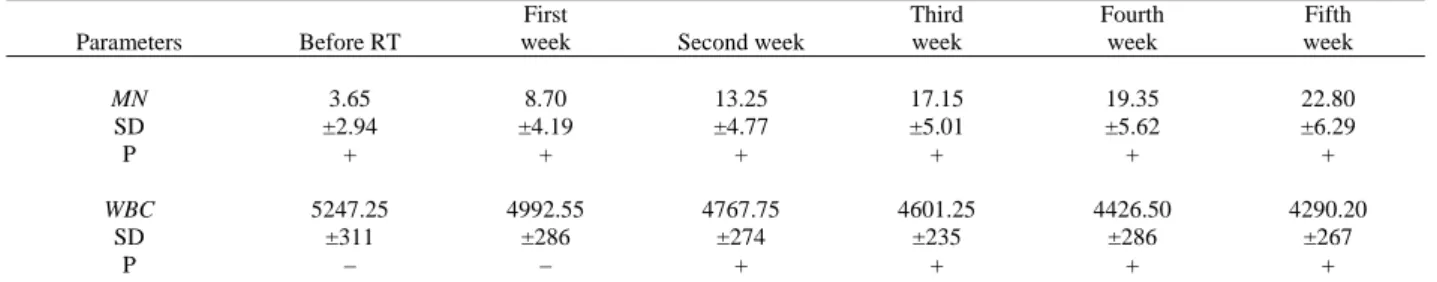
The frequency of micronuclei and morphological effects on white blood cells following radiotherapy
Tam metin
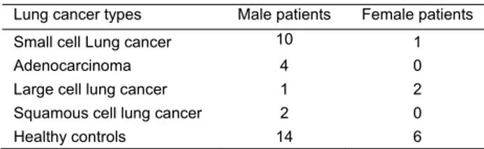
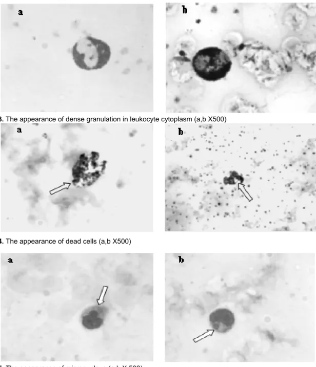
Şekil
Benzer Belgeler
Bu bulguya göre yaratıcı drama yöntemiyle verilen çevre eğitimi etkinliklerinin uygulandığı deney grubunun çevre eğitimine yönelik farkındalıkları ile çevre eğitimi
All of two intermediate-level vancomycin-re- sistant enterococci and five vancomycin-suscepti- ble isolates were also analysed for the presence of vancomycin resistance genes
All types of silk tofu significantly reduced the L/B value; ALT activity, total cholesterol, hepatic MDA and PC levels, beside, liver vitamin C content increased compared to CCl 4
7) Eslem ilk gün 31 sayfa, ikinci gün ise birinci gün okuduğunun 2 fazlası sayfa kitap okumuştur. Eslem iki günde toplam kaç sayfa kitap okumuştur??. 14) Ece' nin yaşının
İncelemede “Osmanlı tasfiyeciliği meselesi” bağlamında dikkatimizi çeken hususlara gelin- ce, Michot’nun daha ziyâde Akhisârî’nin bazı görüşleri ile kimi
O zamana kadar Turklerde vatan diye bir kavram yok, gogebe bir kavim oldugu igin Turklerde vatan degil yurt vardi.. Yurt da gadir
borderline-hypercholesterolemic subjects (N=31, 200 ≦TC< 240mg/dl) and control subjects (N=25,TC<200mg /dl).Total cholesterol、TG and LDL-C levels appeared to be higher
Sonuç olarak; uranyum ve toryum iyonlarının topo reaktifi ile hem kesikli hem de sürekli ekstraksiyon işlemlerinde ortam sıcaklığı, donör ve akseptör faz pH’ı,
