See discussions, stats, and author profiles for this publication at: https://www.researchgate.net/publication/293082662
A new species of Sarcophaga (Sarcophaga) (Diptera: Sarcophagidae) from
Turkey
Article in Turkish Journal of Entomology · June 2011
CITATIONS
4
READS
193
4 authors, including:
Some of the authors of this publication are also working on these related projects:
The determination of fruit fly (Diptera: Tephritidae) fauna in Adıyaman, Kilis, and Şanlıurfa provinces with a new record for Turkish faunaView project Gamze Pekbey Bozok University 12PUBLICATIONS 36CITATIONS SEE PROFILE Rustem Hayat Ataturk University 52PUBLICATIONS 390CITATIONS SEE PROFILE René Richet 20PUBLICATIONS 119CITATIONS SEE PROFILE
Türk. entomol. derg., 2011, 35(2): 285-293 ISSN 1010-6960
Orijinal araştırma (Original article)
A new species of Sarcophaga (Sarcophaga)
(Diptera: Sarcophagidae) from Turkey
Gamze PEKBEY1* Rüstem HAYAT1,2 René RICHET3 Ruth M. BLACKITH4
Summary
A new species Sarcophaga (Sarcophaga) trabzonensis n. sp. is described from Turkey. The male and female terminalia are illustrated and the species is compared with the similar Sarcophaga (Sarcophaga) apsuarum Rohdendorf, 1937 and Sarcophaga (Sarcophaga) pagensis Baranov, 1939.
Key words: Diptera, Sarcophagidae, Sarcophaga (Sarcophaga) trabzonensis, new species, Turkey
Anahtar sözcükler: Diptera, Sarcophagidae, Sarcophaga (Sarcophaga) trabzonensis, yeni tür, Türkiye
Introduction
The genus Sarcophaga Meigen, 1826 belongs to the subfamily Sarcophaginae (Pape, 1987, 1996). Members of this subfamily are easily recognized from the combination of a thorax with three strong black stripes and a speckled or checker-board patterned abdomen (Pape et al., 2009). Also, it is a well supported monophyletic group that includes about 1800 described species worldwide, divided into 51 genera (Pape, 1996). Although flies vary greatly in size (from 3–18 mm), externally they are very uniform in appearance. By contrast, the male genitalia, especially the phalli, are highly distinctive at the species level and have long been used in species recognition (Giroux et al., 2010).
The genus Sarcophaga in the broad sense contains more than 800 species (Pape, 1996). To deal with this large number of species and the phylogenetic patterns they are considered to form, named partitions or subdivisions are necessary. The relative inclusiveness of these subdivisions differs considerably among authors (Pape & Bänziger, 2003).
1 Department of Plant Protection, Faculty of Agriculture, Ataturk University, 25240, Erzurum, Turkey 2
Ardahan University Rectorate, 75100, Ardahan, Turkey
3
Pontificia 1 16 Grande Rue, F-03220 Jaligny-sur-Besbre, France
4
Zoology Department, Trinity College, Dublin 2, Ireland
*
Corresponding author e-mail: [email protected]
Studies of Turkish flesh flies are very limited, several researches were performed by Süreyya (1931), Rohdendorf (1937), Nizamlıoğlu (1954), Collart (1962), Merdivenci (1966), Verves (1986), Pape (1996) and Ebejer (2000). Kara & Pape (2002) listed 81 species with 43 new records. Civelek & Tezcan (2005) added some new records to the Dipteran fauna of Turkey but Sarcophaga
lehmanni (Mueller, 1922) was given as Sarcophaga lasiostyla Macquart, 1843
(Hayat et al., 2008). Since then, eight new records have been added to the Turkish fauna (Aslan, 2006; Hayat et al., 2008; Pekbey & Hayat, 2010). There are now 89 flesh fly species listed for Turkey including four species belonging to
Sarcophaga (Sarcophaga). These are Sarcophaga (Sarcophaga) bergi
Rohdendorf, 1937, Sarcophaga (Sarcophaga) croatica Baranov, 1941,
Sarcophaga (Sarcophaga) lehmanni Muller, 1922 and Sarcophaga (Sarcophaga) variegata (Scopoli, 1763) (Kara & Pape, 2002 Aslan & Çalışkan 2009).
A new species of Sarcophaga (sensu stricto) in material of Sarcophagidae from North-Eastern and Eastern Turkey is described below and compared with two similar species, Sarcophaga (Sarcophaga) apsuarum Rohdendorf, 1937 and Sarcophaga (Sarcophaga) pagensis Baranov, 1939.
Material and Method
The specimens were collected, using an insect sweep net, from the provinces Trabzon, Artvin and Kars. The areas swept were of meadow and pasture containing a variety of flowering plants. The male was obtained in 1993 and the females in 2009.
Text on labels is given in verbatim quotation; individual lines are separated by a slash (/). Extra information is given in square brackets.
The following abbreviations are used in the figures: d=dorsal plate,
h=harpes, j=juxta, s=stylus (lateral), v=vesica, vm=ventral membrane, vp= ventral plate, ST=sternite, TG=tergite.
Results
Sarcophaga (Sarcophaga) trabzonensis n. sp.
Holotype, 1♂, Trabzon [North-eastern Turkey] / Yıldız, Vakfıkebir [41o01’38.49’’N, 39o18’06.64’’E, 285 m] / 28.VII. 1993 / [leg.] C. KUDU. Paratypes, Artvin [North-eastern Turkey] / 1♀, Borçka, Demirciler Village [40o22’38’’N, 042o23’40’’E, 157 m] / 04.VIII. 2009 / [leg.] G. PEKBEY. 2♀, Kars [Eastern Turkey] / Sarıkamış [40o19’02’’N, 042o38’01’’E, 2101 m] / 23.VII.2009 /
[leg.] G. PEKBEY.
The specimens are deposited in Entomology Museum, Erzurum, Turkey (EMET).
Description
Male
Length: Approximately 14 mm. Colour. Head black, with dense microtrichosity on parafacials and fronto-orbital plate, changing from gold to brown with the incidence of light. Frontal vitta black. Genae, frontal plate and occiput with silver-grey and some gold microtrichosity. Antenna, prementum and palpi black. Ground colour of thorax black, grey-microtrichose with three longitudinal dark vittae; legs black; tegula and basicosta yellow. Abdomen black, densely grey-microtrichose, with typical checkered pattern changing with the incidence of light. Protandrial segment with fine grey dusting; epandrium shining black. Cercus black; surstylus brown; pre- and postgonites brown. Head: Antennae: Postpedicel 0.80mm, 1.3X length of pedicel. Frons at its narrowest point 0.70mm, width across eyes 3.50 mm in dorsal view, ratio 0.20. Frontal vitta 0.35mm, 0.50X width of frons at its narrowest point, visibly widening towards antennal insertion. Ocular and vertical setae broken. Twelve frontal setae, not descending below level of middle of pedicel. Fronto-orbital plate with a row of fine setulae. Parafacial plate with scattered fine setulae. Parafacial at its narrowest point 0.40mm, eye width in strict lateral view 1.10mm, ratio 0.36. Lower facial margin only just visible in lateral view below vibrissal angle. Facial ridge above vibrissa with a few decumbent setulae. Gena in profile 0.80mm, 0,42X vertical height of eye; genal setulae black; postgenal setulae white. Two to three irregular rows of black occipital setulae behind postocular setae, remaining occipital setulae white. Thorax. Setae on the scutum and scutellum mostly broken. Legs. Fore tibia with 3 small anterodorsal setae proximally. Mid femur with 4 subapical posterodorsal setae, several anteroventral setae near the middle and moderately dense, long, fine setulae posteroventrally. Mid tibia with 4 (1 very small) anterodorsal setae, 2 posterodorsal setae and 1 posterior seta (anteroventrals obscured). Hind trochanter posteroventrally with a brush of fairly short, strong setae (Figure 1 A). Hind femur with a row of anterodorsal, anterior and anteroventral setae and fairly dense, short, fine setulae ventrally. Hind tibia with 1 long and 1 short anterodorsal setae and a few smaller anterodorsal setae, 1 long and 1 short posterodorsal setae and dense, long, fine setulae ventrally. Wing. Costal spine probably small (wing broken at base). Vein R1 without setae. Vein R4+5 with setae on the dorsal surface at the base only. Second costal section 1.5mm, 1.4X fourth costal section. Small spines on costa reaching about 1/3 of the way across fourth costal section. Cell r4+5 open at wing margin. Abdomen. Syntergite 1+2 without median marginal setae. Both terga 3 and 4 with a pair of strong median marginal (only 1 unbroken) setae and 1–3 lateral marginal setae. Tergite 5 with a complete row of marginal setae. Terminalia. Sternite 5 with long fine setulae (Figure 1 B). Protandrial segment with a row of long setulae along
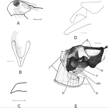
posterior margin. Epandrium with long setulae on the exposed surface. Cercus (Fig. 3 A) in lateral view, straight on the dorsal margin, sinuous on the ventral margin, sloping steeply on the dorsal margin to a tapered point; length of tip, about 0.8X times height of cercus at level of inflexion (Figure, 1C). Surstylus (Figures, 3A and D) elongate. Pregonite (Figure, 1D) almost straight, the tip slightly curved and rounded. Postgonite (Figure, 1D) broad and almost straight. Distiphallus (Figures, 3B and E). Lateral styli large, strongly curved, toothed along part of the ventral margin, the tip greatly expanded, directed ventrally well below the vesica, and posteriorly, towards the base of the vesica. Juxta thin, membranous, with fine ridges, closely pressed to and covering most of the lateral styli leaving only a small part of the tip exposed, anterodorsally developed into a projecting membrane with points and ridges. Dorsal plate raised into sclerotised ridges antero-laterally; below the ridge a sclerotised area covered in microscopic warts extends ventrally. Ventral plate of moderate size about 0.6X height of dorsum above it, its lower margin more or less level with that of the vesica. The vesica is strongly sclerotised, curved up at the tip and much longer than wide (length about 2.2X width); the, membrane lying between the right and left halves of the vesica on the ventral surface of the distiphallus (‘ventral membrane’ in Figure1 E), projects below the vesica and is clearly visible in lateral view. The harpes is hidden by this membrane except for the tip.
Figure 1. Male of Sarcophaga (Sarcophaga) trabzonensis Pekbay, Hayat, Richet, Blackith n.sp., A. Hind trochanter. Scale bar = 0.30 mm, B. Sternite 5. Scale bar = 0.30 mm, C. Cercus tip. Scale bar =0.45 mm, D. Surstylus and pre- and postgonites. Scale bar = 0.10 mm, E. Distiphallus in lateral view. Scale bar = 0.10 mm (Original).
Female:
Length. Approximately 17-18 mm. The genitalia of this species are similar to those of other Sarcophaga (sensu stricto) but are unique in the wide membranous space between the two halves of TG1 (Figure, 2A, C, E). The space is wider than the distance between the anterior and posterior margins of a half part. This membranous space has a fairly regular row of long setae on the posterior margin and a second more or less complete row of weak setae. TG2 is absent. ST7 is clearly wider than long (Figure, 2B, D, F) (width = 1,93–2,33X the length). ST6 is slightly wider than long (width = 1,21–1,32X the length). The spermathecae are long, twisted once and with the extremity slightly inflated (Figure, 2G). As in other species of Sarcophaga (sensu stricto), the cerci are finger-shaped and the hypoproct is subtriangular with microscopic setae (Figure, 2H).
Figure 2. Females of Sarcophaga (Sarcophaga) trabzonensis, Pekbey, Hayat, Richet, Blackith n. sp. A and B specimen 1 from Turkey, Artvin, Borçka; C and D specimen 2 from Turkey, Kars, Sarıkamış; E and F specimen 3 from Turkey, Kars, Sarıkamış; G specimen 2; H specimen 3. A, C, E. TG1; B, D, F. ST6, ST7 and ST8; G. Spermathecae; H. Cerci and hypoproct (Original).
Etymology: The species epithet is derived from Trabzon Province located in the Black Sea Region of Turkey from where the holotype was collected.
Habitat: The specimens were collected from areas of meadow and pasture containing a variety of flowering plants.
Differential diagnosis
Sarcophaga (Sarcophaga) trabzonensis (Figures, 3B and 1E) most
closely resembles S. (S.) apsuarum Rohdendorf, 1937 and Sarcophaga (S.)
pagensis Baranov, 1939 (Figure, 3). To help in the comparison of the two
species the distiphallus of a male of S. (S.) apsuarum from Gruzia (examined by Ruth Blackith) is illustrated (Figure, 3D). The species differ in the following respects. In S. (S.) trabzonensis in lateral view, the ventral plate is larger and the vesica lies dorsal to a horizontal line touching the tip of the ventral plate. In
S. (S.) apsuarum, in lateral view, the ventral plate is smaller and at least half the
vesica lies ventral to a horizontal line touching the tip of the ventral plate. In S. (S.) trabzonensis the vesica is narrow with no visible internal ridge though the ventral membrane between the two halves of the vesica is visible in lateral view. In S. (S.) apsuarum the vesica is wide and has an internal ridge the tip of which is just visible but the ventral membrane is not visible in lateral view. In S. (S.)
trabzonensis the lateral styli are almost straight on the outer margin but strongly
curved on the inner margin so that the tip is very wide and extends beneath the vesica reaching almost to its base. In S. (S.) apsuarum the lateral styli widen more evenly on both margins and the tip is narrower and extends only a short distance beneath the vesica. Anteriorly the median point of the dorsal plate is tiny in S. (S.) trabzonensis compared with the large knob in S. (S.) apsuarum; also the anterolateral ridges on the dorsal plate in S. (S.) trabzonensis are broader and less prominent than in S. (S.) apsuarum. Both S. (S.) trabzonensis and S. (S.) apsuarum differ from S. (S.) pagensis (Figure, 3E) in having a thinner, flatter juxta compared with the thick, broad juxta in S. (S.) pagensis.
Key to separate the three species
1 (2) Vesica in lateral view less than twice as long as wide (length about 1.6X width); dorsal plate anteriorly with a prominent knob.
. . . Sarcophaga (Sarcophaga) apsuarum Rohdendorf, 1937 2 (1) Vesica in lateral view more than twice as long as wide (length about 2.2X width); dorsal plate anteriorly with, at most, a small point.
3 (4) Juxta thin and flattened against the lateral styli; lateral stylus projecting along the ventral surface of the distiphallus, its tip extending only to about the level of the midpoint of the vesica.
4 (3) Juxta thick and projecting anteriorly away from the lateral styli; tip of lateral stylus reaching, or extending beyond, the level of the base of the vesica.
. . . Sarcophaga (Sarcophaga) pagensis Baranov, 1939.
Figure 3. Males of Sarcophaga (Sarcophaga) trabzonensis, Pekbey, Hayat, Richet, Blackith n. sp.
Sarcophaga (Sarcophaga) apsuarum and Sarcophaga (Sarcophaga) pagensis. A. Sarcophaga (Sarcophaga) trabzonensis, terminalia in lateral view; B. Sarcophaga
(Sarcophaga) trabzonensis distiphallus in lateral view; C. Sarcophaga (Sarcophaga)
trabzonensis distiphallus in lateroventral view to show opening of lateral styli; D. Sarcophaga
(Sarcophaga) apsuarum distiphallus in lateral view; E. Sarcophaga (Sarcophaga) pagensis distiphallus in lateral view (Original).
Discussion
Sarcophaga (Sarcophaga) trabzonensis, Sarcophaga (Sarcophaga) apsuarum and Sarcophaga (Sarcophaga) pagensis can be separated from most
other species of Sarcophaga (Sarcophaga) by having at least two of the following character states: - the tip of the lateral stylus is very wide and directed ventrally; the ventral plate is small; the base of the lateral stylus is either directed ventrally or slightly anteroventrally, but lacks a mainly anteriorly directed section.
Özet
Türkiye’den yeni bir Sarcophaga (Sarcophaga) (Diptera: Sarcophagidae) türü
Türkiye’den yeni bir tür Sarcophaga (Sarcophaga) trabzonensis n. sp. tanımlan-mıştır. Erkek ve dişi terminalyası resimlenmiş ve benzer türler olan Sarcophaga (Sarcophaga) apsuarum Rohdendorf, 1937 ve Sarcophaga (Sarcophaga) pagensis Baranov, 1939ile karşılaştırılmıştır.
Acknowledgements
We are grateful to Thomas PAPE, Zoological Museum University of Copenhagen, Denmark (ZMUC), for the loan of Sarcophaga (Sarcophaga)
apsuarum. Also, we would like to dedicate this study to the memory of the late
Cemal KUDU who collected the holotype.
References
Aslan, A. & H. Çalışkan, 2009. Eskişehir Sarcophagidae (Insecta, Diptera) faunası ve Türkiye için yeni kayıtlar. Sakarya Üniversitesi Fen Edebiyat Dergisi, 2: 15-27. Civelek, H. S. & S. Tezcan, 2005. Some new records for Diptera fauna of Turkey and
additional notes on the dipterous fauna of cherry orchards. Türkiye Entomoloji Dergisi, 29 (1): 11- 16.
Collart, A., 1962. Sarcophagidae (Diptera) recueillis en Turqois. Bulletin et Annales de la Société royale Belge d’Entomologique, 98: 437.
Ebejer, M. J., 2000. Description of third instar larva and puparium of Blaesoxipha calliste Pape (Diptera: Sarcophagidae). Studia dipterologica, 7: 121–124.
Giroux, M., T. Pape & T. A. Wheeler, 2010. Towards a phylogeny of the flesh flies (Diptera:Sarcophagidae): morphology and phylogenetic implications of the acrophallus in the subfamily Sarcophaginae. Zoological Journal of the Linnean Society, 158: 1- 39.
Hayat, R., R. Richet, N. Bayrak & G. Pekbey, 2008. Contributions to the knowledge of flesh flies (Diptera: Sarcophagidae) from Turkey, with a new record. Turkish Journal of Zoology, 32 (4): 385–390.
Kara, K. & T. Pape, 2002. Check list of Turkish Sarcophagidae (Insecta, Diptera) with
new records. Mitteilungen aus dem Museum für Naturkunde in Berlin. Deutsche
Entomologische Zeitchrift, 49 (2): 291–295.
Merdivenci, A., 1966. The systematics of the parasites of Turkey. İstanbul Üniversitesi Fen Fakültesi Mecmuası, 31: 73–108 (in Turkish).
Nizamlıoğlu, K., 1954. A few parasites new to Turkey. Tomurcuk, (3): 8–9.
Pape, T., 1987. The Sarcophagidae (Diptera) of Fennoscandia and Denmark. Scandinavian Science Press. Ltd., Leiden, Copenhangen, 181 pp.
Pape, T., 1996. Catalogue of the Sarcophagidae of the World (Insecta, Diptera). Memoirs on Entomology International, 8: 1-558.
Pape, T. & H. Bänziger, 2003. Three new species of Sarcophaga Meigen, found during ecological studies on flesh flies (Diptera: Sarcophagidae) in Thailand. Entomological Science, 6, 49–56.
Pape, T., G. Dahlem, C. A. Mello Patiu & M. Giroux, 2009. The world of flesh flies (Diptera: Sarcophagidae). (Web page: http://www.zmuc.dk/entoweb/sarcoweb/ sarcweb/Intro/Intro.htm. (Date accessed: October, 2010).
Pekbey, G. & R. Hayat, 2010. Erzurum ili Sarcophagidae (Diptera) türleri üzerinde faunistik çalışmalar. Türkiye Entomoloji Dergisi, 34 (2): 263–275
Rohdendorf, B. B., 1937. Fam. Sarcophagidae (Part 1). Fauna of the USSR, New Series, 19: 1-500, Moscow & Leningrad (In Russian and German).
Süreyya, M., 1931. Turkey: Natural enemies of the desert and Italian locusts. International Bulletin of Plant Protection, (3): 41–42.
Verves, Yu. G., 1986. Family Sarcophagidae. In: Soós Á. and Papp L. (eds.), Catalogue of Palaearctic Diptera, 12. Akadémiai Kiadó, Budapest, Elsevier, Amsterdam, 58-193.