KEYWORDS Streptomyces
Total Cell Protein Profile SDS-PAGE
Numerical Analysis
ABSTRACT
Present study has been conducted for finding out the total protein profile of bacterial strain Streptomyces sps by sodium dodecyl sulphate polyacrylamide gel electrophoresis. Total 139 isolates of Streptomyces have been isolated from the soil. Amongst all isolated strain, total 20 isolates were used for getting protein profile by SDS PAGE. Amongst all isolates, 20 isolates were selected for protein profiling and these were divided in two groups. Two strains of Streptomyces i.e. S. violaceus and S. albidoflavus were selected as a reference strain for both groups. Band profile were analyzed and assessed by computer added program BioRad Quantity with the use of Unweighted Pair Group Method of Analysis (UPGMA). As a result of this computer assisted numeric analysis study, approximately 40 different types of protein bands were reported between 10 or 100 kD molecular weight. Analysis of acquired dendogram on the basis of similarities ratios, all 40 proteins can be divid ed in to 7 groups. In addition, the isolates A4B3G, D145B, S5036.6 and reference isolate S. violaceus were available in the same group, while 805A, C804B, F1705 isolates and reference sample S. albidoflavus were detected in the same group. The test organisms which were similar to each other in terms of morphological and biochemical characters delivered the same protein bands. SDS-PAGE method is an effective method in terms of determining taxonomical relations between the various species of genus Streptomyces.
Özdemir K
1*. Berber İ
2. Öğün E
1and Atalan M
3 1Yüzüncü Yıl University, Faculty of Science, Department of Biology, Van, Turkey2
Sinop University, Faculty of of Arts and Science, Department of Biology, Sinop, Turkey
3İnönü University, Faculty of Arts and Sciences, Department of Biology, Malatya, Turkey
Received: June 05, 2013; Revision: July 05, 2013; Accepted: July 12, 2013 Available Online July 20, 2013.
DETERMINATION OF TOTAL CELL PROTEIN PROFILES OF
Streptomyces SPECIES
E-mail: kerem@yyu.edu.tr (Kerem ÖZDEMİR)
Peer review under responsibility of Journal of Experimental Biology and Agricultural Sciences.
* Corresponding author
Journal of Experimental Biology and Agricultural Sciences, July - 2013; Volume – 1 (3)
Journal of Experimental Biology and Agricultural Sciences
http://www.jebas.org
ISSN No. 2320 – 8694
1 Introduction
Soil is the most frequent habitat for the members of the genus Streptomyces and the presence of this genus was reported from the all types of soils (Williams et al., 1989). The members of this genus have substantial role in decomposing of various components in the soil and are dependent on plant wastes or fungus mycelium (Kutzner 1986; Goodfellow and Simpson 1987). These are reported from the all habitats those are rich of organic substances (Hagedorn 1976). Amongst all available soil microflora, up to 20% generally belongs to Streptomyces. Amongst all isolated actinomyecete from soil, 64-97% were belongs to the Streptomyecetes (Kutzner 1986; Xu et al., 1996; Wang et al., 1999). The enzymes produced by the members of genus Streptomyces have degradation capacity to cellulose, silicon, cotton, PVC plastic, wool, wood, hay, cereals, jute fibrile and natural rubber materials and various other substances (Morosoli et al., 1997; Jendrossek et al., 1997). Jendrossek et al. (1997) have reported that amongst 50 discovered rubber decomposer 33 were belongs to the genus Streptomyces.
Protein profiling is an effective method for identifying the various species of genus Streptomyces and it will help to get information regarding the phylogenetic relationship. For protein profiling dissolution of cell protein is an important step. As compared to the one way electrophoresis of total cell proteins, two-way electrophoresis were giving better results (O’Farrell 1975). Recently, cellular protein profiles acquired with SDS-PAGE method and non-cell protein profiles have been successfully employed for differentiating the various types and subtypes of microorganisms. In the diagnosis and classification of various bacteria, cellular protein profilin g with SDS-PAGE were used affectively (Berber et al., 2003, Berber et al., 2004; Berber and Yenidünya 2005). Protein profile and SDS-PAGE enable to Berber and Cokmus (2001) to differentiated Bacillus sphearicus strains into six subtypes. Manchaster et al. (1990) analyzed the cellular proteins of 37 strains belongs to S. albidoflavus 1A, S. anulatus 1B; S. cyaneus 18; S. rimosus 42 and S. griseocarneium 55 taxons. On the behalf of results acquired, different species of taxons S. albidoflavus and S. anulatus formed (Manchester et al., 1990). Information regarding the use of total cellular protein profile in establishment the phylogenetic relationship between various species of genus Streptomyces is in scarcity. Therefore, present investigation had been undertaken for the finding out the importance of SDS-PAGE based protein profile in establishment of phylogenetic relationship between various species of genus Streptomyces.
2 Materials and Methods
2.1 Isolation and culturing of bacteria
Streptomyces were isolated from the plant root and rhizosphere soils. These strains were maintained on Bennet’s broth on 150 rpm at 25C in an incubator. Afterwards, 1.3 ml sample of each strain were transferred in to eppendorf tubes and centrifuge at 1200 rpm for 3 minutes. Following to the centrifuge, cells rinse by sterilized water for 3 times. For maintaining to the cells, 25 µl SDS-sample buffer was added and later on mixed 0.06M Tris-HCl + 2.5% Glycerol + 0.5% SDS + 1.25% β mercaptoethanol. Now tube with this all mixture was boiled for 5 minutes, this process denatured the total protein available in bacterial cell (Laemmli 1970). 2.2 SDS-PAGE
Denatured proteins were exposed to SDS PAGE electrophoresis (V16-2BRL Gaithersburg MD, USA) as described by Laemmli (1970). Afterwards, bromphenol blue an indicator was processed at 30 mA current until the gel ending. Finally, gel was stained by Coomassie Brillant Blue R-250 stain for giving various colors to bacterial protein. 2.3 Analysis
The resulted gel was examined to analyze the protein profile on the basis of bands. The acquired data were transferred to electronic environment by the use of UPGMA in the BioRad Quantity One Program. The relation and similarity between the Streptomyces isolates and total cellular protein profiles were presented as dendogram with UPGMA.
3 Results and Discussion
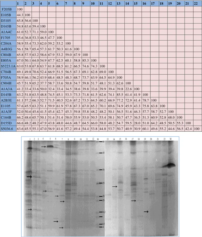
The protein profile image of various isolates, acquired by polyacrylamide gel electrophoresis has been illustrated in Figure 1. Similarly, table 1 represents the similarity ratios of various isolates analyzed by UPGMA in the BioRad Quantity One Program. As illustrated in Figure 1, approximately 40 different types of protein bands were available on gel and the molecular weight of these proteins varies between 10 to 100 kD. The bands those are specific among strains were represented by arrows in figure 1.
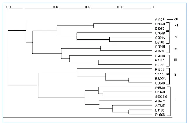
All protein profiles of each isolate were analyzed numerically in BioRad Quantity One program, by the use of UPGMA and the acquired dendogram is illustrated in Figure 2. Results of analysis represents that 60% similarity ratio were based on the total 7 groups. Group 1st contains total 7 members including the reference strain S. violaceus. Other members of this group were D155D, E1105, A2B3E, A1A4C, D145B and A4B3G. Group 2nd includes the reference strain S. albidoflavus with C804B, E805A and F1705. 3rd group included F205B, F705A and C704B isolates while the 4th group contained only tow isolates i.e. C904H and A1A3A. Isolate D3105, C204A and C164B belongs to the group 5th.
187 Özdemir et al.
Table 1 Similarity matrix based on SDS-PAGE protein profiles of isolates of Streptomyces. 1 2 3 4 5 6 7 8 9 10 11 12 13 14 15 16 17 18 19 20 21 22 F205B 100 E105B 44.3 100 D3105 65.8 56.6 100 D165B 54.8 63.6 59.4 100 A1A4C 61.0 52.7 71.1 59.0 100 F1705 55.6 36.8 53.3 46.5 47.7 100 C204A 58.9 55.4 73.3 62.0 59.2 53.2 100 A4B3G 56.1 58.7 65.4 57.3 61.7 50.3 61.6 100 C804B 65.8 57.5 63.2 58.6 67.9 53.2 59.0 67.9 100 E805A 67.0 50.1 64.0 54.9 67.7 62.5 60.1 58.8 85.3 100 S5223.1A 63.0 53.8 67.8 63.7 61.8 68.5 61.2 66.5 74.6 74.3 100 C704B 69.1 49.8 70.6 52.4 66.9 51.5 56.5 67.3 69.1 62.8 69.0 100 F705A 58.9 46.1 56.2 43.9 68.4 48.5 48.3 68.7 73.7 63.9 64.5 64.9 100 C904H 45.7 51.9 65.2 37.7 58.7 33.6 50.8 54.7 59.8 51.7 48.1 51.3 62.6 100 A1A3A 41.2 33.4 33.6 50.0 32.4 33.4 34.5 38.6 39.8 33.6 39.9 39.4 39.8 22.6 100 D145B 63.2 51.8 63.5 48.8 74.5 45.1 53.3 73.3 71.8 61.5 62.6 74.1 85.5 61.4 41.9 100 A2B3E 61.1 57.2 66.3 52.7 71.5 40.5 52.6 67.2 73.5 64.5 60.2 66.9 77.2 72.9 41.4 78.7 100 E1105 57.6 45.5 63.2 51.1 59.9 61.9 57.8 67.3 67.0 65.2 70.1 69.6 74.9 45.9 43.3 75.8 63.8 100 A1A3F 52.0 50.6 45.6 41.5 45.4 42.7 45.3 59.8 55.8 48.2 48.2 50.1 56.5 51.6 46.3 57.7 58.7 52.7 100 C164B 66.2 48.6 65.7 50.1 51.4 51.4 58.0 55.9 53.0 50.5 53.4 58.1 50.7 47.7 36.5 51.3 60.9 52.8 68.0 100 D155D 66.6 48.2 48.2 47.9 43.8 48.0 44.6 48.7 64.5 66.0 58.0 48.2 54.7 59.5 28.0 51.0 64.2 48.5 50.5 55.3 100 S5036.6 63.6 45.5 55.1 47.0 56.9 41.6 57.2 49.4 54.4 53.8 44.8 53.7 50.7 40.9 30.9 60.1 49.6 55.2 44.6 56.5 42.4 100
Figure 1 SDS PAGE protein profile of Streptomyces.
[Molecular Marker: bovine albumin 66kD; egg albumin 45 Kd, pepsin 34,7 Kd, trypsinogen 24 Kd, -laktoglobin 18,4 Kd ve lysozyme 14,2 Kd. 1. F205B; 2. E105B; 3. D3105; 4. D165B; 5. A1A4C; 6. F1705; 7. C204A; 8. A4B3G; 9. C804B; 10. E805A; 11. S5223.1A; 12. C704B; 13. F705A ; 14. C904H; 15.
A1A3A; 16. D145B; 17. A2B3E; 18. E1105; 19. A1A3F; 20. C164B; 21. D155D; 22 S5036.6.]
Figure 2 UPGMA dendogram SDS-PAGE protein profiles of Streptomyces isolates. Similarly, isolates D165B and E105B belongs to the group 6th
while group 7th have single member i.e. A1A3F. Types of amino acids available in protein and the total molecular weight of protein affect the net charge available on particular protein. So, these factors are playing an important role at the time of electrophoresis (Smithies 1955).
Although, One way protein electrophoresis is relatively simple, cheap and reliable method but two way electrophoresis is giving better results as compare to one way (Vauterin et al. 1993). Electrophoretic separation of cells and cell wall proteins generally provide sufficient information about the type and subtype of bacterial strains. Genetic relation between all cellular proteins comply the results obtained by DNA: DNA hybridization. Therefore, interspecies population shows less difference between protein profiling. Association of numeric analysis with electrophoresis profiles provides better results and taking less time than DNA:DNA hybridization studies. Electrophoretic protein profiles of a bacterium cells can be scanned and copied to computer and following the analysis by appropriate software, can help to establish a data bank. In present study, SDS-PAGE associated protein profiles were determined for the Streptomyces isolated from some plant roots and rhizosphere. An appropriate distribution was reported between different isolates of Streptomyces and various color groups. The findings of present study are in conformity with the findings of Manchaster et al. (1990). Similarly, Boynukara
et al. (2004) also prepared total cellular protein profile for bacterial strain Aeromonas by SDS-PAGE method and identify the similarities between various types and subtypes of bacteria (Manchester et al. 1990; Boynukara et al. 2004). In the analyzed dendogram results, 60% similarity ratio was reported from the only 7 groups. Amongst these 6 groups had numerous members only 7 group had only one member i.e. A1A3F. The tested organisms those similar to each other in terms of morphological and biochemical characters provided the same pattern protein bands. In conclusion, it is apparent that the combinations of cellular protein profiles obtain by SDS-PAGE method with the specific software added computer analyzer helps in determining the taxonomic and phylogenetic
relationship between various species of genus Streptomyces. Acknowledgments
This work was supported by the Scientific and Technological Research Council of Turkey (TUBITAK) Grant Number = TBAG-2344(103T156).
References
Berber I, Atalan E, Cokmuş C (2004) The influence of pesticides on the spore viability, toxin stability and larvicidal acitivity of Bacillus sphearicus 2362 starin. Fresenius Environmental Bulletin 13: 424-429.
Berber I, Cokmus C (2001) Characterization of Bacillus sphaericus strains by Native-PAGE. Bulletin of Pure & Applied Sciences 20: 17-21.
Berber I, Cokmuş C, Atalan E (2003) Characterisation of Staphylococcus species by SDS-PAGE of whole cell and extracelluler proteins. Microbiology 72: 42-47.
Berber I, Yenidünya E (2005) Identification of Alkaliphilic Bacüllus Species Isolated from Lake Van and Its Surroundings by Computerized Analysis of extracellular Protein Profiles. Turkish Journal of Biology 29: 181-188.
Boynukara B, Korkoca H, Senler NG, Gulhan T, Atalan E (2004) The characterisation of protein profiles of the isolated Aeromonas sobria strains from animal feces by SDS-PAGE. Indian Veterenary Journal 81: 245-249.
Goodfellow M, Simpson KE (1987) Ecology of Streptomyces. Frontiers in Applied Microbiology 2: 97-125.
Hagedorn C (1976) Influences of soil acidity on Streptomyces populations inhabiting forest soils. Applied and Environmental Microbiology 32: 368-375.
Jendrossek D, Tomasi G, Kroppenstedt RM (1997) Bacterial degradation of natural rubber. A privilege of actinomycetes. FEMS Microbiology Letters 150: 179-188.
Kutzner KJ (1986) The family Streptomycetaceae. In: Starr MP, Stolp H, Tr_per HG, Balows A, Schlegel HG (eds) The prokaryotes, A Handbook on Habitats, Isolation, and Identification of Bacteria, vol. 2, Springer-Verlag, New York, pp 2028-2090.
Laemmli UK (1970) Cleavage of structural proteins during the assembly of the head of bacteriophage T4. Nature 227: 680-685.
Manchester L, Pot B, Kersters K, Goodfellow M (1990) Classification of Streptomyces and Streptoverticillium species by numerical analysis of electrophoretic protein patterns. Systematic and Applied Microbiology 13: 333–337.
Morosoli R, Shareck F, Kluepfel D (1997) Protein secretion in Streptomyces. FEMS Microbiology Letters 146: 167-174. O'Farrell PH (1975) High resolution two dimensional electrophoresis of proteins. Journal of Biological Chemistry 250: 4007-4021.
Smithies O (1955) Zone electrophoresis in starch gels: Group variations in the serum proteins of normal human adults. The Biochemical Journal 61: 629-641.
Vauterin L, Swings J, Kersters K (1993) Protein electrophoresis and classification. In: Goodfellow M, O'Donnell AG (eds) Handbook of New Bacterial Systematics, Academic Press, London, pp. 251-281.
Wang Y, Zhang ZS, Ruan JS, Wang YM, Ali SM (1999) Investigation of actinomycete diversity in the tropical rainforests of Singapore. Journal of Industrial Microbiology and Microbiology 23: 178-187.
Williams ST, Goodfellow M, Alderson G (1989) Genus Streptomyces Waksman and Henrici 1943, 339AL. In: Williams ST, Sharpe
ME, Holt JG (eds) Berys’s Manual of Systematıc Bacteriology, Volume 4, Baltimore, USA, pp: 2452-2492.
Xu LH, Li QR, Jiang CL (1996) Diversity of soil actinomycetes in Yunnan, China. Applied and Environmental Microbiology 62: 244-248.

