Cukurova Medical Journal
Cukurova Med J 2016;41(Suppl 1):88-90ÇUKUROVA ÜNİVERSİTESİ TIP FAKÜLTESİ DERGİSİ DOI: 10.17826/cutf.254646
Yazışma Adresi/Address for Correspondence: Dr. Elif Karadeli, Baskent University, Faculty of Medicine, Department of Radiology, Adana, Turkey E-mail: [email protected]
Geliş tarihi/Received: 09.05.2015 Kabul tarihi/Accepted: 27.06.2015
OLGU SUNUMU/CASE REPORT
Late pancreatic metastasis of renal cell carcinoma with absence of
FDG-uptake in PET-CT
PET-BT'de FDG-tutulumu göstermeyen renal hücreli karsinomun geç pankreas
metastazı
Elif Karadeli
1, Sermin Tok
1, Gürcan Erbay
1, Mehmet Reyhan
21Başkent Üniversitesi, Tıp Fakültesi, Radyoloji Anabilim Dalı, 2Nükleer Tıp Anabilim Dalı, Adana, Turkey
Cukurova Medical Journal 2016;41(Suppl 1):88-90.
Abstract Öz
The primary tumors, which raise isolated pancreas metastases are frequently of renal origin, where colorectal cancer, melanoma, breast and lung cancers and sarcoma are the following causes of metastatic pancreas cancer . In this article, we present a case of pancreas-metastatic renal cell carcinoma with its radiological features, which did not exert anF-18 FDG-uptake in the whole-body positron emission tomography (PET).
Renal hücreli karsinom metastatik tümörler pankreas kanserlerinin yaklaşık %2’sini oluşturur. İzole pankreas metastazlarının kaynaklandıkları primer tümörler renal orjinli, takiben kolorektal kanser, melanom, meme, akciğer kanserleri, sarkomlardır. Bu makalede, PET-BT’de F18-FDG tutulumu göstermeyen renal hücreli karsinomun pankreas metastazının radyolojik bulguları sunulmuştur . Key words: Renal cell carcinoma, pancreatic metastasis,
PET-CT. Anahtar kelimeler: Renal hücreli karsinom, pankreas metastazı, PET-BT.
INTRODUCTION
Renal cell carcinoma (RCC) lesions typically metastasize to lung, bones, liver and brain and less frequently to thyroid, subcutaneous fatt issue and pancreas1. Metastatic tumors consist about 2%
percent of pancreas cancers. The primary tumors, which raise isolated pancreas metastases are frequently of renal origin, where colorectal cancer, melanoma, breast and lung cancers and sarcoma are the following causes of metastatic pancreas cancer2.
In this report, we present a case of pancreas-metastatic renal cell carcinoma with its radiological features, which did not exert anF-18 FDG-uptake in the whole-body positronemissiontomography (PET).
CASE
A 55 years old female patient, who underwent
nephrectomy due to a renal cell carcinoma on her left kidney 10 years ago, admitted to the department of general surgery with abdominal pain since last month. No pathological sign was encountered in her physical examination. Her laboratory findings were: Hb: 11,5 g/dL, AST: 39 IU/L, ALT: 23 IU/L, ALP: 120 IU/L, Amylase: 62 U/L. In the abdominal USG, the pancreas can not be evaluated due to gas distension and no pathological USG finding was witnessed in the patient who underwent left nephrectomy. However, in the iv-oral contrast abdominal CT, a dilation of the pancreatic channel was observed which extended from tail to head of the pancreas with an anteroposterior diameter of 6 mm.
A nodularity with an approximate size of 26 mm featuring contrast enhancement was observed at the level of the pancreatic head on the arterial phase images (figure 1). In the MR images, signs of pancreatic dilation were observed. A focal lesion
Karadeli et al. Cukurova Medical Journal
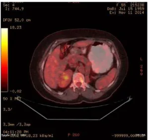
suspicious of malignancy was witnessed at the uncinate process of the pancreas adjacent to duodenum, which demonstrated heterogenous diffusion restriction, dense contrast uptake at the early arterial phaseand a heterogenous signal intensity (figure 2). F-18 FDG PET was performed to the patient, who underwent a previous surgery due to RCC. No pathological contrast uptake was witnessed in the PET-CT examination (figure 3). Whipple operation was performed in the department of general surgery and the pathological analysis outcoma indicated an RCC metastasis.
Figure 1. Nodularity demonstrating contrast uptake at the level of the pancreatic head during the arterial phase of the abdominal CT
Figure 2. A focal lesion suspicious of malignancy at the uncinate process of the pancreas adjacent to duodenum with heterogenous diffusion restriction, dense contrast uptake at the early arterial phase and a heterogenous signal intensity.
Figure 3. No pathological FDG uptake was detected in the PET-CT examination.
DISCUSSION
RCC can metastasize to pancreas via both haematogenous and lymphatic routes, yet its direct metastatic spread to pancreas is rare. Commonly, a single and asymptomatic pancreas metastasis is observed3. The metastasis frequency is 25% percent
at the RCC diagnosisand 40% percent during the disease progress. An average interval of 6 years could be encountered between detection of the primary tumor and the metastatic lesion1.
In our case, a 10 years gap had occured between the pancreatic metastasis and the nephrectomy performed due to RCC;and the patient presented with abdominal pain at her recent admission. Three types of metastatic involvement were defined for the metastatic tumors of pancreas: Single localized nodule (50%-73%), diffuse pancreatic enlargement (%15-%44) and multiple pancreatic nodules (%5-%10). In the contrast-free CT, single lesions are generally hypodense relative to the normal pancreas;and in the contrast-enhanced CT, their contrast uptake ocur commonly during the arterial phase.
In the MRI scanning, they present as hypointense at the T1A and heterogenously hyperdense at the T2A images, respectively. If thelesion has a large size, a rim can be seen around the lesion at the contrast images. Homogenous contrast uptake generally occurs in small lesions4.
Cilt/Volume 41 Yıl/Year 2016 Renal cell carcinoma
In our case, the iv-oral contrast abdominal CT examination revealed a prominent dilation of the pancreatic channel from the pancreatic tail to head and a nodularity with contrast enhancement at the level of the pancreatic head on the arterial phase images. Pancreatic dilation signs were also seen in MRI scanning. A focal lesion suspicious of malignancy was witnessed at the uncinate process of the pancreas adjacent to duodenum, which demonstrated heterogenous diffusion restriction, dense contrast uptake at the early arterial phase and a heterogenous signal intensity. Malignant cells exert an enhanced glucose consumption;and hence, the elevated glucose metabolism can be demonstrated via PET analysis4. Although, the PET-CT could give
negative results in some malignancies. Bronchoalveolar carcinomas and carcinoid tumors of the lung, hepatoma, colon cancer, mucinous tumors of the gastrointestinal system and low-grade lymphnodes could be among the FDG-PET-negative malignant lesions. Also in our case, any pathological FDG-uptake was not demonstrated. In conclusion, RCC is a malignancy, which could make late metastasis to organs including pancreas. It should be kept in mind that the FDG-PET
resultscould be negative in primary and metastatic RCC lesions; and hence, CT findings shall be cautiously interpreted in patients with a history of RCC.
REFERENCES
1. Facy O, Angot C, Guiu B, Al Samman S, Matte A, Rat P et al. Interest of intraoperative ultrasonography during pancreatectomy for metastatic renal cell carcinoma. Clin Res Hepatol Gastroenterol. 2013;37:530-4.
2. Sperti C, Moletta L, Patanè G. Metastatic tumors to the pancreas: The role of surgery. World J Gastrointest Oncol 2014; 15; 6: 381-92.
3. Markinez I, Jiménez R, Ruiz I, Villarreal E, Lizarazu A, Borda N et al. Pancreatic metastases due to renal carcinoma. Our cases and a literature review. Cir Esp. 2013;91:90-5.
4. Ascenti G, Visalli C, Genitori A, Certo A, Pitrone A, Mazziotti S. Multiple hypervascular pancreatic metastases from renal cell carcinoma: dynamic MR and spiral CT in three cases. Clin Imaging. 2004;28:349-52.
5. Long NM, Smith CS. Causes and imaging features of false positives and false negatives on F-PET/CT in oncologic imaging. Insights Imaging. 2011;2:679-98.