Scandinavian Journal of Surgery 103: 167 –174, 2013
Operating On penetrating trauma tO the mediastinal
Vessels
t. h. Yilmaz
1, t. evers
2, m. sussman
3, p. Vassiliu
4, e. degiannis
4and d. doll
4,5,61 Department of Surgery, Baskent University, Izmir, Turkey
2 Department of Anaesthesiology and Intensive Care, Military Hospital Ulm, Teaching Hospital of the
University of Ulm, Ulm, Germany
3 Department of Cardiothoracic Surgery, Milpark Hospital, Johannesburg, South Africa
4 Directorate for Trauma and Burns, Chris Hani Baragwanath Academic Hospital, University of
Witwatersrand Medical School, Johannesburg, South Africa
5 Department of Surgery, St Marien Hospital Vechta, Teaching Hospital MHH Hannover University, Vechta,
Germany
6 Vechta Institute for Research Promotion & Interdisciplinary Research (Vechtaer Institut für
Forschungsförderung VIFF e.V.), Vechta, Germany
aBstraCt
Background and Aims: patients with penetrating trauma of the major vessels of the chest
are infrequently encountered. this is due to the fact that the majority of these patients
die on scene, as well as due to the overall dramatic decline in the incidence of penetrating
trauma in the Western world. a certain proportion of survivors are physiologically stable
and can be transferred to adequate care. patients who are physiologically unstable must
be dealt with by the surgeons available without delay. rapid diagnosis and operation
can salvage patients who would otherwise be lost, and all general surgeons should be
capable of recognizing these injuries and intervening if a trauma and/or cardiothoracic
surgeon is not immediately available.
Material and Methods: technical description of practical emergency surgery approaches
to patients bleeding to death from penetrating mediastinal vessel injuries.
Results: the scope of this review familiarizes the “uninitiated” surgeon with the operative
management of this rare and lethal type of injuries. technical aspects are described, and
pitfalls as well as tips and tricks of the trade are discussed.
Conclusions: patients with penetrating injuries to the mediastinal vessels can be saved
by swift and knowing operative management of this rare and lethal type of injuries,
even if a trauma and/or cardiothoracic surgeon is not immediately available.
Key words: Major mediastinal vessels, gunshot, gunshot wound chest, mortality, resus room mortality, emergency room surgery
Correspondence:
Dietrich Doll, M.D., Ph.D. Department of Surgery St Marien Hospital Vechta
Teaching Hospital MHH Hannover University Marienstr. 6
D-49377 Vechta Germany
Email: [email protected]
reVieW
would otherwise be lost (5, 6), and all general sur-geons should be capable of recognizing these injuries and intervening if a trauma and/or cardiothoracic surgeon is not immediately available. Transfer or referring expertise might be hampered in the military trauma setting, where strategic and logistic reasons enforce care under austere conditions (7). This leads into the fields of Emergency Room Thoracotomy (ERT) for patients arriving in extremis and to Damage Control Surgery (DCS), using abbreviated surgical maneuvers of bleeding and contamination control before swift transport to the intensive care unit (ICU) and recovery of the physiological injury can be initi-ated to overcome the so-called “deadly triad” of hypothermy, lactacidosis, and coagulopathy (7–9). nevertheless, damage control procedures in the chest are scarce, as cardiac and vascular injuries need to be addressed immediately and definitely (10, 11). The scope of this review is to familiarize the “uninitiated” surgeon with the operative management of this rare and lethal scope of injuries.
InCISIonS
EMERGEnCY DEPARTMEnT THoRACoToMY
Emergency department thoracotomy is recommended in moribund patients with penetrating trauma to the chest (6, 12, 13). Although an injury to the major ves-sels of the chest is unlikely to be repaired through this incision, it is useful to discuss this approach as the sur-geon will choose it as a desperate procedure in patients with torrential chest bleeding of unknown origin. A supine anterolateral thoracotomy is the accepted and swift approach for emergency department thoracot-omy. A left-sided approach (left anterolateral thora-cotomy) is used in all patients in traumatic arrest and with injuries to the left chest.
The skin incision extends from the lateral border of the left sternocostal junction, inferior to the nipple to the midaxillary line laterally, using the 5th inter-costal space. In females, a submammary incision is performed (the breast is retracted cranially and the incision is made along the submammary fold). Elevation of the arm allows further extension of the incision laterally. The pectoral fascia, pectoralis, and serratus muscles (anteriorly and laterally) are divided with a scalpel or high coagulation electrocautery in the same line underneath the skin incision, and then
the intercostal muscles are divided along the upper margin of the 6th rib. Particular attention is paid not to injure the lung when opening the pleura. A rib spreader is inserted, with its horizontal part parallel to the sternum, and opened quickly. This always almost results in rib fracture. The margins of the inci-sion should be covered now with abdominal swabs (your hands should be inserted in the rib cavity with great care to avoid injury from rib spikes).
If there is any bleeding from the right heart side or in case the right chest side has to be accessed as well, then the sternum is split transversely (“clamshell” incision; Fig. 1) at the level of the skin incision. Crossing the sternum from left to right, the incision should be directed upward to the 3rd intercostal space, thus to stay above the right nipple, facilitating expo-sure of the upper mediastinal structures (e.g. innomi-nate artery). The sternum is divided by a Gigli saw, a power saw, or—if these are not available—the Lebsche knife, always making sure that upward traction is applied when the latter is hit with the hammer (14). A bone cutter is another alternative, albeit more time-consuming. The internal mammary arteries will be transected, and must be ligated afterward even if there is no obvious bleeding from them at the time of tran-section. In the hypovolemic patient, they can retract and not bleed when first divided. It is important to look for them within the areolar tissue posteriorly and immediately bilaterally to the sternum and ligate them before closing the sternal incision.
When a left anterolateral thoracotomy is extended to the right, the pericardium should be inspected to exclude cardiac tamponade, unless there is active bleeding from another anatomical area (in this case, direct digital pressure or a Satinsky clamp may be applied onto the bleeding tissues). In addition to the identification of a bulging tense pericardium, cardiac
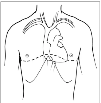
Fig. 1. The line of a clamshell (bilateral chest) incision.
Source: Reproduced with permission from Springer Publishers and courtesy of Velmahos et al. (15)).
tamponade can be notified by a white bluish color, produced by an underlying clot. A classic error of inexperience is to leave the pericardium unopened because it looks “normal” from the outside (16). The pericardium should always be opened even if there is no “obvious” hemopericardium. Surprisingly, often a significant amount of blood can be evacuated from the pericardial sack (from an injury to the heart or the intrapericardial mediastinal vessels), which is not vis-ible as long as the pericardium is left intact.
Bleeding from a large vessel near the pulmonary hilum or uncontrollable bleeding from the lung paren-chyma can be best dealt with by clamping of the hilum. The lung is initially retracted anteriorly and cranially, and the inferior pulmonary ligament is divided. It is generally advised that this should be per-formed by sharp dissection, but in our experience, blunt separation of the ligament from the diaphragm using the left hand is more practical in the scenario of an emergency department thoracotomy. In both instances, it is important to remember that the liga-ment terminates at the inferior pulmonary vein, and care should be exercised when approaching the hilum. Following the division of the inferior pulmonary liga-ment, the left hand is advanced around the lower pole of the lung, and the hilum is controlled between the index finger and third finger. A large Satinsky clamp is then taken with the right hand, and the hilum is clamped from the opposite direction of the hand already in place (the clamp is inserted from the sur-geons’ right to the left, and the hilum is clamped just medially to the left fingers of the surgeon; this can be seen in Fig. 2). If clamping of the hilum is not possible, a pulmonary hilar “twist” can produce the same results. Ventilation is stopped, and the upper and lower pulmonary poles are held with the right and left hand of the surgeon (abdominal swabs on both palms ease this maneuver as the lung is slippery). Then the lung is twisted by 180°, resulting in occlusion of the
pulmonary vascular supply at the hilum. The lung tends to return to its original position, and it is impor-tant to fill the upper thoracic cavity with abdominal swabs in order to prevent this. Some surgeons have abandoned the pulmonary twist as hilar bleeding con-trol is achieved faster and more easily—with two fingers.
If clamping of the descending aorta is required for the preservation of all blood supply to vital organs (heart, brain), this is accomplished by elevating the left lung medially and dividing the parietal pleura superficial to the aorta with scissors in a transverse plane (Fig. 3). Application of the vascular clamp to the aorta without opening of the periaortic pleura is not advisable as the clamp may easily slip. Care should be taken not to clamp the esophagus beneath.
Closure of the incision takes place in the operating theater, after all injuries are dealt with. Meticulous hemostasis is important, as toward the end of the operation there is a lot of bleeding from the chest wall mostly due to the fact that the blood pressure of the patient is usually established to acceptable levels or due to coagulopathy. Two intercostal drains are always inserted: one in the anterior axillary line anterior to the lung and extending up to the apex, and the second one in the posterior axillary line, directed toward the dia-phragm. In some patients where the pericardium has been opened, a third intrapericardial drain is required. The intercostal incision is closed by approximating the ribs with three number 2 Ethibond or Vicryl sutures. If the sternum has been transversely divided, it is easily approximated with metal wire stitches. The overlying muscles are closed in layers.
MEDIAn STERnoToMY
A midline incision is performed, and diathermy is used to cut the skin down to the sternum, ensuring that the sternal midline is marked. A right angle Lahey clamp is inserted posteriorly to the sternal notch by pushing rostrally. There is a feeling of “
Fig. 2. Control of pulmonary hilum using a finger, followed by the insertion of a clamp. Please note that other than a soft clamp may crush hilar structures.
Source: Reproduced with permission from Springer Publishers and courtesy of Velmahos et al. (15)).
Fig. 3. Aortic clamping after a horizontal incision of the pleura above and beneath the descending aorta in order to avoid the slipping of the clamp.
Source: Reproduced with permission from Springer Publishers and courtesy of Velmahos et al. (15)).
the operation to place a sandbag or an infusion bag between the patient’s shoulders and extend the neck, as during the splitting of the proximal sternum it is possible to injure the face with the hammer. The ster-nal incision follows the midline marks already done by diathermy. In the heat of the moment, it is easy to divert from the midline and split through the costal cartilages, rendering subsequent closure problematic and resulting in wound healing complications. Bleeding from the sternal margins is controlled either by electrocautery or by using bone wax (the latter should be used sparingly as excessive use can predis-pose to infection (17)). A sternal retractor is subse-quently inserted and opened gradually, ensuring that the sternum is not fractured.
At the end of the operation, the sternotomy is closed by approximating the two halves of the sternum with wire sutures. It can be very difficult for a surgeon unfamiliar with the procedure to insert the needle through the hard sternum. This is facilitated by apply-ing the needle holder at the junction of the front and middle third of the needle and advancing it in a verti-cal direction, rather than in the usual rotational move-ment. As the sternum may yield suddenly to the needle, a sterile spoon (with a concavity facing upward) can be placed below the sternum to avoid inadvertent injury to the underlying structures.
PoSTERoLATERAL THoRACoToMY
An incision is made from the anterior axillary line run-ning backward (about 1 cm below the tip of the scap-ula), cutting midway between the medial edge of the scapula and the vertebral column. The soft tissues are divided, using electrocautery, up to the level of the ribs. At this stage, the intercostal space to be opened is identified. The 5th space is usually suitable for most trauma cases. The scapula should be elevated and the ribs counted down from the uppermost rib, using the finger tips. The 1st rib is sometimes difficult to pal-pate, but even if the chest is entered via the 6th space instead of the 5th, the superior rib is easily removed if necessary to improve any exposure needed. once the space is identified, the intercostal muscles are divided at the level of the superior border of the rib to avoid the neurovascular bundle. Prior to entering the pleu-ral cavity, the lung is collapsed by the anesthetist (a double-lumen endotracheal tube should be used; this is usually possible even in the emergency situation,
is easier to make the initial cut with a blade and then continue the incision with scissors. The pericardium is opened longitudinally, with the incision made in the midline (middle of surgical field). Extending the peri-cardial incision as an inverted T along the diaphragm enhances exposure. Clots and blood are evacuated from around the heart and the site of injury to the aorta as well as the cardiac rhythm ascertained. If the heart is in asystole or fibrillation, internal massage is commenced using rapid but gentle compression, tak-ing care not to distract the heart or damage it (18). For a non-perfusing ventricular arrhythmia, start with a shock of 20 J, with one internal paddle behind the heart, and one in front (18). Defibrillation should be repeated if required with shocks up to 50 J. Complete cardiac “standstill” should not be defibrillated, as this can further damage the myocardium (18); systemic doses of adrenaline according to current resuscitation guidelines should then be administered. Close com-munication with the anesthetist is essential.
Initial control of bleeding from the anterior aspect of the ascending aorta is achieved by digital pressure or side-clamping. This can be facilitated by intermit-tent aortic occlusion (partial or complete) and con-comitant caval occlusion; these maneuvers are useful both during exposure and subsequent suturing (by reducing blood pressure). It is important to note that aortic and caval occlusion can also be utilized in cases of proximal injuries to the branches of the aortic arch that cannot be controlled by side-clamping (2, 19). Complete aortic occlusion results in a profound increase in afterload and peripheral resistance, neces-sitating careful management of the patient’s hemody-namic parameters by the anesthetic team. Aortic cross-clamp time should be kept to a minimum and should be restricted to 20–30 min (20).
In most instances, the hole in the aorta is small and lateral aortorrhaphy is sufficient. Direct suturing with 3.0 or 4.0 monofilament nonabsorbable sutures is eas-ily accomplished. The best closure technique for larger (2–3 cm) aortic tears is a running horizontal mattress suture, which everts 2–3 mm of aortic wall on each side, followed by simple “over-and-over” suture across the everted edges. This should be done with the injury isolated by side-clamping or total clamping of the involved aorta. When possible, intraluminal pres-sure should be kept low, while sutures are tied; if the patient does not tolerate a reduced systemic pressure, the surgeon can sometimes temporarily apply a
side-clamp around the involved aorta. For large wounds and gunshot injuries, large Teflon felt, pericardial pledgets, or patches of prosthetic material (e.g. Dacron or Gortex) are used. nonabsorbable monofilament sutures are suitable for all these repairs.
When encountering injury to the anterior aortic wall, it is important to look for a possible correspond-ing injury to the posterior aortic wall and the heart. Exposure of the posterior aspect of the ascending aorta is achieved by sharp dissection between the pulmo-nary artery and the aorta. Although primary repair of anterior lacerations is usually accomplished without adjuncts, cardiopulmonary bypass may be necessary in the case of posterior injury. The possibility of peripheral bullet embolus must be considered in all patients with gunshot injury to the aorta (or to major veins) irrespective of their anatomical site (21).
InJURIES To THE AoRTIC ARCH
Exposure of the aortic arch (and its branches) neces-sitates dissection of the thymus and the left innomi-nate vein as soon as the sternum is opened. There is a filmy layer of pleura enveloping the thymus, and this is divided with scissors. The thymus itself can be divided or even pushed out of the way with a swab on the surgeon’s hand. The left innominate vein then presents itself in the mid operation field. If necessary, which is often, it is divided and transfixed to improve exposure.
The methods of repair of aortic arch injuries are similar to those for the ascending aorta (19). These— as well as proximal injuries to the aortic arch branches—can present as a hematoma of the anterior mediastinum. If the latter is encountered, it should never be tackled “head on.” Anterior mediastinal hematomas never extend into the pericardial sack, because the pericardium is adherent around the large vessels which originate from the heart. As they exit the pericardial sac, they thus act as an anatomical barrier. Therefore, it is safer to start the dissection of the anterior aspect of the aorta from its intrapericar-dial portion and proceed distally. The pericardium is opened and umbilical tapes are applied around the ascending aorta and the cava veins (superior and inferior cava), thus being prepared for a possible temporary partial occlusion of the aorta or a cardiac inflow control, respectively. Distal control of the branches of the arch must be established, and this may require extension of the median sternotomy incision up to the neck in order to expose the carotid sheath. The dissection proceeds from the intraperi-cardial anterior aspect of the ascending aorta, and the hematoma is entered at its base following the ante-rior surface of the aortic arch. Digital compression, rather than blind application of clamps, can control the bleeding until the area of injury has been dis-sected and a vascular clamp can be applied.
Dissection of the posterior and concave surface of the arch should also start from inside the pericardium, as the fibrous layer of the pericardium fuses with the aortic wall and forms the aortic adventitia. Dissection starting outside the pericardium can lead to an appar-ent cleavage plane, which may actually be within the
aortic wall, potentially resulting in disastrous iatro-genic injury (19).
InJURIES To THE BRAnCHES oF THE AoRTIC ARCH
The surgeon treating injuries to branches of the aortic arch must be aware of the vascular anomalies of the region, as their presence may complicate diagnosis, control, and repair. In 5% of the Caucasian population and up to 30% of the African American population, the innominate and left carotid arteries have a com-mon origin from the aortic arch (Fig. 4).
InnoMInATE ARTERY
Injuries to the innominate artery are approached through a median sternotomy with extension to the neck (15). In selected patients with only partial tears, the innominate artery may be primarily repaired using 4.0 polypropylene sutures. More often, injuries to this vessel require repair via the bypass exclusion technique, as described by Johnston et al. (22). This technique does not require cardiopulmonary bypass, hypothermia, or systemic anticoagulation. Bypass grafting is performed from the ascending aorta to the distal innominate artery using a Dacron tube graft. The area of injury is carefully avoided until the bypass is completed. The proximal anastomosis connects the graft to the ascending aorta away from the origin of the innominate artery, which is accomplished by a partially occluding, side-biting clamp to the ascend-ing aorta. The distal anastomosis now requires proxi-mal and distal control of the innominate artery. If the
Fig. 4. In 5% of the Caucasian and up to 30% of the African (and Afro-American) population, the innominate and left carotid arteries have a common origin from the aortic arch.
proximal portion of the artery is injured, a partially occluding clamp can be placed across the adjacent aorta. For distal control, a vascular clamp is placed proximal to the bifurcation of the innominate artery, in order to allow collateral flow from the right subcla-vian artery to perfuse the right carotid artery. now the artery is divided between the two clamps. Care must be taken not to injure the right vagal nerve as it crosses in front of the proximal right subclavian artery. The repair is completed by an “end-to-end” anastomosis of the graft to the distal innominate artery and by oversewing the native origin of the artery. In case of injuries to the innominate vessels, care should be taken to rule out concomitant injuries to the distal trachea (Fig. 5).
CoMMon CARoTID ARTERIES
The operative approach is similar to the innominate artery injuries repair (23). A cervical extension of the midline sternotomy incision can be added if necessary. In the case of a cervical extension, the strap muscles should be divided near their insertion to the sternum to expose the carotid sheath. As with other great ves-sels originating from the aorta, the use of shunts or pumps is unnecessary. If the artery is transected near its origin, repair using a bypass (5, 22) is preferred over a primary end-to-end anastomosis. Care should be taken to identify the left recurrent laryngeal and vagal nerves before clamping. Exposure of the right common carotid artery is carried out in a similar fash-ion. The innominate artery is dissected from its origin toward its bifurcation into the right subclavian and right common carotid artery.
InJURIES To THE DESCEnDInG THoRACIC AoRTA
Penetrating injuries to the descending thoracic aorta are extremely rare in survivors reaching the hospital (24). If encountered, a left posterolateral thoracotomy is performed (4). Injuries to this part of the aorta result in large mediastinal hematomas, making identification
of the exact source of injury very difficult. Preoperative angiography is very useful if hemodynamics allow for any diagnostics (25).
The current standard technique of repair involves vascular clamping and direct reconstruction (4). The pleura overlying the proximal left subclavian artery is opened, and the artery is mobilized by blunt dissec-tion. The aorta is then mobilized between the left sub-clavian and left carotid arteries, using a combination of sharp and blunt dissection (Fig. 6). Enough space is created to accommodate a clamp there. Care should be taken to avoid injury to the left recurrent laryngeal nerve. To obtain proximal control, vascular clamps are applied to the aortic arch between the left subclavian and left common carotid artery and to the left subcla-vian artery. Similarly, distal control is obtained by mobilizing the distal descending thoracic aorta above the diaphragm, and applying a vascular clamp. It is imperative to achieve adequate proximal and distal control prior to entering any hematoma in the area of the injury, as it may avoid torrential bleeding. Close communication between the surgical and anesthetic teams is essential in order to maintain hemodynamic stability of the patient. When the hematoma is entered, it is important to limit ligation of intercostal vessels to the area required for adequate aortic repair. The proxi-mal and distal ends of the descending thoracic aorta are completely transected and dissected away from the esophagus. The injury is then repaired by “end-to-end” anastomosis or by Dacron graft interposition.
The most significant complication of injury to the descending thoracic aorta is paraplegia (24). An aortic cross-clamp time of less than 30 min has been argued to provide a safe margin against paraplegia (26), and the use of protective adjuncts (passive ascending to descending aorta bypass shunts (27) and pump-assisted left heart bypass (left atrium to femoral artery) (28)) has been recommended for longer cross-clamp times. However, no prospective, randomized trial has identified the superiority of any single method.
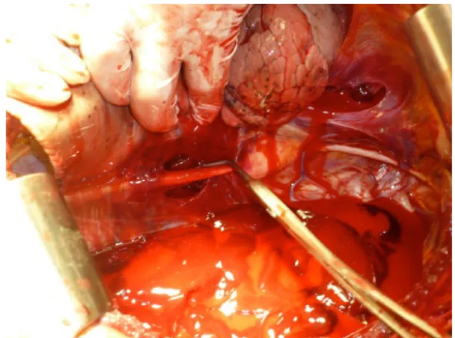
Fig. 6. Sternotomy and a supraclavicular incision for injuries of the proximal left subclavian artery. The left common carotid and left subclavian artery can be seen with a PTFE graft starting behind the left internal jugular vein.
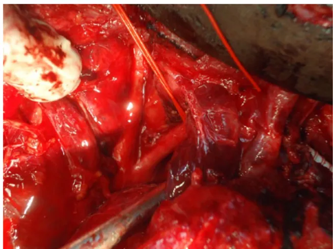
PTFE: polytetrafluoroethylene. Fig. 5. Sternotomy with a retractor between the right common
carotid and the brachiocephalic trunk (innominate artery) showing the distal trachea to exclude injury of it.
InJURIES To THE PULMonARY ARTERY
Patients with injuries to the intrapericardial pulmo-nary arteries do present with cardiac tamponade. Such injuries are best approached via a median sternotomy, as this facilitates their exposure with minimal dissec-tion. The right intrapericardial pulmonary artery is exposed by dissecting between the superior vena cava and the ascending aorta. Repair of anterior surface injuries is performed using a side-clamp to get best possible control and 4.0 or 5.0 monofilament sutures. However, cardiopulmonary bypass may be required for repair of posterior surface injuries (29). Pneumonectomy is performed in the case of extensive injuries, necessitating ligation of any of the two pul-monary arteries.
Distal (extra-pericardial) pulmonary artery injuries usually present as massive hemothorax and are approached through a posterolateral thoracotomy. This injury can be encountered in patients undergoing emergency department thoracotomy due to exsan-guinating hemothorax. In the presence of extensive injury in a physiologically unstable patient, a quick pneumonectomy can be lifesaving.
VEnoUS InJURIES
Control of a venous bleed is often difficult than arte-rial bleeding, but the principles of proximal and distal control are essential as well. A device which appears to be useful in this setting is the incomplete ring (“Dr. Franceschi’s haemostat”) available in different sizes (Delacroix Chevalier, France). This enables one to apply pressure to the injured vein and creates a rela-tively bloodless field within which one can then suture
the vein. Lateral repair (direct suturing) should be attempted in all venous injuries. If this is technically impossible or time-consuming, division and ligation can be well tolerated in most cases.
An injured innominate vein can be ligated without impunity. However, if possible, one innominate vein should be left intact to reduce the likelihood of a supe-rior vena cava syndrome developing postoperatively (Fig. 7). Injuries of subclavian veins can be extremely difficult to repair, even with excellent exposure (30). Ligation proximal and distal to the injury is usually much safer than a difficult repair. Injuries to the major pulmonary veins are very unusual because of their pos-terior position, and repairing these is very difficult without bypass. There is also the risk of air embolism, particularly when associated with a major airway injury in parallel. If such an injury is encountered, bet-ter control is obtained by clamping the affected side’s pulmonary artery, thus reducing inflow. occlusion of the entire hilum may be necessary to facilitate repair. If a pulmonary vein is ligated, the corresponding lobe needs to be resected.
Superior vena cava injuries present with mediastinal hematoma and/or a large right hemothorax. Injury to the intrathoracic portion of the inferior vena cava can cause cardiac tamponade, as it is situated within the pericardium. The surgical approach will be deter-mined by the presentation (tamponade or hemotho-rax). In case of pericardial tamponade, the incision of choice is a median sternotomy; if a massive hemotho-rax is present, an anterolateral thoracotomy should be performed. For all major venous injuries, initial con-trol of the bleeding is achieved by compression with large abdominal swabs, as attempts to aspirate the blood will be associated with continuous bleeding, which may be aggravated by the decompression of opening the chest cavity. once the cavity is packed and control is obtained, serially removing the packs will expose the source of bleeding. In exceptional cir-cumstances when there is torrential bleeding, proxi-mal and distal clamping of the superior cava vein in the chest is necessary. In this case, the sudden severe reduction in venous return can cause cardiac arrest particularly in hypovolemic patients. However, if the descending thoracic aorta is clamped, not as much preload is required, and clamping of the intrathoracic cava is generally much better tolerated. It has to be found that successful clamping of the descending tho-racic aorta from either a median sternotomy or a thor-acotomy is exquisitely difficult and thus should not be attempted unless absolutely necessary. Injuries to the anterior aspect of both cava veins are closed by lateral venorrhaphy (direct suturing) using 4.0 or 5.0 nonab-sorbable monofilament sutures. occasionally, patch repair/angioplasty are required, using autologous or bovine pericardium, and in exceptional circumstances, an interposition tube graft (Dacron or ringed polyte-trafluoroethylene) may be inserted. Exposure of the posterior aspect of the intrathoracic inferior vena cava is extremely difficult and usually requires the patient to be placed on cardiopulmonary bypass. Ligation of the cava vessels is performed only as an attempt to save the patients’ life.
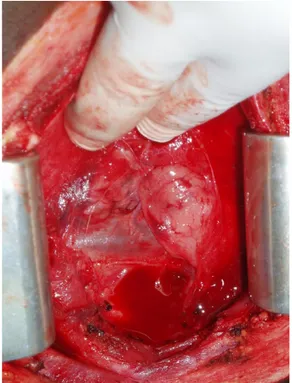
Fig. 7. The innominate vein is the gatekeeper of the upper mediastinum, but it can be ligated with impunity.
ACKnoWLEDGEMEnT
Figures 1 to 3 are published with courtesy from Springer-Verlag: Velmahos G, Degiannis E & Doll D (Eds) Penetrating Trauma: A Practical Guide on operative Technique and Peri-operative Management. Springer-Verlag, Berlin; Heidelberg; London; new York, 2012.
DECLARATIon oF ConFLICTInG InTERESTS
This article has neither been submitted in general, in parts, or in parallel. There is no conflict of interest.
FUnDInG
This research received no specific grant from any funding agency in the public, commercial, or not-for-profit sectors.
REFEREnCES
1. Mattox KL, Feliciano DV, Burch J et al: Five thousand seven hundred sixty cardiovascular injuries in 4459 patients. Epidemiologic evolution 1958 to 1987. Ann Surg 1989;209: 698–705.
2. Demetriades D: Penetrating injuries to the thoracic great ves-sels. J Card Surg 1997;12:173–179.
3. Krug EG, Mercy JA, Dahlberg LL et al: The world report on violence and health. Lancet 2002;360:1083–1088.
4. Wall MJ Jr, Hirshberg A, LeMaire SA et al: Thoracic aortic and thoracic vascular injuries. Surg Clin north Am 2001;81: 1375–1393.
5. Mattox KL: Approaches to trauma involving the major vessels of the thorax. Surg Clin north Am 1989;69:77–91.
6. Velmahos GC, Degiannis E, Souter I et al: outcome of a strict policy on emergency department thoracotomies. Arch Surg 1995;130:774–777.
7. Eiseman B, Moore EE, Meldrum DR et al: Feasibility of dam-age control surgery in the mandam-agement of military combat casualties. Arch Surg 2000;135:1323–1327.
8. Fulton Jo, de Groot KM, Buckels nJ, et al: Penetrating inju-ries involving the intrathoracic great vessels. S Afr J Surg 1997;35:82–86.
etrating trauma to the heart: a heuristic approach for the non-initiated. Injury 2013;44:146–150.
15. Velmahos G, Degiannis E, Doll D (Eds): Penetrating Trauma: A Practical Guide on operative Technique and Peri-operative Management. Springer-Verlag, Berlin; Heidelberg; London; new York, 2012.
16. Breaux EP, Dupont JB Jr, Albert HM et al: Cardiac tamponade following penetrating mediastinal injuries: improved survival with early pericardiocentesis. J Trauma 1979;19:461–466. 17. Ulicny KS Jr, Hiratzka LF: The risk factors of median
sternot-omy infection: a current review. J Card Surg 1991;6:338–351. 18. Degiannis E, Bowley DM, Westaby S: Penetrating cardiac
injury. Ann R Coll Surg Engl 2005;87:61–63.
19. Pate JW, Cole FH Jr, Walker WA et al: Penetrating injuries of the aortic arch and its branches. Ann Thorac Surg 1993;55:586–592. 20. Hoyt DB, Coimbra R, Potenza BM et al: Anatomic exposures for
vascular injuries. Surg Clin north Am 2001;81:1299–1330, xii. 21. Mattox KL, Beall AC Jr, Ennix CL et al: Intravascular
migra-tory bullets. Am J Surg 1979;137:192–195.
22. Johnston RH Jr, Wall MJ, Jr, Mattox KL: Innominate artery trauma: a thirty-year experience. J Vasc Surg 1993;17:134–139. 23. Wall MJ Jr, Granchi T, Liscum K et al: Penetrating thoracic
vas-cular injuries. Surg Clin north Am 1996;76:749–761.
24. Von oppell Uo, Dunne TT, De Groot MK et al: Traumatic aor-tic rupture: twenty-year metaanalysis of mortality and risk of paraplegia. Ann Thorac Surg 1994;58:585–593.
25. LeBlang SD, Dolich Mo: Imaging of penetrating thoracic trauma. J Thorac Imaging 2000;15:128–135.
26. Mattox KL, Holzman M, Pickard LR et al: Clamp/repair: a safe technique for treatment of blunt injury to the descending tho-racic aorta. Ann Thorac Surg 1985;40:456–463.
27. Verdant A: Traumatic rupture of the thoracic aorta. Ann Thorac Surg 1990;49:686–687.
28. Pate JW, Fabian TC, Walker WA: Acute traumatic rupture of the aortic isthmus: repair with cardiopulmonary bypass. Ann Thorac Surg 1995;59:90–98.
29. Clements RH, Wagmeister LS, Carraway RP: Blunt intraperi-cardial rupture of the pulmonary artery in a surviving patient. Ann Thorac Surg 1997;64:258–260.
30. Degiannis E, Bonanno F, Titius W et al: Treatment of penetrat-ing injuries of neck, chest and extremities. Chirurg 2005;76: 945–958.
Received: May 11, 2013 Accepted: September 16, 2013