Lipoblastoma in the Inguinal Region of an Infant: Review of the
Literature
Bir Bebeğin İnguinal Bölgesindeki Lipoblastom: Literatür İncelemesi
Gönül Küçük
1, Ufuk Ateş
1, Gülnur Göllü
1, Saba Kiremitçi
2, Hüseyin Dindar
11 Ankara University Faculty of Medicine Department of Pediatric Surgery 2 Ankara University Medical Faculty Medical Pathology Department
Lipoblastoma, a rare benign adipose tumor is primarily seen under the age of five years. Common sites of this tumor are extremities and trunk. The most common symptom is usually painless, rapidly growing soft mass. The usual recommended treatment of both lipoblastoma and lipoblastomatosis is complete surgical excision with clean surgical margins without compromising function or damaging adjacent structures. The aim of this paper is to present a case of 13-month old female infant who presented with a mass in the ingui-nal region and was diagnosed as lipoblastoma following surgical excision of the mass.
Key Words: adipose tissue neoplasm; infant; lipoblastoma
Lipoblastom, primer olarak 5 yaş altında görülen seyrek bir adipoz doku tümörüdür. En sık yerleşim yerleri gövde ve ekstremitelerdir. En sık semptomu ağrısız hızlı büyüyen yumuşak dokudur. Lipoblastom ve lipo-blastomatozisin önerilen olağan tedavisi, fonksiyon kaybına yol açmadan ve komşu yapılara zarar vermeden gerçekleştirilen temiz cerrahi sınırlı tam cerrahi eksizyondur. Bu çalışmanın amacı, inguinal bölgede kitle ile başvuran ve lipoblastoma olarak tanı alıp kitlesi cerrahi olarak çıkarılan 13 aylık kız çocuğunun sunulmasıdır.
Anahtar Sözcükler: adipoz doku tümörleri; bebek; lipoblastoma
Lipoblastoma primarily occurs in children under the age of five years. This rare benign tumor of adipose tissue arises from embryonic white fat (1-10). Adipose tissue tumors compose 6% of all soft tissue tumors in children and lipomas account for the majority of these adipose tissue tumors. The remaining of these tumors are lipoblastoma, liposarcoma and hibernoma (3).
Lipoblastoma with common presenting symptom as painless, rapidly growing mass are most commonly found on extremities, trunk and rarely in the inguinal region (2-7). Lipoblastomas are composed of immature mesencymal tissue which has a variable composition of immature to maturing adipocytes. They have a well-circumscribed fibromembranous pseudocapsule, lobulated with prominent septae, consist of myxoid stroma and occur superficially in the subcutis(1-5).
Lipoblastoma was first described by Jaffe in 1926, cited by Kirkham et al (1). in the groin of a child and since then there have been limited number of cases of lipoblastoma located in the inguinal region. In this paper, we aimed to present a case of 13-month old female infant presented with a mass in the inguinal region and was diagnosed as lipoblastoma following surgical excision.
Case
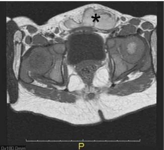
Thirteen-month old girl with a non-tender, palpable mass noticed two months ago in her left inguinal region was consulted. Physical examination revealed a non-reductable inguinal mass of 2 cm in diameter. Ultrasound examination revealed two masses of which the bigger was 2cm in diameter in the left inguinal region and the other, 9 mm in diameter in the right inguinal region not compatible with gonad and incarcerated inguinal hernia. Magnetic resonance imaging scans revealed lobulated mass of 3x1.8 cm with smooth borders subcutaneously located at the superior of symphisis pubis on left side and a mass of 12x7 mm with similar characteristics extending towards right side (Figure 1). In fat suppressed scan series, the masses were partially suppressed. Radiologically differential diagnosis included lipomatous lesions. The mass was excised with the left transverse inguinal incision whose histopathological examination revealed lipoblastoma. Histopathologic evaluation revealed a cellular tumor exhibiting various stages of adipocyte differentiation in which immature fat cells were admixed with mature fat cells. The tumor was seperated in to lobules by prominent fibrous septa and the stroma of the tumor Received :June 25,2015 • Accepted: March 09,2016
Corresponding Author: Yrd. Doç Dr. Gülnur Göllü E-mail: [email protected] Phone: +90 (312) 595 84 34 Fax: +90 (312) 595 65 63
Ankara Üniversitesi Tıp Fakultesi, Cocuk Cerrahisi Anabilim Dalı, 06100 Cebeci-Ankara, Türkiye
Ankara Üniversitesi Tıp Fakültesi Mecmuası 2016, 69 (3)
DOI: 10.1501/Tıpfak_000000955 CERRAHİ BİLİMLERİ/ SURGICAL SCIENCES
Ankara Üniversitesi Tıp Fakültesi Mecmuası 2016, 69 (3)
Lipoblastoma in the Inguinal Region of an Infant: Review of the Literature 254
Figure 1: Magnetic resonance imaging scans showing the mass with asteriks
Figure 2: A. The lobular appearance of the tumor with fibrous septa is prominent.
He-matoxylen-Eosin x 100 . B. The tumor is composed of spindle shaped adipocytes ad-mixed with multi-vacuolated or signet ring lipoblasts in the myxoid stroma with plexiform vascular network. Hematoxylin- Eosin x 400.
was myxoid with plexiform vascular network. (Figure 2). However, six months later, there was recurrence in the left inguinal region extending to the right groin and the mass was totally re-excised by bilateral groin incisions. In four months of follow-up after the second surgery the patient is doing well without any recurrence.
Discussion
Lipoblastoma arising from embryonic white fat is a rare, benign, encapsulated tumor. These lesions most commonly occur in early childhood and infancy (2, 6). Though lipoblastoma was described in the
inguinal region, this region is very rare for lipoblastoma (1, 2,4-6). This pathology can be confused with other inguinal entities. These tumors usually present as rapidly enlarging, non-tender, soft masses. Radiological examinations aid in the diagnosis and management of these lesions. Magnetic resonance imaging scans are usually valuable and helpful. The lesion is hypointense on T1 relative to subcutaneous fat which is most likely because of increased cellularity and immaturity of cells when compared to mature adipose cells. Imaging is especially useful in determining the degree and depth of the lesion. Computerized tomography and ultrasonography may also aid in differential diagnosis but with limited benefits (2).
Lipoblastomas are superficially located, well-circumscribed with fibromembranous pseudocapsule, lobulated with prominent septae and consist of myxoid stroma. More mature adipocytes are located centrally within the tumor and immature cells are peripherally located (1). Their features of being superficially located, well-circumscribed and encapsulated differentiate them from lipoblastomatosis which are multicentric, infiltrative, diffuse and deeper located (3).
The usual recommended treatment of both lipoblastoma and lipoblastomatosis is complete surgical excision with clean surgical margins without compromising function or damaging adjacent structures. When not completely excised, there is 14-25% risk of recurrence of both (1, 3, 4). Local re-excision is the choice of treatment in recurrences. Metastases haven't been reported in cases of lipoblastoma (1, 3). Though follow-up period for lipoblastomatous
tumors is recommended at least two years, recently authors propose five years follow-up (1, 6, 7).
The most important point of benign lipoblastoma is to differentiate from myxoid liposarcoma. Myxoid liposarcomas grossly resemble lipoblastomas, however they are malignant and have a tendency to metastasize with poor prognosis and extremely rare in patients under 10 years of age (1,2,4). Histologically, myxoid liposarcoma are also lobulated but not seperated by septae, display nuclear atypia and hyperchromasia. The maturing adipocytes of myxoid liposarcoma are located at the periphery of the lobules instead of the center as seen in lipoblastomas.
Most recently, karyotype analyses of lipoblastomas and liposarcomas have demonstrated chromosomal involvement however lipoblastomas have rearrangements affecting long arm of chromosome 8 spesifically (1-4).
The literature review revealed only 14 cases of inguinal lipoblastoma. Nine of the
cases were male and five of them were female. Seven of the cases were right-sided, six of the cases were left-sided and one case was located in the suprapubic region. All of the cases were surgically treated. Of the reported cases, three cases recurred (1-4, 11-17).
Conclusion
Although lipoblastoma is a rare entity in inguinal masses, it is important to consider them in the differential diagnosis of a prepubertal child with inguinal mass. Complete surgical excision is important to prevent recurrence and a minimum two years of follow-up is recommended in these patients. Recently, karyotype analyses have also been used in the differential diagnosis of lipoblastomas and liposarcomas.
Journal of Ankara University Faculty of Medicine 2016, 69 (3)
Gönül Küçük, Ufuk Ateş, Gülnur Göllü, Saba Kiremitçi, Hüseyin Dindar 255
REFERENCES
1. Kirkham YA, Yarbrough CM, Pippi Salle JL, et al. A rare case of inguinolabial lipoblastoma in a 13-month-old female. J Pediatr Urol 2013;9:64-67.
2. Chien AL, Song DH, Stein SL. Two young girls with lipoblastoma and a review of the literature. Pediatr Dermatol 2006;23:152-156.
3. Hicks J, Dilley A, Patel D, et al. Lipoblastoma and lipoblastomatosis in infancy and childhood: histopathologic, ultrastructural, and cytogenetic features. Ultrastruct Pathol 2001;25:321-323.
4. Collins MH, Chatten J. Lipoblastoma/ lipoblastomatosis: a clinicopathologic study of 25 tumors. Am J Surg Pathol 1997;21:1131-1137.
5. Mohta A, Anand RK. Lipoblastoma in infancy. Indian Pediatr 2006;43:78-79. 6. Kok KY, Telisinghe PU. Lipoblastoma:
clinical features, treatment, and
outcome. World J Surg 2010;34:1517-1522.
7. McVay MR, Keller JE, Wagner CW, et al. Surgical management of lipoblastoma. J Pediatr Surg 2006;41:1067-1071.
8. Speer AL, Schofield DE, Wang KS, et al. Contemporary management of lipoblastoma. J Pediatr Surg 2008;43: 1295-1300.
9. Kloboves-Prevodnik VV, Us-Krasovec M, Gale N, et al. Cytological features of lipoblastoma: a report of three cases. Diagn Cytopathol 2005;33:195-200. 10. Gökçe A, Albayrak AL, Kulaçoğlu S. Lipoblastoma in a 62 Years Old Female: Case Report]. Turkiye Klinikleri J Med Sci 2009;29:750-752.
11. Reinders J, Noyez L, Munting J, et al. A benign inguinal lipoblastoma in a 14-year-old girl. A case report. Acta Chir Belg 1983;83:427-429.
12. Sarsu SB, Karakus SC, Belen B. Lipoblastoma mimicking inguinal hernia. APSP J Case Rep 2015;6:7.
13. Russell ST, Gettman MT, Arndt CA, et al. Inguinal lipoblastomatosis in a male infant. J Urol 1998;160:2204.
14. Kerkeni Y, Sahnoun L, Ksia A, et al. Lipoblastoma in childhood: About 10 cases. Afr J Paediatr Surg 2014;11:32-34. 15. Jaffe RH. Recurrent lipomatous tumors
of the groin: liposarcoma and lipoma pseudomyxomatodes. Arch Pathol 1926;1: 381–387.
16. Jung SM, Chang PY, Luo CC, et al. Lipoblastoma/lipoblastomatosis: a clinicopathologic study of 16 cases in Taiwan. Pediatr Surg Int 2005;21:809-812. 17. El-Dhuwaib YZ, Izzidien AY.
Lipoblastoma in an 11-month old infant. Saudi Med J 2004;25:1109-1110.