Ankara Üniv Vet Fak Derg, 54, 2007 61 Ankara Üniv Vet Fak Derg, 54, 61-64, 2007
Short Communication / Kısa Bilimsel Çalışma
Spirocercosis in a dog
M. Ziynet YILDIRIM1, Osman KUTSAL1, Hamza AVCIOĞLU2
1 Department of Pathology, Faculty of Veterinary Medicine, Ankara University, Ankara, 2Department of Parasitology, Faculty of
Veterinary Medicine, University of Atatürk, Erzurum.
Summary: Pathological findings in spirocercosis encountered in a female, 7 months old, mongrel dog which was found dead
in a heavily forested area and brought to the Pathology Department of Veterinary Medicine Faculty of Ankara University to be necropsied with toxication suspicion by the Directorship Branch Office of Nature Protection and National Parks of Ankara Province are described. At necropsy, 23 independent, 0.8-1 cm diametered nodules which were hard in consistency and showed ash-gray whitish color and had red string-like material in the center of the cut surface that were diffusely located on mesenterium mainly close to curvatura major of the stomach are observed. At jejenum, under serosa another nodule with the same characteristics is seen. Another three masses that were able to be seen even from the serosa of the stomach are encountered. At the part of aorta that is close to the apertura thoracis cranialis numerous nodules with the same characteristics are found. In histopathological examination of the nodules that were taken from aorta, mesenterium, stomach and the intestine, granulomas consisted of parasite (Spirocerca lupi) in the center and was surrounded by necrotic tissue and cellular infiltrations of mainly eosinophil and neutrophil leukocytes with plasma cells, macrophages and lymphocytes are observed in tunica adventitia of aorta, under serosa in the intestine and mesenterium as solid nodules. It is also observed that cellular infiltration was surrounded by connective tissue.
Key words: Dog, Spirocerca lupi, spirocercosis
Bir köpekte spiroserkozis olgusu
Özet: Ormanlık bir alanda ölü olarak bulunmuş ve toksikasyon şüphesi ile Ankara Valiliği, Doğa Koruma ve Milli Parklar
Şube Müdürlüğü tarafından Ankara Üniversitesi Veteriner Fakültesi Patoloji Anabilim Dalı’na nekropsisi yapılmak üzere getirilen, 7 aylık, dişi sokak köpeğinde Spirocercosis olgusu patolojik bulguları ile tanımlandı. Nekropside, mezenterium üzerine yayılmış ve özellikle mezenteriumun, midenin kurvatura major’una yakın kısmında yoğunlaşmış; 0.8- 1 cm çapında, sert kıvamlı, kesit yüzü boz beyaz renkte olan ve kesitin merkezinde kırmızı renkte iplik benzeri yapılar bulunduran, birbirinden bağımsız 23 adet kitleye rastlandı. Jejenum üzerinde, seroza altında benzer özellikte bir adet kitle görüldü. Mide serozası üzerinden de seçilebilen yine 3 adet kitle dikkati çekti. Apertura thoracis cranialis’e yakın bölgede aorta üzerinde çok sayıda benzer kitlelere rastlandı. Aorta, mezenterium, mide ve bağırsaklardan alınmış kitlelerin yapılan histopatolojik incelemelerinde aorta’nın adventisya tabakasında, bağırsak serozası altında ve mezenteriumda solid nodüller şeklinde görülen kitlelerin merkezlerinde parazit (Spirocerca lupi)’e ait yapılar ile etrafında nekrotik doku ve çoğunluğunu nötrofil ve eozinofil lökositlerin oluşturduğu, aralarında plazma hücresi, makrofaj ve lenfositlerin de bulunduğu hücre infiltrasyonuna rastlandı. Hücre infiltrasyonunun etrafının bağ doku hücrelerince çevrelenmiş olduğu dikkati çekti.
Anahtar sözcükler: Köpek, Spirocerca lupi, spiroserkozis.
Spirocercosis is a parasitic disease encountered in all wild felidae and canidae mainly dogs and foxes and caused by nematoda Spirocerca lupi which is a member of Thelaziidae family (2, 5, 8, 18). It is widely distributed throughout the world and is most prevalent in tropical and subtropical countries. Caprophageus beetles (Scrabeus) play an important role as intermediate hosts while animals such as lizards, snakes, frogs, birds, rodents and rabbits as paratenic hosts in the disease (5, 8, 18). L1 formed embryonated eggs found in the feces of definitive hosts are taken by intermediate hosts;
caprophageus beetles and transformed into L3 form. In definitive hosts that were fed with infected beetles or paratenic hosts which had hunted infected beetles before, larvae penetrate into the stomach wall; pass to the gastric arters and migrate until they reach to the thorasic aorta. Parasitic larvae reaching to the aorta in 1- 2 weeks after taken to the organism, develop especially in the thorasic part of the aorta in 90 days; migrate to the esophagus wall and transform into mature parasites in cystic nodules (5, 8, 10, 17, 18). Mature male and female parasites are red colored, 30- 54 mm and 54- 80 mm in lenght
M. Ziynet Yıldırım - Osman Kutsal - Hamza Avcıoğlu 62
respectively. Cystic nodules are opened to the esophagus lumen with a fistula; eggs of the female parasites pass to the esophagus lumen with this fistula and discarded with the feces of the definitive host (2, 5). Although the prepatent period of the disease is reported as 5- 6 months (8,18) there are still questions to be answered on the life-cycle of the parasite and the pathogenesis of the disease as the disease is also encountered in 3- 4 months old dogs (11).
Clinical findings show variations with the severity and localization of the disease and may not be observed if the disease occurs in mild form. Due to the obstructions in the esophagus, anorexia, vomiting and loss in body weight may be seen. In blood analysis leucocytosis and normocytic or microcytic anemia may be observed. Increase in alkaline phosphatase (ALP), creatine kinase (CK), amylase (AMYL) and lactate dehydrogenase (LDH) levels are observed biochemical changes. Sudden death may develop if a rupture occurs during the migration of the parasite in the aorta wall (6, 7).
Macroscopical findings are mainly encountered in aorta and esophagus where the parasite migrate and settle down (1, 2, 5, 8). Ectopic localizations to the organs and tissues such as organs in the thorasic cavity, kidney, and skin with the migration of the parasite in blood circulation are also reported (2, 16). Tumor like nodules that may extend to a diameter of 4 cm are found in submucosa of the esophagus and adventitia or media of the aorta. These nodules are composed of neutrophils and macrophages around the parasite in the middle and surrounded by a thick capsule made of connective tissue (2, 6, 7, 8, 13). Fibrosarcoma, osteosarcoma and undifferentiated sarcoma cases are associated in animals with Spirocercosis. Lung, kidney, stomach, adrenal glands, regional lymph nodes, heart and lingual metastases of these tumors are encountered (2, 8, 14). Another typical lesion of the disease is spondylitis which is thought to occur due to parasite migration and cyst formation in the caudal thorasic vertebrae and can be diagnosed with radiography (2, 8, 10). Clinical diagnose can be performed from anemnesis and considering nodular formations in the esophagus with endoscopy. Definite diagnosis of the disease can be put by finding embryonated eggs in feces (8, 9, 17).
Several studies were performed on incidence, distribution and pathology of the disease in Turkey (1, 3, 4, 12, 15).
Spirocercosis cases are encountered rarely as Spirocerca lupi can only complete its development in caprophageus beetles. The case is reported for further studies as mature parasites are not found in the esophagus lumen; parasitic granulomas are encountered
on mesenterium as well as esophagus, stomach and aorta and as the case shows the pathogenesis of the disease step by step.
Case History
A female, 7 months old, mongrel dog which was found dead in a heavily forested area and brought to the Pathology Department of Veterinary Medicine Faculty of Ankara University to be necropsied with toxication suspicion by the Directorship Branch Office of Nature Protection and National Parks of Ankara Province is constituted the case material. After necropsy the tissues are examined macroscopically and tissue samples are fixed in 10 % neutral formaldehyde solution. Tissue samples are processed routinely, embedded in paraffin, five µ sections are prepared and stained with hematoxylin and eosin (H&E).
Parasites are collected from opened parasitic granulomas, put in lactophenol and diagnosed as Spirocerca lupi under light microscope (9) in Department of Parasitology, Faculty of Veterinary Medicine, Ankara University.
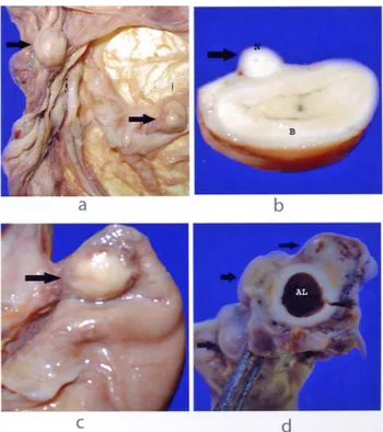
Figure 1. Parasitic granulomas (arrows) in spirocercosis a. Mesenterium, b. Jejenum; N: Nodular formation, B: Intestine (jejenum), c. Cardia of stomach; Stomach., d. Aorta; AL: Aorta lumen.
Şekil 1. Spiroserkozis’ te görülen parazitik granulomlar (oklar) a.Mezenteriyum, b. Jejenum; N: Nodüler yapılar, B: Bağırsak (jejenum), c. Midenin kardiyası; Mide. d. Aorta; AL: Aorta lümeni.
In macroscopical examination, hard in consistency, 23 independent, 0.8- 1 cm diametered nodules that were diffusely located on mesenterium mainly close to
Ankara Üniv Vet Fak Derg, 54, 2007 63
curvatura major of the stomach are observed (Figure 1a). On cut surface of the nodules it is realized that red colored string like structures in the center were surrounded by a large whitish colored area. More masses with the same characteristics are found located one on jejenum under serosa (Figure 1b), three on stomach, one on esophagus where enters the stomach (Figure 1c) and numerous on aorta (Figure 1d).
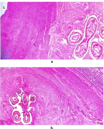
Upon histopathological examinations of the masses; sections of the parasite Spirocerca lupi in the center of nodules in adventia of the aorta (Figure 2a), under serosa of esophagus, stomach, intestine (Figure 2b) and mesenterium are encountered. These parasite sections were surrounded by necrotic tissue, cellular infiltrations mainly composed of eosinphil and neutrophil leucocytes, plasma cells, macrophages and lymphocytes and by fibroblasts and fibrocytes in the outer layer.
Figure 2. Microscopical appreance in spirocercosis.
a. Parasitic granuloma developed in the adventitia of Aorta; L: Lumen. x40
b. Parasitic granuloma in serosa of intestine. x40. Şekil 2. Spiroserkozis’ te mikroskobik görünüm.
a. Aorta’nın adventisya tabakasında şekillenmiş parazitik granülom; L: Lümen. x 40
b. Bağırsağın serozasında parasitik granülom. x 40.
Spirocercosis with long prepatent duration is mainly known to occur in aged dogs (8, 10) and one year old or younger dogs are considered to be in minimum risk group (10). As reported in only a few cases (6, 11), it is found interesting that the dog is 7 months old in the case.
When the pathogenesis of the disease is considered, the parasitic nodules are mainly localized on stomach, esophagus and aorta and if the nodules are encountered in other locations it is called ectopic (2, 8, 16). In this case numerous (23 unit) nodules were observed to show ectopic localization on mesenterium. We thought that the etiologic agent might have come to mesenterium through the gastric arters.
As the case material was found dead and brought to our department with the suspicion of toxication, clinical examination of the dog could not be performed. That’s why we were not able to make any comments on clinical or laboratory findings.
Mature parasites were not encountered in the lumen of esophagus and there were no lesions on the mucosa. This showed us that the disease had not completed its development yet.
References
1. Atasever A, Yazar S, Yıldırım A (2005): Köpeklerde
spirocercosis olguları. Ankara Üniv Vet Fak Derg, 52,
127-130.
2. Bailey WS (1972): Spirocerca lupi: a continuing inquiry. J. Parasitol 58, 3-22.
3. Doğanay A (1983): Ankara köpeklerinde görülen helmint
türleri, bunların yayılışı ve halk sağlığı yönünden önemi.
Ankara Üniv Vet Fak Derg, 30, 550-561.
4. Güralp N, Dinçer Ş, Kemer C, Cantoray R, Taşan E (1977): Elazığ yöresi köpeklerinde görülen iç ve dış
parazitler ile bunların yayılış oranları üzerinde araştırmalar. Fırat Üniv Vet Fak Derg, 5, 7-11.
a
5. Güralp N (1981): Helmintoloji. Ankara Üniversitesi Veteriner Fakültesi Yayınları, Ders Kitabı, Ankara Üniversitesi Basımevi, Ankara.
6. Hamir AN (1984): Perforation of thoracic aorta in a dog
associated with Spirocerca lupi infection. Aust Vet J, 61, 65.
7. Ivogli B (1977): Fatal aortic aneurysm and rupture caused
by Spirocerca lupi in a dog. JAVMA, 170, 834.
8. Jones TC, Hunt RD, King NM (1997): Diseases caused
by parasitic helminths and arthropods. 622-624. In: Cann
C (Ed), Veterinary Pathology. Sixth Edition, Williams & Wilkins, Philadelphia.
b 9. Levine ND (1968): Spirurorids. 397-435. In: Levine ND
(Ed), Nematode Parasites of Domestic Animals and of
Man: Burgess Publishing Company, Minneapolis.
10. Mazaki-Tovi M, Baneth G, Aroch I, Harrus S, Kass PH, Ben-Ari T, Zur G, Aizenberg I, Bark H, Lavy E (2002): Canine spirocercosis: clinical,diagnostic, pathologic, and epidemiologic characteristics. Vet
Parasitol, 107, 235-250.
11. Melendez RD, Suarez-Pellin C (2001): Spirocerca lupi
and dogs: the role of nematodes in carcinogenesis. Trends
Parasitol, 17, 516.
12. Özer H, Metin N, Karadaş E (1989): Köpeklerin
özefagus ve aorta’larında saptanan Spirocerca lupi nodülleri ve bu nodüllerin morfolojik özellikleri. Fırat Üniv
M. Ziynet Yıldırım - Osman Kutsal - Hamza Avcıoğlu 64
13. Pence DB, Stone JE (1978): Visceral lesions in wild
carnivores naturally infected with Spirocerca lupi. Vet
Pathol, 15, 322-331.
14. Ranen E, Lavy E, Aizenberg A, Perl S, Harrus S (2004): Spirocercosis-associated esophageal sarcomas in
dogs. A retrospective study of 17 cases (1997-2003). Vet
Parasitol, 119, 209-221.
15. Taşan E (1985): Elazığ kırsal yöresi köpeklerinde
helmintlerin yayılışı ve insan sağlığı yönünden önemi.
Doğa Bilim Derg, 8, 160-167.
16. Turk RD (1960): Occurance of the nematode Spirocerca
lupi in unusual locations. JAVMA, 137, 721-722.
17. URL 1-Spirocerca lupi in Dogs and Cats.
Erişim: http://www.goldenvetlab.co.za/spirocerca%20lupi.htm Erişim tarihi: 08.02.2004
18. URL 2- Spirocerca lupi.
Erişim:http://cal.vet.upenn.edu/dxendopar/parasitepages/ filariidsandspirurids/s_lupi.html
Erişim tarihi: 08.02.2004
Geliş tarihi: 28.02.2006 / Kabul tarihi: 22.03.2006
Address for correspondence
M. Ziynet Yıldırım Ankara Üniversitesi Veteriner Fakültesi Patoloji Anabilim Dalı, 06110 Dışkapı, Ankara.