Extraskeletal Ewing Sarcoma of the
Diaphragm Presenting With
Hemothorax
Atilla Erog˘lu, MD, I˙brahim Can Ku¨rkc¸u¨og˘lu, MD, Nurettin Karaog˘lanog˘lu, MD, Fatih Alper, MD, and Cemal Gu¨ndog˘du, MD
Departments of Thoracic Surgery, Radiology, and Pathology, Atatu¨rk University, Medical Faculty, Erzurum, Turkey
Ewing sarcoma is a relatively uncommon malignant bone neoplasm that usually occurs in children and young adults and involves the major long bones, pelvis, and ribs. Primary diaphragmatic Ewing sarcoma is extremely rare. To the best of our knowledge, only three cases of primary Ewing sarcoma of the diaphragm have been reported. A 12-year-old girl presented spontaneous oc-currences of the right hemothorax. After drainage, a roentgenogram film, computed tomography, ultrasonog-raphy, and magnetic resonance image showed a giant mass on the right diaphragm. Primary diaphragmatic tumor was resected totally by right posterolateral thora-cotomy, and histologically, an extraskeletal Ewing sar-coma was identified. The patient received adjuvant radio-chemotherapy, and there was no evidence of disease 10 months after the operation. Although extremely rare, extraskeletal Ewing sarcoma should be kept in mind in the differential diagnosis of diaphragmatic soft tissue tumors.
(Ann Thorac Surg 2004;78:715–7) © 2004 by The Society of Thoracic Surgeons
E
xtraskeletal Ewing sarcoma (EES) is simply Ewing sarcoma arising in soft tissues, which is now re-garded as a member of the family of small, round cell neoplasms of bone and soft tissue, including primitive neuroectodermal tumor and neuroblastoma. The most frequent sites of occurrence are the chest wall, lower extremities, and paravertebral region. Less frequently, the tumor occurs in the pelvis and hip region, the retroperitoneum, and the upper extremities [1]. It occurs predominantly in adolescents and young adults between the ages of 10 and 30 years [1]. Extraskeletal Ewing sarcoma of the diaphragm presenting with hemothorax has not been reported until now. We describe a case of primary diaphragmatic Ewing sarcoma and its clinic, radiologic, and histopathologic findings.A 12-year-old girl presented with dyspnea for 1 month. On admission, physical examination revealed decreased
breath sounds in her right chest. Heart rate was 106 beats per minute, blood pressure was 100/70 mm Hg, and temperature was 37.1°C. Laboratory studies revealed a hemoglobin level of 10.8 g/dL and a platelet count of 360.000/mL. A chest radiograph showed a right pleural effusion (Fig 1A). Thoracentesis was performed and non-coagulated bloody fluid, with a hematocrit of 28%, was removed. No tumor cells were present. Gram stain of the fluid sediment showed no microorganisms, and cultures for bacteria and fungi produced no growth. A right intercostal drain was inserted and 1,600 mL of blood was evacuated. A computed tomography (CT) scan of the thorax after drainage showed a 9 ⫻ 6 ⫻ 5-cm solid extraparenchymal mass in the right recessus costodia-phragmaticus and a large pleural effusion (Fig 1B). Mag-netic resonance imaging (Fig 1C) showed an extensive lesion, with the largest diameter of 10 cm, at the base of the right hemithorax compressing the liver and extend-ing to the right thoracic wall. A CT scan– guided tru-cut biopsy was performed. The histology was quite charac-teristic of Ewing sarcoma.
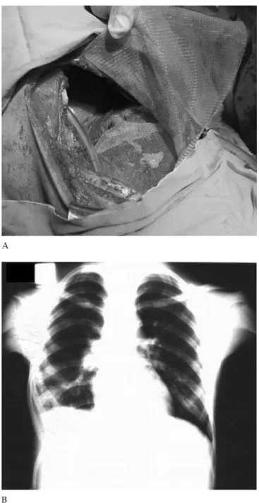
At thoracotomy, the pleura-covered tumor was found completely apart from the liver and lung. Further explo-ration showed the tumor to be fixed to the diaphragm. A hard mass, 9 ⫻ 6 ⫻ 5 cm, arising from the right dia-phragm had invaded the seventh, eighth, and ninth ribs and pleura, and no invasion of the liver was found. Because of the extent of the lesion, the main tumor, seventh, eighth, and ninth ribs, and right diaphragmatic muscle partially were resected. The diaphragmatic and thorax wall defects were closed with a Prolene patch (Fig 2A).
Microscopically, the lesion was composed of sheets of fairly uniform round-to-oval cells. The cytoplasm was relatively clear and indistinct, but contained large quan-tities of periodic acid-Schiff (PAS)-positive, diastase-digestible material, indicating high concentrations of glycogen (not shown). The tumor cells showed diffuse, intense membrane reactivity for CD99 (MIC 2) on immu-nohistochemical staining. The diagnosis was thus EES. The margins of resection were free of tumor and the following staging examinations showed no evidence of metastatic disease.
The postoperative course was uneventful, and the patient was discharged from the hospital on the 9th postoperative day. One month after discharge, chemo-radiotherapy was carried out. Our patient received MAID (mesna, doxorubicin, ifosfamide, and dacarbazine) chemotherapy postoperatively, and the patient has had no recurrence of disease within 10 months of follow-up (Fig 2B).
Comment
Primary tumors of the diaphragm are rare. They can occur at any age but most cases occur in the fourth and fifth decades of life. Primary tumors may be benign or malignant. The most recent summary was published in 1998 and describes 106 cases, including cysts, benign neoplasms, and malign tumors [2]. Benign tumors are Accepted for publication June 19, 2003.
Address reprint requests to Dr Erog˘lu, Department of Thoracic Surgery, Faculty of Medicine, Atatu¨rk University, 25240 Erzurum, Turkey; e-mail: [email protected].
715
Ann Thorac Surg CASE REPORT EROG˘ LU ET AL
2004;78:715–7 EWING SARCOMA OF THE DIAPHRAGM
© 2004 by The Society of Thoracic Surgeons 0003-4975/04/$30.00
Published by Elsevier Inc doi:10.1016/S0003-4975(03)01418-8
CASE
somewhat more frequent than malignant tumors, with fibrosarcoma being the most common malignant tumor. In a study of 71 primary tumors of the diaphragm, only five affected children below 15 years of age [3].
Sarcomas with diaphragmatic origin are extraordinar-ily rare in pediatric patients, with fibrosarcoma, rhabdo-myosarcoma, and leiomyosarcoma representing the ma-jority of cases. Raney and associates published a comprehensive review of soft tissue sarcomas arising in the diaphragm, which comprised 15 cases, three of which were EES [4]. The largest series of Ewing sarcoma origi-nating from soft tissues was composed of 130 cases recorded from 1972 to 1991 [5]; all of the patients were aged less than 21 years. To the best of our knowledge, Fig 1. (A) Chest radiograph showed pleural effusion on the right
hemithorax. (B) Contrast-enhanced thorax computed tomography scan showed an extrahepatic localization of the mass with compres-sion of adjacent liver parenchyma and a centrally located area of
necrosis. (C) Coronal T2-weighted magnetic resonance imaging scan
revealing an extrahepatic mass, growing from the diaphragm towards the thorax wall. The tumor compresses the liver parenchyma, which is indicated by the rounded edges of the liver adjacent to the tumor.
Fig 2. (A) The reconstruction of diaphragm and thorax wall defects with Prolene patch. (B) Chest radiograph after 10 months operation.
716 CASE REPORT EROG˘ LU ET AL Ann Thorac Surg
EWING SARCOMA OF THE DIAPHRAGM 2004;78:715–7
CASE
only 3 patients with primary Ewing’s sarcoma of the diaphragm have been reported [4], of whom, only 2 were children (aged 15 years; the third was 20 years old). Our patient is the youngest so far.
Almost 20% of patients with diaphragmatic tumors may be asymptomatic. Respiratory symptoms may in-clude chest pain, cough, and dyspnea. Gastrointestinal symptoms are caused by left-sided tumors, which com-press the gastric cardia and lead to anorexia, nausea, vomiting, and difficulty swallowing. Physical examina-tion may reveal an abdominal or a chest wall mass, atelectasis, or pleural effusion. Our case presented with progressively increasing dyspnea due to right hemotho-rax. Diaphragmatic tumors presenting with hemothorax have not been published previously.
A certain preoperative discrimination of diaphrag-matic tumors from other thoracic and abdominal neo-plasms is difficult [6]. Sometimes, a typical diaphragmatic “hump” may be observed [6]. The sharp border to the liver and the close relation to the diaphragm in the upper parts of the neoplasm may lead to the suspicion of a primary diaphragmatic neoplasm, but these signs are also possible in other large abdominal or retroperitoneal lesions. Radiographs may show irregularity of the dia-phragm [3]. The frequent occurrence of the normal vari-ant of diaphragmatic lobulation, localized diaphragmatic eventration, and juxtadiaphragmatic abnormalities caus-ing irregular diaphragmatic contour make discovery of diaphragmatic tumors difficult on plain radiographs. Tu-mors of the diaphragm may be difficult to differentiate from liver lesions in diagnostic imaging when located at the border of the liver. In the case presented, the MRI was the diagnostic tool showing precisely the rounded edges of the liver at the lateral margins of the lesion, indicating the growth of the depicted tumor from extra-hepatic towards the liver with compression of liver parenchyma.
Ewing’s sarcoma commonly arises from skeletal bone, but rarely may have an extraskeletal origin. Extraskeletal Ewing sarcoma can be confused with other small, round, blue cell tumors, including embryonal rhabdomyosar-coma, lymphoma, and neuroblastoma. This tumor shares histologic, immunohistochemical, and molecular find-ings with Ewing sarcoma of bone [7]. The classic histo-logic description of EES is of a tumor composed of small, uniform, round or oval cells in solid sheets divided by fibrous strands. Its cytoplasm is scanty, pale staining, and vacuolated because of the presence of glycogen, and the nuclei are round with “salt and pepper” chromatin and small nucleoli. Because no skeletal involvement was demonstrable in the present case, the diaphragm was considered the origin of this tumor. A sensitive and relatively specific antigen, CD99/MIC2, and a character-istic chromosomal translocation, t (11, 22)(q24; 12), have been identified in skeletal Ewing’s sarcoma and EES [7].
The treatment of EES is aggressive because most patients with Ewing sarcoma die 2 or 3 years after diagnosis. The most effective treatment is surgery with combination chemotherapy and high-dose radiation therapy. When resectable, the surgical excision must be wide in accordance with the classic oncologic principles. The defect may be repaired by direct approximation of diaphragmatic edges, or this may require prosthetic repair of the diaphragm. Adjuvant chemotherapy and radiation have been used with variable results. When the tumor is unresectable, preoperative chemotherapy to cause complete or partial reduction of tumor is tried. Three prior cases with diaphragmatic EES [4] had been treated with debulking and postoperative radiotherapy and conbination chemotherapy with vincristine, actino-mycin D, and cyclophosphamide and one additional doxorubicin. One patient survived more than 10 years and the other patients survived 2 years. In our patient, MRI and CT indicated the site of origin of the abdominal tumor, which was found resectable at thoracotomy. Our patient was treated successfully with surgery and multi-ple-agent, long-range chemotherapy, and high-dose ra-diation therapy, and there has been no recurrence during 10 months of follow-up. Survival has improved signifi-cantly in recent years, and current relapse-free survival is 55% at 5 years [8].
Extraskeletal Ewing sarcoma is a rare tumor and sen-sitive to multimodality treatment. Early surgical treat-ment followed by chemo- and radiotherapy has greatly improved the prognosis of this entity.
References
1. Rud NP, Reiman H, Pritchard DT, Frassica FJ, Smithson WA. Extraosseous Ewing’s sarcoma: a study of 42 cases. Cancer 1989;64:1548 –53.
2. Weksler B, Ginsberg RJ. Tumors of the diaphragm. Chest Surg Clin North Am 1998;8:441–7.
3. Eustace S, Fitzgerald E. Primary rhabdomyosarcoma of the diaphragm: an unusual cause of adolescent pseudo-achalasia. Pediatr Radiol 1993;23:622–3.
4. Raney RB, Anderson JR, Andrassy RJ, Crist WM, Donaldson SS, Maurer HM. Soft-tissue sarcomas of the diaphragm: a report from the Intergroup Rhabdomyosarcoma Study Group from 1972 to 1997. J Pediatr Hematol Oncol 2000;22:510 –4. 5. Raney RB, Asmar L, Newton WA, et al. Ewing sarcoma of soft
tissues in childhood: a report from the Intergroup Rhabdo-myosarcoma Study, 1972 to 1991. J Clin Oncol 1997;15:574 –82. 6. Midorikawa Y, Kubota K, Mori M, et al. Rhabdomyosarcoma of the diaphragm: report of an adult case. Jpn J Clin Oncol 1998;28:222–6.
7. Guiter GE, Gamboni MM, Zakowski MF. The cytology of extraskeletal Ewing sarcoma. Cancer 1999;87:141–8.
8. Cotterill SJ, Ahrens S, Paulussen M, Ju¨rgens HF, Vouˆte PA, Gadner H, Craft AW. Prognostic factors in Ewing’s tumor of bone: Analysis of 975 patients from the European Intergroup Cooperative Ewing’s Sarcoma Study Group. J Clin Oncol 2000;18:3108 –14.
717
Ann Thorac Surg CASE REPORT EROG˘ LU ET AL
2004;78:715–7 EWING SARCOMA OF THE DIAPHRAGM
CASE