DOI: 10.5455/annalsmedres.2019.11.739 2020;27(2):664-9
Evaluation of clinical outcomes of 271 patients undergoing
lumbar microdiscectomy in the light of literature
Ozkan Ozger1, Necati Kaplan2
1Istinye University, Faculty of Medicine, Department of Neurosurgery, Istanbul, Turkey 2Istanbul Rumeli University, Corlu Reyap Hospital, Clinic of Neurosurgery, Tekirdag, Turkey Copyright © 2020 by authors and Annals of Medical Research Publishing Inc.
Abstract
Aim: Lumbar microdiscectomy (LMD) is considered as a gold standard surgical technique for the treatment of patients with lumbar
disc hernia (LDH) although various types of treatment are being developed. This study aimed to investigate the safety and efficacy of LMD in our clinic.
Material and Methods: The study exclusively included 271 patients undergoing LMD due to LDH. Preoperative and postoperative
(10th day, 1st month, and long-term) Visual Analog Scale (VAS) and Oswestry Disability Index (ODI) scores of the patients were compared. Clinical outcomes were analyzed retrospectively based on modified Macnab criteria. Complications were noted. SPSS Statistics 22.0 software package was used to analyse the data for pre- and postoperative VAS and ODI scores. A p value of <0.05 was considered statistically significant.
Results: The mean length of stay in the operating room and hospital was 165.04±36.99 min and 1642.02±574.90 min, respectively.
There was a significant decrease in postoperative VAS and ODI (%) scores compared to preoperative scores. Preoperative VAS and postoperative long-term VAS scores were 8.99±0.62 and 1.38±0.95, respectively whereas preoperative ODI (%) and postoperative long-term ODI (%) scores were 85.33±6.74 and 12.96±9.58, respectively (p<0.001). The success rate for excellent or good outcomes according to the modified Macnab criteria was 87.45%. During surgery, cerebrospinal fluid (CSF) leak and dura defect developed in four patients (1.48%) and there was only dura defect in two patients (0.73%). After the surgery, recurrent LDH, spinal infection, and spinal epidural hematoma developed in seven (2.58%), three (1.11%), and one patient (0.37%), respectively.
Conclusion:LMD is an effective and safe method in the treatment of LDH as it has low complication rates and high success rates. Keywords: Complication; efficacy; lumbar disc herniation; lumbar microdiscectomy; safety
Received: 17.11.2019 Accepted: 11.12.2019 Available online: 10.03.2020
Corresponding Author: Ozkan Ozger, Istinye University, Faculty of Medicine, Department of Neurosurgery, Istanbul, Turkey
E-mail: [email protected]
INTRODUCTION
Low back pain is a very common condition in societies. About 60–80% of people suffer from low back pain and 35% experience sciatica pain at least once throughout their lives. Surgical intervention may be required in 10% of patients with lumbar disc hernia (LDH). Therefore, low back pain and LDH is a major problem for the community. Pain caused by LDH usually generally heals over six weeks with medical and physical therapy. Epidural steroid injections can be tried for pain. Severe and long-lasting pain, neurological deficit and patient preferences may require surgery. Surgical treatment has been reported to be more beneficial than conservative treatment in
patients with severe symptoms. The classical discectomy technique for the treatment of sciatica pain caused by LDH was first described by Mixter and Barr in 1934. At the present time, minimally invasive techniques are in the foreground (1,2).
One of these techniques is lumbar microdiscectomy (LMD). This method, which has been used for many years, is the surgical removal of the damaged portion of a herniated disc causing pressure on the nerve root under the microscope. In recent years, nerve surgeons have gained great experience in this method. Satisfactory outcomes have been obtained in 60–80% of patients after LMD (3).
The aim of this study was to evaluate the efficacy and safety of LMD in light of clinical parameters and demographic data.
MATERIAL and METHODS
Study Design
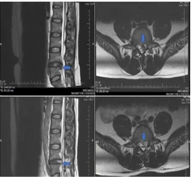
Patients who underwent LMD in a single center and were operated by a single surgeon between 2015 and 2019 were retrospectively analyzed. All data were collected from patient files. All patients were diagnosed by preoperative magnetic resonance imaging (MRI) of the lumbar spine (Figure 1). The surgical technique to be applied to the patients was explained in detail before the operation and their consent was obtained.
Figure 1. Preoperative sagittal and axial lumbar MRI images and
early postoperative sagittal and axial lumbar MRI images of a 37-year-old male patient. Extruded disc fragment appears to be removed after LMD (blue arrows)
Inclusion and Exclusion Criteria
The study included adult patients who were diagnosed with single or multiple levels of LDH, had a neurological deficit or pain that did not respond to medical treatment within six weeks, underwent LMD, and whose informed consent was obtained.
Patients with preoperative spondylolisthesis, spine fracture, spine tumor, severe scoliosis (Cobb angle 40 degrees or greater) (23), and spine or disc infection besides LDH despite LMD and whose follow-up duration was less than one-month were excluded from the study.
Surgical Technique
The operations were performed under a microscope in a single center by a single neurosurgeon. Single-dose of antibiotic (1 g cefazolin intravenously) prophylaxis was administered to all patients before the surgery. The technique used was LMD. The operations were performed under general or spinal anesthesia in the prone position.
The distance to be operated was determined with scopy. An incision of 1.5–3 cm was made for a single distance depending on whether the patient’s waist area was rich or poor from adipose tissue and the depth between the lamina and the skin surface. Interlaminar approach was preferred. Classical operation, i.e. hemilaminectomy, microdiscectomy, and foraminotomy, was performed. After the nerve root freedom and hemostasis check, the tissues were closed in accordance with their anatomy using resorbable sutures. The skin was combined with sterile strips. Sutures that were non-absorbable and required to be removed later were not used.
Evaluation Criteria
Postoperative clinical results of all patients were evaluated under four categories according to modified Macnab criteria (Table 1) (22). Preoperative and postoperative 10th-day and first-month VAS (for leg pain) and ODI scores of all patients were recorded. However, long-term (about 11 months) VAS and ODI scores were recorded in 71 patients since the follow-up period was between two months and three years.
Table 1. Modified Macnab criteria for the assessment of clinical outcomes after LMD
Outcome Characteristic
Excellent No pain; no restriction of mobility; return to normal work and level of activity
Good Occasional nonradicular pain; relief of presenting symptoms; able to return to modified work
Fair Some improved functional capacity; still handicapped and/or unemployed
Poor additional operative intervention needed at index level Continued objective symptoms of root involvement; irrespective of length of postoperative follow-up
Statistical Analysis
In this study, preoperative and postoperative 10th-day and first-month VAS and ODI scores and scores measured as follow-up post-test were evaluated. Statistical and visual analyses were utilized in the analysis of the data. Repeated measures analysis of variance (ANOVA) was used to analyze the changes in VAS and ODI scores over time. Statistical analysis was performed using SPSS version 22.0 software. A p value of <0.05 was considered statistically significant.
RESULTS
Patients’ Characteristics
A total of 271 patients and 300 vertebral levels were operated. Of the patients, 144 were male and 127 were female. Male to female ratio was 1:13. The patients were in the 25–88 age range with a mean age of 52.27±14.27 years. All patients had at least one neurological deficit or
leg pain that did not respond to non-surgical treatment within six weeks (radiculopathy).
Of the 300 LDH levels treated with LMD, eight (2.67%) were L1-2, 12 (4.00%) were L2-3, 35 (11.67%) were L3-4, 139 (46.33%) were L4-5, and 106 (35.33%) were L5-S1. Of the levels, 243 (89.67%) were single, 27 (9.96%) were two and one (0.37%) was three levels. Of the patients, 154 (51.33%) were operated for left LDH, 132 (44.00%) for right LDH and 14 (4.67%) for bilateral LDH.
The mean time between entry and exit from the operating room was calculated as 165.04±36.99 min (60–240 min). The mean length of hospital stay was 1.14±0.40 days (0.34–4.01 days).
Postoperative Health Status Analysis
According to the modified Macnab criteria, LMD results were excellent in 69.37% (n=188), good in 18.08% (n=49), fair in 7.38% (n=20), and poor in 5.17% (n=14) of the patients.
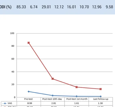
Table 2 shows the pre-test and post-test mean and standard deviation values of VAS and ODI values and Figure 2 shows the graph of these values.
Table 2. Pre-test and post-test mean and standard deviation values of VAS and ODI values
Pre-test (n=271) Post-test 10th day (n=271) Post-test 1st month (n=271) Last follow-up (n=71)
Mean SD Mean SD Mean SD Mean SD
VAS 8.99 0.62 2.81 1.24 1.61 1.05 1.38 0.95
ODI (%) 85.33 6.74 29.01 12.12 16.01 10.70 12.96 9.58
Figure 2. Mean values of VAS and ODI values
The mean VAS score of the patients before the intervention was 8.99±0.62 and after the intervention was 2.81±1.24 at the 10th day, 1.61±1.05 in the first month and 1.38±0.95 in the long term post-test. All three measurements showed a decrease in the VAS scores of the patients after the surgery. According to the results of repeated measures ANOVA, this decrease in VAS values was statistically significant, F(1.70)=1231.628 (p<0.001) (Table 3). The comparisons made between the groups were examined with Bonferroni correction to determine the difference between the measurements and the difference between all measurements was found to be statistically significant.
Table 3. Repeated measures ANOVA results of pre-test and post-test scores of VAS values
Source of variance SquaresTotal (SS) SD SquaresMean (MS)
F p
Subjects within 4380.750 71
Measurement 4145.158 1 4145.158 1231.628 0.000
Error 235.592 70 3.366
The mean ODI (%) score of the patients before the intervention was 85.33±6.74 and after the intervention was 29.01±12.12 at the 10th day, 16.01±10.70 in the first month and 12.96±9.58 in the long term post-test. All three measurements showed a decrease in the ODI scores of the patients after the surgery. According to the results of repeated measures ANOVA, this decrease in ODI values was statistically significant, F(1.70)=1288.824 (p<0.001) (Table 4). The comparisons made between the groups were examined with Bonferroni correction to determine the difference between the measurements and the difference between all measurements was found to be statistically significant.
Table 4. Repeated measures ANOVA results of pre-test and post-test scores of ODI values
Source of variance SquaresTotal (SS) SD SquaresMean (MS) F p Subjects within 416178.000 71 Measurement 394738.535 1 394738.535 1288.824 0.000 Error 21439.465 70 306.278 Complications
The encountered complications in 271 patients undergoing LMD were as follows: recurrent LDH requiring revision surgery in five (1.84%), recurrent LDH not requiring revision surgery in two (0.73%) (total recurrent LDH rate was 2.58%), postoperative spinal infection in three (1.11%), dura defect alone in two (0.73%), dura defect and CSF
leak in four (1.48%), and spinal epidural hematoma in one patient (0.37%). The ratio of all complications developed in 17 patients to the total number of patients was 6.27%. There were no surgery-related deaths (Table 5).
Table 5. Distribution of complications in patients undergoing LMD
Complications Number of patients (n) Rate (%) Recurrent LDH 7 2.58
CSF leak and dura defect 4 1.48
Spinal infection 3 1.11
Dura defect alone 2 0.73
Spinal epidural hematoma 1 0.37
Total 17 6.27
In all patients with recurrent LDH, the mean LDH recurrence time was 14.43±7.02 (5–24) months after the first operation. In two patients with CSF leak, tissue glue was used during surgery whereas muscle and adipose tissue were used in other two patients. Spinal epidural hematoma was operated urgently on the same day. In one of the cases where spinal infection was developed, the agent pathogen could not be detected although culture was taken twice. Wound discharge and laboratory and clinical findings of the patient regressed with empirical antibiotic therapy two months later. The other patient had complaints of severe low back pain and sweating one month after the operation. No specimens were taken from the surgical site for culture. According to the blood test and lumbar MRI report, spondylodiscitis diagnosis was made and antibiotic treatment was applied for a total of three months. At the end of the third month, the complaints of the patient regressed. In the last patient, destructive osteomyelitis started on the 15th day of the operation, particularly in the vertebrae adjacent to the operated vertebral level. The clinical condition of the patient was good until that time but began to worsen gradually. He/she refused sampling from the surgery site. In our laboratory tests, Quantiferon-TB Gold test positivity and control lumbar MRI results were consistent with tuberculosis (Tbc). Therefore, empirical Tbc treatment was initiated. Six months later, the clinical status was better than the preoperative period but worse than the 10th postoperative day. The patient, who refused to undergo surgery, returned to work at the end of the first year.
DISCUSSION
Although there is much discussion about the treatment of LDH, the effectiveness of the surgical intervention has been reported to be 49–90% in some studies. In a study involving 45 patients undergoing LMD, which was conducted in Italy in 2014, good and excellent results were found to be 90% according to the modified Macnab criteria (4,13). In the present study, the success rate according
to the modified Macnab criteria was found to be 87.45%, which is compatible with the literature.
In patients with LDH, surgical treatment has been shown to be superior to conservative treatment in the recovery of symptoms and functional capacity at the end of the four-year follow-up (5).
Standard microdiscectomy is still used as a gold standard in the treatment of LDH with minor changes in surgical technique. In recent years, some studies have reported successful results with microendoscopic discectomy (MED). However, its superiority to LMD has not been proved yet (6).
A retrospective study of 49 patients in 2018 showed that VAS and ODI scores were significantly reduced after the surgery. Similar results were obtained in a prospective study of 100 patients carried out in the same year (3,7). The significant decrease in VAS and ODI scores after LMD in the present study is compatible with the literature. This supports that LMD is an effective method for the treatment of radicular leg pain.
The most common levels affected by LDH are L4-5 and L5-S1 segments. In a recent study, the level of herniation has been reported to be L4-5 in 84 (55.26%) of 152 patients who underwent LMD (8). In the present study, L4-5 level is the most common level undergoing LMD, with a rate of 46.33%. In a recent study, the mean age of the patients undergoing LDH was 44.85±12.26 years (20–70 years) and the male to female ratio was 1:8 (9). Our study is parallel to this study in terms of male to female ratio, mean age, and age range.
In a study conducted in 2009, 25 patients undergoing percutaneous endoscopic lumbar discectomy (PELD) and 29 patients undergoing open LMD (OLMD) were compared in terms of clinical outcomes. The mean VAS score of the undergoing OLMD has been reported to be 8.6±1.7 and 3.5± 3.1 before and after the surgery, respectively and the mean ODI (%) score has been reported to be 63.1±22.3 and 18.2±15.4 before and after the surgery, respectively. The decrease in values has been found to be significant, but there was no significant difference between PELD and OLMD in terms of VAS and ODI values (10). Although the number of patients included in the present study is approximately 10 times higher than those included in this study, our clinical results are similar.
LMD is still the most commonly used surgical technique for the treatment of LDH in the USA. The rate of good outcomes is 90–95%. The most common complication is recurrent LDH, with an incidence rate of 5–15% in the literature. In a study conducted in 2017, where LMD was performed in 177 LDH patients, recurrent LDH developed in 30 (16%) patients and 27 of these patients were operated (11). The rate of patients who underwent re-operation due to recurrent LDH was reported to be 4.5% in the transforaminal endoscopic discectomy (TFED) series of 134 cases with a mean age of 38, a success rate of 89%, and a mean follow-up duration of eight months (12). The
outcomes obtained with TFED have been reported to be equivalent to those obtained with LMD. In our series, the rate of recurrent LDH was 2.58%, which was slightly lower than in the literature. This low rate is probably due to our increased experience in LMD.
In a study comparing LMD and MED complication rates in 2012, no significant difference was found between the two methods in terms of dural injury, the rate of which was reported to be 5.94% for LMD and 5.40% for MED (14). In another LMD series of 122 patients, dural injury was reported in two patients (1.63%) (15). In our study, dural injury was seen in a total of six patients (2.21%) and was similar to the literature findings.
Spondylodiscitis is a very rare complication of lumbar discectomy. Its incidence is reported to be around 0.1– 18.8% by many different authors. The most common pathogen is Staphylococcus aureus. In a recent study, spondylodiscitis was observed in 12 (1.03%) of 1154 patients and Staphylococcus aureus grew in three (25%), Staphylococcus epidermidis grew in four (33%) and Escherichia coli grew in three (25%) patients whereas no causative pathogen was observed in two patients (16). In a study published in 2018, the wound infection rate was reported to be 3.3% in 243 patients undergoing LMD (17). In our study, there were no infectious agents produced in three patients, but the spinal infection was diagnosed based on the clinical and laboratory findings. The spinal infection rate in our study was 1.11%, compatible with the literature.
In the postoperative period, asymptomatic epidural hematoma can be seen in 33–100% of patients undergoing spine surgery while the incidence of symptomatic epidural hematoma is 0.1–0.24%. In a study that retrospectively examined 15562 patients undergoing spinal surgery in 2015, the spinal epidural hematoma requiring re-operation was detected in 25 patients after surgery (18). This ratio is 0.37% in our study and compatible with the literature. In a new article published in 2019, LMD was compared retrospectively with lumbar sequestrectomy (LST). In this study, the duration of postoperative analgesic use and the time required to return to daily life were less in the LST group. Preoperative VAS values were decreased in both groups after a mean follow-up of 7 months. There was no significant difference between the two groups in terms of VAS (19). Reduction of leg pain in patients has similar features to our study. LST may be more appropriate in selected patients.
In a manuscript published in 2016, duration of surgery and length of hospital stay for PELD and OLMD were studied and the duration of surgery was reported to be 67.8 min for PELD and 136.7 min for OLMD and the length of hospital stay was reported to be 1.5 days for PELD and 7.2 days for OLMD (20). In a study conducted in France in 2017, patients undergoing LMD were compared in two groups: receiving inpatient and outpatient treatment. The mean length
of hospital stay was calculated as 36 hours 48 minutes (about 1.5 days) in patients receiving inpatient treatment (21). In our study, the length of hospital stay was 1.14 days, which is good for inpatient LMD operations. Most of the studies in the literature have studied the duration of surgery. However, we calculated the difference between the time of entry and exit from the operating room instead of the duration of surgery, meaning that all the time the patient spent in the operating room was included. This time was found to be about 165.04 minutes.
In this study, we discussed the postoperative outcomes of patients who were treated for LDH and needed LMD. As discussed above, our results and complication rates are in line with the literature. Although it has been reported to be disadvantageous in terms of length of hospital stay compared to other newly introduced methods, similar results have been obtained with endoscopic methods in our study.
CONCLUSION
In conclusion, LMD has been compared with many methods in patients with LDH and is still considered the gold standard for the treatment of LDH. Lumbar microdiscectomy is still an effective and safe treatment option for suitable patients with LDH since the length of stay in hospital and operating room is short, clinically satisfactory outcomes are obtained, and complication rates are low.
Competing interests: The authors declare that they have no competing interest.
Financial Disclosure: There are no financial supports.
Ethical approval: Permission from Chief Physician to use data (approval number: 22.10.2019/450).
Ozkan Ozger ORCID: 0000-0001-7257-8379 Necati Kaplan ORCID: 0000-0001-5672-0566
REFERENCES
1. Campbell P, Wynne-Jones G, Muller S, et al. The influence of employment social support for risk and prognosis in nonspecific back pain: a systematic review and critical synthesis. Int Arch Occup Environ Health 2013;86:119-37.
2. Dey PC, Nanda SN. Functional Outcome after Endoscopic Lumbar Discectomy by Destandau’s Technique: A Prospective Study of 614 Patients. Asian Spine J 2019;13:786-92.
3. Gelalis ID, Papanastasiou EI, Pakos EE, et al. Clinical outcomes after lumbar spine microdiscectomy: a 5-year follow-up prospective study in 100 patients. Eur J Orthop Surg Traumatol 2019;29:321-7.
4. Veresciagina K, Spakauskas B, Ambrozaitis KV. Clinical outcomes of patients with lumbar disc herniation, selected for one-level open-discectomy and microdiscectomy. Eur Spine J 2010;19:1450-8. 5. Weinstein JN, Lurie JD, Tosteson TD, et al. Surgical
herniation: four-year results for the Spine Patient Outcomes Research Trial (SPORT). Spine (Phila Pa 1976) 2008;33:2789-800.
6. Lau D, Han SJ, Lee JG, Lu DC, Chou D. Minimally invasive compared to open microdiscectomy for lumbar disc herniation. J Clin Neurosci 2011;18:81-4. 7. Cetin A, Gokdemir TM. Evaluation of pain status and
quality of life in patients with lumbar disc hernia who underwent microdiscectomy. Medicine Science 2018; 7:745-7.
8. Ahn Y, Lee SG, Son S, et al. Transforaminal Endoscopic Lumbar Discectomy Versus Open Lumbar Microdiscectomy: A Comparative Cohort Study with a 5-Year Follow-Up. Pain Physician 2019; 22: 295-304. 9. Zahid H, Ali I, Choudhary N, Rashid J, Mehmood K. Microscopic Discectomy Outcomes in Lumbar Disc Herniation Patients. Pak J of Neurol Surg 2019;23: 142-5.
10. Lee DY, Shim CS, Ahn Y, et al. Comparison of percutaneous endoscopic lumbar discectomy and open lumbar microdiscectomy for recurrent disc herniation. J Korean Neurosurg Soc 2009;46:515-21. 11. Camino Willhuber G, Kido G, et al. Factors
associated with lumbar disc hernia recurrence after microdiscectomy. Rev Esp Cir Ortop Traumatol 2017;61:397-403.
12. Tzaan W. “Transforaminal percutaneous endoscopic lumbar discectomy.” Chang Gung Med J 2007;30: 226-34.
13. Galarza M, Gazzeri R, De la Rosa P, et al. Microdiscectomy with and without insertion of interspinous device for herniated disc at the L5–S1 level. J Clin Neurosci 2014;21:1934-9.
14. Martín-Láez R, Martínez-Agüeros JÁ, Suárez-Fernández D, et al. Complications of endoscopic microdiscectomy using the EASYGO! system: is there
any difference with conventional discectomy during the learning-curve period? Acta Neurochir (Wien) 2012;154:1023-32.
15. Singhal A, Bernstein M. Outpatient lumbar microdiscectomy: a prospective study in 122 patients. Can J Neurol Sci 2002;29:249-52.
16. Diren F, Can H, Onal MB, et al. The rate of spondylodiscitis in patients with lomber discectomy using single dose prophylactic antibiotics. JAREM 2019;9:1-6.
17. Calikoglu C, Cakir M. Open Discectomy vs. Microdiscectomy: Results from 519 Patients Operated for Lumbar Disc Herniation. Eurasian J Med 2018;50: 178-81.
18. Kao FC, Tsai TT, Chen LH, et al. Symptomatic epidural hematoma after lumbar decompression surgery. European Spine Journal 2015;24:348-57.
19. Somay H,Karaarslan N.Sequestrectomy or microdiscectomy in patients with lumbar disc herniation.Ann Med Res 2019;26:753-78. 20. Choi KC, Kim SJ, Park CK. Percutaneous endoscopic
lumbar discectomy as an alternative to open lumbar microdiscectomy for large lumbar disc herniation. Pain physician 2016;19:291-300.
21. Debono B, Sabatier P, Garnault V, et al. Outpatient lumbar microdiscectomy in France: from an economic imperative to a clinical standard-an observational study of 201 cases. World Neurosurg 2017;106:891-7. 22. Hubbe U, Franco-Jimenez P, Klingler JH, et al.
Minimally invasive tubular microdiscectomy for recurrent lumbar disc herniation. J Neurosurg Spine 2016;24:48-53.
23. Nada D, Julien C, Rompré PH, et al. Association of Circulating YKL-40 Levels and CHI3L1 Variants with the Risk of Spinal Deformity Progression in Adolescent Idiopathic Scoliosis. Sci Rep 2019;9:1-13.