Research Article
Comparison of Platelet-Rich Plasma-Impregnated Suture
Material with Low and High Platelet Concentration to Improve
Colonic Anastomotic Wound Healing in Rats
Mehmet Akif Aydin
,
1Eray Metin Guler,
2Ahu Senem Demiroz,
3Muhammet Fatih Aydin,
4and Gulcan Saglam
51Department of General Surgery, Altinbas University Faculty of Medicine Bahcelievler Medical Park Hospital, Istanbul, Turkey 2Department of Medical Biochemistry, Bezmialem Vakif University Faculty of Medicine, Istanbul, Turkey
3Department of Pathology, Istanbul University Cerrahpasa Faculty of Medicine, Istanbul, Turkey
4Department of Gastroenterology, Altinbas University Faculty of Medicine Bahcelievler Medical Park Hospital, Istanbul, Turkey 5Department of Medical Biochemistry, Istinye University Faculty of Medicine, Istanbul, Turkey
Correspondence should be addressed to Mehmet Akif Aydin; [email protected] Received 21 December 2019; Revised 14 March 2020; Accepted 7 April 2020; Published 26 May 2020 Academic Editor: Tatsuya Toyokawa
Copyright © 2020 Mehmet Akif Aydin et al. This is an open access article distributed under the Creative Commons Attribution License, which permits unrestricted use, distribution, and reproduction in any medium, provided the original work is properly cited.
Objective. This study was designed to investigate the impact of using suture material impregnated with platelet-rich plasma (PRP) in different platelet concentrations on colonic anastomotic wound healing in rats. Methods. A total of 24 Sprague Dawley female rats were separated into 3 groups (n = 8 for each) including the control group (CON; standard vicryl suture repair), the low platelet concentrate PRP group (L-PRP; suture material impregnated with PRP containing average 2.7-fold (range, 2.0 to 3.1) higher amount of platelets vs. control), and the high platelet concentrate PRP group (H-PRP; suture material impregnated with PRP containing average 5.1-fold (range, 4.8 to 5.4) higher amount of platelets vs. control). Rats were sacrificed on the postoperative 7th day for analysis of colonic anastomosis region including macroscopic observation, measurement of anastomotic bursting pressure (ABP), and the hydroxyproline levels and histopathological findings in colon tissue samples. Results. Total injury scores were significantly lower in the L-PRP and H-PRP groups than those in the control group (median (range) 13.00 (7.00) and 11.50 (6.00) vs. 15.50 (4.00),p < 0:05 and p < 0:01, respectively). ABP values (180.00 (49.00) vs. 124.00 (62.00) and 121.00 (57.00) mmHg,p < 0:001 for each) and tissue hydroxyproline levels (0.56 (0.37) vs. 0.25 (0.17) and 0.39 (0.10)μg/mg tissue, p < 0:001 and p < 0:05, respectively) were significantly higher in the L-PRP group as compared with those in the control and H-PRP groups. Conclusion. In conclusion, ourfindings revealed PRP application to colonic anastomosis sutures to promote the anastomotic healing process. The platelet concentration of PRP seems to have a significant impact on the outcome with superior efficacy of L-PRP over H-PRP in terms of bursting pressures and collagen concentration at the anastomotic site.
1. Introduction
Despite marked advances in preoperative management and suture techniques and materials, gastrointestinal anastomotic leakage or dehiscence remains a common complication in colorectal surgery being associated with an increased risk of perioperative morbidity and mortality [1–4]. This led to a
continuing search for innovative methods or technical mod-ifications to avoid anastomotic leakage [5, 6].
Platelet concentrates such as platelet-rich plasma (PRP) are known to act as a natural fibrin clot and to promote wound healing by delivering high quantities of growth fac-tors that regulate cell proliferation, matrix remodeling, and repair process such as platelet-derived growth factor (PDGF) Volume 2020, Article ID 7386285, 8 pages
controversial results [5, 6, 12–14].
The use of different PRP preparation methods resulting in different platelet concentrations is suggested to have a potential role in this controversy between studies [6]. The opposite impact of low PRP (stimulatory effect) and high PRP (inhibitory effect) concentrates on intestinal wound healing has also been reported in a recent rat model study [6]. Identification of anastomotic collagen deposition via tissue hydroxyproline levels and of anastomotic strength via anastomotic bursting pressure (ABP) measurement is considered to be the most reliable indicators of anasto-motic wound healing and outcome of gastrointestinal anastomoses [5, 15, 16].
This study was therefore designed to evaluate the impact of using suture repair augmented with different PRP concen-trations on anastomotic wound healing in a rat model of colonic anastomosis based on bursting pressures, tissue hydroxyproline levels, and histopathological examination.
2. Methods
2.1. Animals and Study Protocol. A total of 24 Sprague Daw-ley female rats (weighing 260-310 g) were kept in a light- and temperature-controlled room with a 12 hr light-dark cycle, temperature of 21°C, and relative humidity of 40-60%. The animals were fed standard rat pellets and provided with water ad libitum. This study was carried out in accordance with the National Institutes of Health Guide for the Care and Use of Laboratory Animals, while the study protocol was approved by the Institutional Animal Care and Use Committee (approval number: 2014/14).
The rats were separated into 3 groups (n = 8 for each) including the control group (CON; standard vicryl suture repair), the low platelet concentrate PRP group (L-PRP; suture material impregnated with PRP containing average 2.7-fold (range, 2.0 to 3.1) higher amount of platelets vs. control blood), and the high platelet concentrate PRP group (H-PRP; suture material impregnated with PRP containing average 5.1-fold (range, 4.8 to 5.4) higher amount of plate-lets vs. control blood).
2.2. Preparation of PRP and Impregnated Sutures. Eight donor rats were used to obtain PRP. At the time of surgery, 8.5 mL of intracardiac homologous blood was drawn from each of the eight rats. The blood was aspirated into 10 mL GLO-PRP (Biotrend Medical, Istanbul, Turkey) tubes con-taining 1.5 mL of acid-citrate-dextrose (ACD) and trans-ferred into a centrifugation chamber and centrifuged using
addition of minimum amounts of PPP. In this way, L-PRP and H-PRP were obtained containing average 2.7-fold (2.0-3.1) and 5.1-fold (4.8-5.1) higher platelet concentrates, respectively, as compared with blood samples used to prepare PRP. Average platelet concentration in blood sample was 0:614 × 106μL (range, 0.545 to 0:740 × 106μL) and white
blood cell count (WBC) 11:2 × 10³μL (range, 8.1 to 14:3 × 10³μL), whereas platelet concentration was 1:676 × 106μL
(range, 1.44 to 1:96 × 106μL) and WBC count 1:6 × 10³μL (range, 0.9 to 2:3 × 10³μL) in the L-PRP group and 3:137 × 106μL (range, 2.78 to 3:72 × 106μL) and WBC count 1:9 ×
103μL (range, 1.3 to 2:5 × 10³μL) in the H-PRP group.
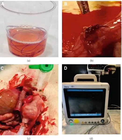
4/0 vicryl sutures were kept in sterile containers involving liquid forms of L-PRP or H-PRP for 3 minutes based on find-ings from a preliminary timeline analysis of weight increase per minute in PRP-impregnated sutures which revealed max-imum saturation (from the baseline value of 0.0640 g to the maximum value of 0.1284 g) to be reached at the 3rdminute (Figure 1(a)).
Afterwards, for preanalysis to determine the platelet con-tent absorbed by the sutures, the amount of PRP was
mea-sured in μL before putting vicryl suture into a PRP
container, and it was found that the suture absorbed 0.7μL PRP within 3 minutes. The PRP-impregnated vicryl sutures were placed in an empty container and added with 2.8μL distilled water which enabled the release of entire PRP content after a 3 min waiting period, while the fluid
was analyzed in the same hemogram device to confirm
the target platelet concentrations. Values obtained from the hemogram device were calculated by taking dilution rate into account. Average platelet concentration in blood sample was 0:580 × 106μL, whereas in platelet
concentra-tions in the fluid obtained from L-PRP- and
H-PRP-impregnated vicryl sutures were 1:334 × 106μL (2.3-fold) and 2:726 × 106μL (4.7-fold), respectively, indicating suffi-cient amount of platelet absorption in the vicryl sutures. 2.3. Surgery. After overnight fasting, the rats were anesthe-tized by intraperitoneal injection of ketamine 35 mg/kg (Ketalar; Parke Davis, Eczacibasi, Istanbul, Turkey) and xylazine 5 mg/kg (Rompun; Bayer AG, Leverkusen, Ger-many). The same surgeon performed all operations. The abdominal skin of the rat was shaved, and a 3 cm midline incision was made under aseptic conditions. The left colon was cut into two 3-4 cm over the peritoneal reflection. A colocolonic single-layer end-to-end anastomosis was per-formed with standard 4/0 and 45 mm length vicryl suture
(polyglactin 910) (Ethicon Inc., NJ, USA) in the control group, whereas with L-PRP-impregnated and H-PRP-impregnated 4/0 vicryl suture in the L-PRP and H-PRP groups, respectively (Figure 1(b)).
Standard length sutures were used for each anastomosis. Midline closure was performed using interrupted 3/0 silk sutures (Ethicon Inc.). No analgesic or antibiotic was admin-istered in the postoperative period, and oral intake was started on the 1st postoperative day in each group of rats. After operation, the animals were fed standard rat pellets and provided with water ad libitum.
2.4. ABP Measurement. Rats were sacrificed on the postoper-ative 7thday via intraperitoneal 2 mL high-dose pentobarbital sodium injection (200 mg/mL, KU life, Copenhagen, Den-mark). Following euthanasia and reopening of the abdominal incision, the peritoneal cavity was assessed for the presence of anastomotic leakage or dehiscence, peritonitis, abscesses, and anastomotic site or other visceral adhesions. Colons were carefully exteriorized, and the anastomotic sites are
identi-fied. Segments containing the anastomosis in the middle were carefully resected and washed with isotonic saline to remove fecal content. An 18 gauge silicone catheter was passed through both ends and attached via 3/0 silk suture. Intraluminal methylene blue-colored isotonic solution infusion (5 m/min) was performed using an infusion pump (Argus Medical AG, Heimberg, Switzerland), while intra-luminal pressure was monitored and recorded through the transducer (Beneview T5, Shenzhen, China) attached to the catheter placed on the other end (Figures 1(c) and 1(d)). The pressure recorded just before the leak was con-sidered to be the ABP. After measurement of ABP, half of the colon segment containing the anastomosis line was used for histopathological analysis and the other half for hydroxyproline analysis.
2.5. Macroscopic Examination and Histopathological
Analysis. Tissue samples involving the colon anastomosis line werefixed in 10% buffered formalin for 48-72 hours and then trimmed and processed for routine histopathological
(a) (b)
(c) (d)
Figure 1: (a) Preparation of PRP-impregnated vicryl suture, (b) complete anastomosis, (c) anastomotic bursting pressure measurement, and (d) anastomotic bursting pressure measurement setting.
examination. Tissue samples perpendicular to the direction of anastomosis line were embedded in paraffin for serial section-ing. 4μm sections were stained with hematoxylin and eosin (HE) and examined under a light microscope by the same pathologist who was unaware of the experimental groups. Semiquantitative scoring of histopathological parameters (necrosis, PMN cells, MN cells, edema, mucosal epithelium, submucosal/mucosal layer, and granulation tissue; each scored from 0 to 3) was performed using the Verhofstad wound heal-ing scale [17]. Lower and higher scores were considered to indicate good and worse healing, respectively, based on the Verhofstad injury scoring system (Table 1).
2.6. Hydroxyproline Measurement. The hydroxyproline level in the tissue was measured colorimetrically with the Hydroxyproline Test Kit (Elabscience, E-BC-K061, Hous-ton, Texas, USA). The principle of measurement was based on the purplish red color occurring upon the reac-tion of dimethylaminobenzaldehyde with the oxidareac-tion
product under the effect of oxidizer. The content of
hydroxyproline was calculated by measuring the OD value at 550 nm.
2.7. Statistical Analysis. Statistical analysis was made using IBM SPSS Statistics for Windows, version 25.0 software (IBM Corp., Armonk, NY, USA). The Kruskal-Wallis test with post hoc Tamhane’s test was used to analyze differences in platelet and wound healing parameters between the study groups. Data were expressed as median (range).p < 0:05 was considered statistically significant. Power of the study was calculated to be 0.99 (alpha 0.05), considering a mean (SD) 4 (0.5) unit difference in mean score (mean control = 16, mean experimental group = 12) between more than two groups (N = 24).
3. Results
3.1. General Characteristics. Except for one rat in the control group which died on the postoperative 3rdday, all rats sur-vived the surgery. Macroscopic evaluation of the colonic anastomosis site revealed no intra-abdominal abscess or leak in rats. No significant difference was noted between study groups in terms of baseline or postoperative body weight. Body weight was slightly decreased from baseline to postop-erative period in each group (Table 1).
3.2. Histopathological Findings on Wound Healing. Total injury scores were significantly lower in the L-PRP and
H-PRP groups than those in the control group (13.00 (7.00) and 11.50 (6.00) vs. 15.50 (4.00), p < 0:05 and p < 0:01, respectively). Specifically, edema score was significantly lower in the L-PRP and H-PRP groups than that in the con-trol group (1.00 (0.00) and 0.00 (0.00) vs. 2.00 (1.00),p < 0:05 and p < 0:01, respectively). Mucosal epithelium scores were significantly lower in the H-PRP group than those in the L-PRP and control groups (1.00 (2.00) vs. 2.50 (1.00) and 3.00 (1.00),p < 0:05 for each). Granulation tissue scores were significantly lower in the L-PRP and H-PRP groups as com-pared with those in the control group (2.00 (1.00) and 2.00 (1.00) vs. 3.00 (1.00),p < 0:01 for each) (Figure 2).
3.3. ABP Values. Median (range) ABP values were signifi-cantly higher in the L-PRP group as compared with those in the control and H-PRP groups (180.00 (49.00) vs. 124.00 (62.00) and 121.00 (57.00) mmHg,p < 0:001 for each), and although ABP values were slightly higher in the H-PRP group compared to the control group, the difference was not statistically significant (124.00 (62.00) vs 121.00 (57.00) mmHg) (Table 2).
3.4. Tissue Hydroxyproline Levels. Median (range) tissue hydroxyproline levels were significantly higher in the L-PRP group as compared with those in the control and H-PRP groups (0.56 (0.37) vs. 0.25 (0.17) and 0.39 (0.10)μg/mg tissue,p < 0:001 and p < 0:05, respectively). Mean (SD) tissue hydroxyproline levels in the H-PRP group were also signifi-cantly higher than levels in the control group (p < 0:001) (Table 2).
4. Discussion
Ourfindings in a rat model of colocolonic end-to-end anas-tomosis support the anastomotic healing effect of PRP, while indicating the likelihood of variation in the efficacy of PRP depending on the platelet concentration used. L-PRP-impregnated sutures and H-L-PRP-impregnated sutures showed improved lesser total injury scores on histopatho-logical assessment, although no significant difference was noted between L-PRP and H-PRP groups in terms of total injury scores related to wound healing when compared to using standard sutures. However, ABP values and tissue hydroxyproline levels were significantly higher in the L-PRP group compared to both H-L-PRP and control groups. ABP values were slightly higher in the H-PRP group com-pared to the those in the control group, although the
difference was not statistically significant. On the other hand, tissue hydroxyproline levels were significantly higher in the H-PRP group than those in the controls. Accordingly, L-PRP rather than H-PRP seems to be associated with
improved anastomotic healing in the present study in terms of the overall criteria assessed including anastomotic strength and integrity (ABP), tissue collagen (hydroxyproline), and tissue regeneration (injury scores).
(a) (b)
(c)
Figure 2: Wound healing area involving (a) marked PMN cell and MN cell infiltration accompanied with mild necrosis, mild edema, and marked granulation tissue with no submucosal or mucosal muscle bridging, H&E ×20 (control group); (b) almost no necrosis along with mild edema, incomplete cubic epithelium, moderate submucosal muscle bridging, and mild granulation tissue, H&E ×40 (L-PRP group); and (c) marked necrosis accompanied with massive PMN and MN cell infiltration and absence of mucosa development, H&E ×100 (H-PRP group).
Table 2: Comparison of parameters in study groups.
Median (range) Control (n = 8) L-PRP (n = 8) H-PRP (n = 8) p value Body weight (g)
Preoperative 277.50 (50.00) 277.50 (40.00) 280.00 (45.00) 0.997 Postoperative 7thday 280.00 (55.00) 275.00 (35.00) 272.50 (35.00) 0.865 Wound healing injury score
Necrosis 1.00 (1.00) 0.00 (2.00) 1.50 (2.00) 0.056
PMN cell infiltration 2.00 (1.00) 2.00 (1.00) 2.00 (2.00) 0.724 MN cell infiltration 2.00 (0.00) 2.00 (0.00) 2.00 (1.00) 0.417
Edema 2.00 (1.00) 1.00 (0.00)∗ 0.00 (0.00)∗∗ <0.001
Mucosal epithelium 3.00 (1.00)q 2.50 (1.00)q 1.00 (2.00) 0.013
Submucosal/mucosal muscle layer 3.00 (0.00) 3.00 (2.00) 3.00 (0.00) 0.159 Granulation tissue 3.00 (1.00) 2.00 (1.00)∗∗ 2.00 (1.00)∗∗ 0.002 Total score 15.50 (4.00) 13.00 (7.00)∗ 11.50 (6.00)∗∗ 0.007 ABP (mmHg) 121.00 (57.00) 180.00 (49.00)∗∗∗,qqq 124.00 (62.00) 0.001 Hydroxyproline (μg/mg tissue) 0.25 (0.17) 0.56 (0.37)∗∗∗,q 0.39 (0.10)∗∗∗ 0.001
ABP: anastomotic bursting pressure; PMN: polymorphonuclear; MN: mononuclear.∗p < 0:05,∗∗p < 0:01, and∗∗∗p < 0:001 compared to control;qp < 0:05, qqp < 0:01, andqqqp < 0:001 compared to HRP. Kruskal-Wallis test with post hoc Tamhane’s test.
PRP concentrations on cell proliferation in osteoblasts and fibroblasts (FBs), the maximum effect was reported to be achieved with a platelet concentration of 2.5x, which was approximately half of the maximal concentrate that could be obtained, while higher concentrations resulted in a reduc-tion of cell proliferareduc-tion [18]. In another study investigating the effect of different platelet concentrations on FBs, among the final platelet concentrations of 8.8%, 17.5%, and 35%, the authors reported that superior proliferation was obtained with the 8.8% and 17.5% preparations as compared with the 35% concentration [19]. The authors also emphasized the association of fibroblast proliferation with maintenance of acid environment and thus improved wound healing [19]. In a study by Vahabi et al., the effects of PRP at concentra-tions of 10, 25, 50, and 75% activated or not activated with calcium gluconate on human gingival fibroblasts (HGFs), and it was reported that the rate of proliferation decreased in both groups as the concentrations increased. In the same study, although the proliferation rate was higher in the acti-vated PRP group, the difference was not statistically signifi-cant [20]. PRP used in our study was not activated.
Unlike these studies, Arpornmaeklong et al. found that proliferation was increased as the concentration increased when rat osteoblastic bone marrow cells were cultured with PRP at different concentrations [21].
Similarly, Kawasumi et al. reported that when rat bone marrow cells were cultured with PRP containing 1.2x, 3.5x, and 10.6x folds higher concentration according to platelet count in the blood sample, PRP containing 10.6x folds higher concentration provided the best proliferation on the 2nd, 4th, and 6thdays [22].
In a study by Yoshida et al., the effects of PRPs contain-ing platelets at 1x concentration same as the blood sample and 3x and 5x folds higher concentrations on in vitro ante-rior crural ligament cells in terms of proliferation, metabo-lism, and production of type 1 and type 3 procollagen were examined, and it was found that PRP at 1x concentration provided higher cellular metabolism, lower cellular apopto-sis, and increased gene expression for collagen that are among the important factors in wound healing. The authors argued that difference results between their study and those reporting increased proliferation as concentra-tions increase might be resulted from cell types [23]. Oste-oblasts andfibroblasts live in different settings in terms of oxygen, nutrition, and peripheral vascular system. Whereas bone injury usually occurs within a well-vascularized bed, ACL injury typically develops in synovial medium, which
anastomosis in rats that underwent hyperthermic
intraper-itoneal chemotherapy (HIPEC) decreased inflammatory
response, increased anastomotic bursting pressure, and increased hydroxyproline levels [24].
Notably, in contrast to topical gel application reported previously, liquid form of PRP was used in our study to impregnate vicryl sutures for thefirst time in the literature, which is a 3 min process versus a 45 min waiting period needed for topical gel application. In a study by Daradka et al. including anastomosis applied in rabbit bowel with a suture material similar to that we used, the suture was first treated with 70% ethanol, kept in PRP containing 6 ± 1:3 × 10⁸/microL platelets gelled with sodium acetate for 30
minutes to provide covering of the suture; the suture was then dried in the room air and used in the anastomosis. In that study, when the suture covered with PRP gel was com-pared with uncovered suture or the suture covered only with sodium citrate, a significant increase was found in tissue hydroxyproline levels and anastomotic bursting pressure [25]. Although the results of that study were consistent with our results, in our technique, much shorter time is needed to cover the suture and the effectiveness of PRP at different con-centrations was compared.
The advantageous biological effects of PRP on bone regeneration was also reported with a platelet concentration of approximately 1,000,000/μL, whereas suboptimal efficacy with lower concentrations and paradoxically inhibitory effect with higher concentrations [26]. Accordingly, ourfindings support the platelet concentration-dependent impact of PRP on the anastomotic healing in rats, with superior efficacy of L-PRP over H-PRP in terms of an increase in ABP and tis-sue hydroxyproline levels, whereas emphasize a milder rather than an inhibitory effect of H-PRP in the healing process.
The PRP preparation technique of the current study revealed PRP concentrates that approximate the appropriate increase over the blood baseline [13, 27], including an increase by 3.7-fold in L-PRP and by 10-fold in H-PRP groups over the average platelet concentrations in the control group. Therefore, higher efficacy of L-PRP vs. H-PRP in improved colonic anastomotic healing in our study seems in accordance with the association of PRP concentrations of a 2.5-fold increase over the original platelet concentration with optimal efficacy with a decrease in efficacy for PRP con-centrations of 4.2- to 5.5-fold increases over the original platelet concentration [18].
ABP is considered to be a reliable marker of early postop-erative anastomotic mechanical strength, particularly within thefirst postoperative week [16, 28]. It is considered to reflect
not only the intestinal physiologic strain but also the indi-rect collagen formation related to collagen deposition and lysis [5, 6, 29]. Therefore, an association of using L-PRP-impregnated sutures for colonic anastomosis with increased ABP values in our study seems important given that ABP is considered not only a composite measure of anastomotic wound healing but also a potential indicator of growing anas-tomotic strength and thus the outcome of gastrointestinal anastomoses [6, 12, 16].
Moreover, as a surrogate of collagen deposition at the anastomosis site with low levels considered to negatively affect the wound healing [12, 16, 30], tissue hydroxypro-line levels were also significantly higher in the L-PRP group than those in the H-PRP and control groups in the present study.
In addition, lack of significant difference between study groups in terms of body weight reduction during the post-operative period in our study also seems notable given the association of body weight reduction with impaired wound healing [6, 14].
4.1. Study Limitations. This study has some limitations. First, homologous PRP was used. We had to use donor animals, because the amount of blood to produce PRP is not sufficient in small animals such as rats. PRP produced from homolo-gous blood is likely to produce an immunity reaction and give faşse results. However, positive results obtained in the H-PRP group and particularly in the L-PRP group may exclude this possibility. Nevertheless, in order to avoid this, we recommend using larger animals from which autologous PRP can be obtained in future studies.
Second, given that postoperative days 3 or 4 of gastrointes-tinal anastomosis have been associated with the lowest value of anastomotic mechanical strength and thus the highest risk of anastomotic leakage [31], L-PRP seems to prevent the risk of anastomotic leak by enabling an increased anastomotic strength starting from the earliest period of inflammatory pro-cess, possibly with acceleration of the stimulation offibroblasts and collagen formation via platelet-derived growth factors [6]. Nonetheless, it should be noted that in the clinical practice, anastomotic leak is a multifactorial phenomenon that is quite difficult to ascribe to a single factor or intervention and most leaks in actual practice occur in the 3- to 5-day period after surgery, while in the current study the rats were assessed rather late (postoperative day 7) for the healing process. Hence, our findings should be interpreted to the extent of the differences observed, within the limitations of an experi-mental animal study.
Third, in our study, we focused on describing an easier and different method of PRP containing platelets at different concentrations for intestinal anastomosis, which can be per-formed in a much shorter time in clinical practice. Further studies are needed to investigate effects of platelet-derived growth factors on anastomotic healing.
Finally, we preferred to use the sutures in the control group without subjecting it to any treatment and this caused us to have knowledge about the control group despite the use of blind manner in ABP measurement, histopathologic eval-uation, and hydroxyproline level measurement. It may be
possible to impregnate the suture with PPR during surgical process in a blinded manner also in control groups.
5. Conclusion
In conclusion, our findings revealed the use of
PRP-impregnated colonic anastomosis suture materials to pro-mote the anastomotic healing process, while with superior efficacy of L-PRP over H-PRP in terms of bursting pressures and collagen concentration at the anastomotic site. To be jus-tified in controlled, randomized, and prospective clinical studies, this emphasizes the potential utility of L-PRP in pre-vention anastomotic leakage in the high-risk period after the operation and thus the achievement of improved wound healing for better outcome of gastrointestinal anastomoses.
Data Availability
Data used in the study are included in the manuscript.
Conflicts of Interest
The authors declare no conflict of interest regarding this study.
References
[1] L. P. Fielding, S. Stewart-Brown, L. Blesovsky, and G. Kearney, “Anastomotic integrity after operations for large-bowel cancer: a multicentre study,” British Medical Journal, vol. 281, no. 6237, pp. 411–414, 1980.
[2] N. P. Rizk, P. B. Bach, D. Schrag et al.,“The impact of compli-cations on outcomes after resection for esophageal and gastro-esophageal junction carcinoma,” Journal of the American College of Surgeons, vol. 198, no. 1, pp. 42–50, 2004.
[3] K. G. Walker, S. W. Bell, M. J. F. X. Rickard et al., “Anasto-motic leakage is predictive of diminished survival after poten-tially curative resection for colorectal cancer,” Annals of Surgery, vol. 240, no. 2, pp. 255–259, 2004.
[4] W. L. Law, H. K. Choi, Y. M. Lee, J. W. Ho, and C. L. Seto, “Anastomotic leakage is associated with poor long-term out-come in patients after curative colorectal resection for malig-nancy,” Journal of Gastrointestinal Surgery, vol. 11, no. 1, pp. 8–15, 2007.
[5] B. Zhou, J. Ren, C. Ding et al.,“Protection of colonic anasto-mosis with platelet-rich plasma gel in the open abdomen,” Injury, vol. 45, no. 5, pp. 864–868, 2014.
[6] R. Yamaguchi, H. Terashima, S. Yoneyama, S. Tadano, and N. Ohkohchi, “Effects of platelet-rich plasma on intestinal anastomotic healing in rats: PRP concentration is a key factor,” The Journal of Surgical Research, vol. 173, no. 2, pp. 258–266, 2012.
[7] R. E. Marx,“Platelet-rich plasma: evidence to support its use,” Journal of Oral and Maxillofacial Surgery, vol. 62, no. 4, pp. 489–496, 2004.
[8] K. Kazakos, D. N. Lyras, D. Verettas, K. Tilkeridis, and M. Tryfonidis, “The use of autologous PRP gel as an aid in the management of acute trauma wounds,” Injury, vol. 40, no. 8, pp. 801–805, 2009.
[12] S. Yol, A. Tekin, H. Yilmaz et al., “Effects of platelet rich plasma on colonic anastomosis,” The Journal of Surgical Research, vol. 146, no. 2, pp. 190–194, 2008.
[13] L. Fresno, D. Fondevila, O. Bambo, A. Chacaltana, F. García, and A. Andaluz,“Effects of platelet-rich plasma on intestinal wound healing in pigs,” Veterinary Journal, vol. 185, no. 3, pp. 322–327, 2010.
[14] Y. K. Daglioglu, O. Duzgun, I. S. Sarici, and K. T. Ulutas, “Comparison of platelet rich plasma versus fibrin glue on colonic anastomoses in rats,” Acta Cirúrgica Brasileira, vol. 33, no. 4, pp. 333–340, 2018.
[15] S. Munireddy, S. L. Kavalukas, and A. Barbul, “Intra-abdomi-nal healing: gastrointesti“Intra-abdomi-nal tract and adhesions,” The Surgical Clinics of North America, vol. 90, no. 6, pp. 1227–1236, 2010. [16] T. Hendriks and W. J. Mastboom,“Healing of experimental intestinal anastomoses. Parameters for repair,” Diseases of the Colon and Rectum, vol. 33, no. 10, pp. 891–901, 1990. [17] M. H. Verhofstad, W. P. Lange, J. A. van der Laak, A. A.
Ver-hofstad, and T. Hendriks, “Microscopic analysis of anasto-motic healing in the intestine of normal and diabetic rats,” Diseases of the Colon and Rectum, vol. 44, no. 3, pp. 423–431, 2001.
[18] F. Graziani, S. Ivanovski, S. Cei, F. Ducci, M. Tonetti, and M. Gabriele,“The in vitro effect of different PRP concentra-tions on osteoblasts andfibroblasts,” Clinical Oral Implants Research, vol. 17, no. 2, pp. 212–219, 2006.
[19] Y. Liu, A. Kalén, O. Risto, and O. Wahlström,“Fibroblast pro-liferation due to exposure to a platelet concentrate in vitro is pH dependent,” Wound Repair and Regeneration, vol. 10, no. 5, pp. 336–340, 2002.
[20] S. Vahabi, Z. Yadegari, and H. Mohammad-Rahimi, “Compar-ison of the effect of activated or non-activated PRP in various concentrations on osteoblast andfibroblast cell line prolifera-tion,” Cell and Tissue Banking, vol. 18, no. 3, pp. 347–353, 2017.
[21] P. Arpornmaeklong, M. Kochel, R. Depprich, N. R. Kübler, and K. K. Würzler,“Influence of platelet-rich plasma (PRP) on osteogenic differentiation of rat bone marrow stromal cells. An in vitro study,” International Journal of Oral and Maxillo-facial Surgery, vol. 33, no. 1, pp. 60–70, 2004.
[22] M. Kawasumi, H. Kitoh, K. A. Siwicka, and N. Ishiguro,“The effect of the platelet concentration in platelet-rich plasma gel on the regeneration of bone,” Journal of Bone and Joint Sur-gery. British Volume (London), vol. 90-B, no. 7, pp. 966–972, 2008.
[23] R. Yoshida, M. Cheng, and M. M. Murray,“Increasing platelet concentration in platelet-rich plasma inhibits anterior cruciate ligament cell function in three-dimensional culture,” Journal of Orthopaedic Research, vol. 32, no. 2, pp. 291–295, 2014.
[27] R. E. Marx, “Discussion,” Journal of Oral and Maxillofacial Surgery, vol. 58, no. 3, pp. 300-301, 2000.
[28] P. Månsson, X. Zhang, B. Jeppsson, and H. Thorlacius, “Anas-tomotic healing in the rat colon: comparison between a radio-logical method, breaking strength and bursting pressure,” International Journal of Colorectal Disease, vol. 17, no. 6, pp. 420–425, 2002.
[29] M. Kerem, A. Bedirli, E. Karahacioglu et al.,“Effects of soluble fiber on matrix metalloproteinase-2 activity and healing of colon anastomosis in rats given radiotherapy,” Clinical Nutri-tion, vol. 25, no. 4, pp. 661–670, 2006.
[30] T. E. Bucknall,“The effect of local infection upon wound heal-ing: an experimental study,” The British Journal of Surgery, vol. 67, no. 12, pp. 851–855, 1980.
[31] F. J. Thornton and A. Barbul,“Healing in the gastrointestinal tract,” The Surgical Clinics of North America, vol. 77, no. 3, pp. 549–573, 1997.