143
Geliş(Recevied) :10/07/2019Kabul(Accepted) :01/10/2019 Araştırma Makalesi/Research Article Doi:10.30708mantar.590274
Morphologic and Molecular Diagnosis of Some Leucoagaricus
Species and Revealing a New Record from Turkey
Ayten DIZKIRICI
1, Aysenur KALMER
2,
İsmail ACAR
3* *Corresponding author: [email protected]1,2Department of Molecular Biology and Genetics, Van Yuzuncu Yil University, Van, Turkey 3Department of Organic Agriculture, Başkale Vocational High School, Van Yuzuncu Yil
University, Van, Turkey
1Orcid ID:0000-0002-0578-5092/[email protected] 2Orcid ID: 0000-0001-6176-8812/[email protected]
3Orcid ID: 0000-0002-6049-4896/[email protected]
Abstract: Members of the genus Leucoagaricus (Agaricaceae) grow in the temperate climatic zone of the world. Only 10 species have been identified up to now in Turkey even though 238 Leucoagaricus species are listed in Mycobank database. In this study, the taxonomic positions of four species, Leucoagaricus subvolvatus, L. barssii, L. subcretaceus and L. leucothites, are discussed by evaluating morphological and molecular evidences and L. subvolvatus is approved as a new record for Turkey mycobiota. Internal transcribed spacer region (ITS) of each studied species is sequenced to resolve phylogenetic relationship and microscopic and macroscopic features of new record are described in detail.
Key words: Leucoagaricus, Molecular taxonomy, New record.
Türkiye’de Bulunan Bazı Leucoagaricus Türlerinin Morfolojik ve Moleküler
Teşhisleri ve Yeni Bir Kaydın Ortaya Çıkarılması
Öz: Leucoagaricus (Agaricaceae) cinsinin üyeleri, dünyanın ılıman iklim bölgesinde yetişirler. Mycobank veri tabanında 238 Leucoagaricus türü listelenmesine rağmen, Türkiye'de şu ana kadar sadece 10 tür belirlenmiştir. Bu çalışmada cinse ait dört türün (Leucoagaricus subvolvatus, L. barssii, L. subcretaceus ve L. leucothites) taksonomik konumları, morfolojik ve moleküler kanıtlar değerlendirilerek tartışılmış ve L. subvolvatus Türkiye mikobiyotası için yeni bir kayıt olarak tanımlanmıştır. Her çalışılan türün iç Internal transcribed spacer bölgesi (ITS) filogenetik ilişkileri çözmek için dizilenmiş ve yeni kayıt türün mikroskobik ve makroskobik özellikleri ayrıntılı olarak açıklanmıştır.
Anahtar kelimeler: Leucoagaricus, Moleküler taksonomi, Yeni kayıt. Introduction
The genus Leucoagaricus (Locq. ex) Singer is widely distributed in both Northern and Southern Hemispheres. Leucoagaricus species are saprophytic, growing on soil, wood chips, grasslands, road verges, and dune vegetation (Vellinga, 2001; Ortiz et al. 2008). In Mycobank database 238 Leucoagaricus species are listed (http://www.mycobank.org, accessed; 02.05.2019) and only 10 of them have been identified up to now in Turkey (Sesli and Denchev 2014). Members of the genus
are characterized by pluteoid basidiomata, fibrillose to squamulose or silky pileal surface, free lamellae, presence of annulus, white to cream or pink spore print, dextrinoid spores and absence clamp connections (Vellinga, 2001; 2004).
According to some researchers Leucoagaricus, as other lepiotoid genera in Agaricaceae, is polyphyletic (Johnson and Vilgalys 1998; Johnson 1999; Vellinga et al. 2003; Vellinga, 2004). Taxonomic and phylogenetic relationships of Leucoagaricus and Leucocoprinus
144
species are unresolved because molecular data for previously described species are limited (Hussain et al., 2018). These two genera were accepted as separate by Vellinga and Davis (2006) and Kumar and Manimohan (2009).In the present study, four Leucoagaricus species (Leucoagaricus subvolvatus (Malençon & Bertault), L. barssii (Zeller) Vellinga, L. subcretaceus Bon and L. leucothites (Vittad.) Wasser) were identified based on morphologically and molecularly. The ribosomal internal transcribed spacers (ITS) comprising ITS1, 5.8S rDNA, ITS2 sub-regions sequences were used to be sure of species delimitation and to determine the phylogenetic relationships of the species within the genus. Moreover, Leucoagaricus subvolvatus are identify as a new record
for Turkey based on microscopic, macroscopic and molecular characters.
Material and Method
Taxon sampling and morphological studies The macrofungus samples were collected from Bingöl and Hakkari province of Turkey (Table 1) and deposited in the Fungarium of Van Yuzuncu Yil University (VANF). Macroscopic and microscopic characters were observed in distilled water and 3% KOH solution under a Leica EZ4 stereo microscope while sections were examined under a Leica DM500 research microscope and they were measured with the Leica Application Suite (version 3.2.0) programme and described based on different studies (Singer, 1986; Breitenbach and Kränzlin, 1991; Vellinga, 2001; Kumar and Manimohan, 2009).
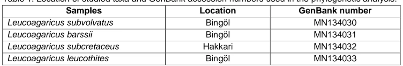
Table 1. Location of studied taxa and GenBank accession numbers used in the phylogenetic analysis.
Samples Location GenBank number
Leucoagaricus subvolvatus Bingöl MN134030
Leucoagaricus barssii Bingöl MN134031
Leucoagaricus subcretaceus Hakkari MN134032
Leucoagaricus leucothites Bingöl MN134033
DNA extraction, PCR amplification and Sequencing
Total DNA was extracted from dried basidiomata using the CTAB method with minor modifications (Doyle and Doyle, 1987). The purity and quantity of extracted DNA were determined by using NanoDrop2000c UV–Vis Spectrophotometer (Thermo Scientific) and 0.8% agarose gel electrophoresis. DNA amplification was performed in a 25 µl volume mixture containing genomic DNA (10 ng/µl), 10X PCR Buffer, MgCl2 (25 mM), dNTP
mixture (10 mM), selected primer pair (10 µM), Taq polymerase (5u/µl) and sterile water. To amplify ITS (ITS1-5.8S-ITS2) region, primer pairs N-nc18S10 5'AGGAGAAGTCGTAACAAG3'/C26A
5'GTTTCTTTTCCTCCGCT3' (Wen and Zimmer, 1996) were used. PCR products were run in a 1.0 % agarose gel and visualized by staining with Gelred dye. Positive reactions were sequenced with forward and reverse PCR primers using ABI 3730XL automated sequencer (Applied Biosystems, Foster City, CA, USA).
Sequence alignment and phylogenetic analysis Forward and reverse sequences were assembled and edited using Alibee Multiple Alignment 3.0 software from the GeneBee website (http://www.genebee.msu.su/genebee.html). Ambiguous
sites were checked manually and corrected by comparing the strands. Sequences of studied samples generated from the present study and additional sequences retrieved from NCBI were analyzed together to see phylogenetic relationships among Leucoagaricus species in the phylogenetic tree. The sequences downloaded from NCBI were selected considering results of BLAST searches. Cystolepiota seminuda (Genbank no: AY176350; Muñoz et al., 2014) was chosen as outgroup taxa. All sequences were aligned with the aid of the program ClustalW (Thompson et al., 1994) and adjusted manually where it was necessary.
Prior to construction of phylogenetic tree, total nucleotide length (bp) and variable sites were calculated using Molecular Evolutionary Genetics Analysis software (MEGA 6.0; Tamura et al., 2013). Phylogenetic tree of ITS region was constructed using two different methods; Maximum Likelihood (ML) and Maximum Parsimony (MP). The sequence data was analyzed by using the Maximum Likelihood (ML) method based on the Tamura-Nei model (Tamura and Tamura-Nei, 1993). To test branch support, bootstrap analysis was used with 1000 replicates (Felsenstein, 1985). In the ML method, initial tree(s) for the heuristic search were obtained automatically by applying Neighbor-Joining and BioNJ algorithms to a matrix of pairwise distances estimated using the
145
Maximum Composite Likelihood (MCL) approach, and then the topology with superior log likelihood value was selected. The Tree-Bisection-Reconnection (TBR) search method was employed with 100 random addition replications to construct the MP trees and the consensus tree inferred from 10 most parsimonious trees was used. All positions containing gaps and missing data were eliminated.Results Taxonomy
Leucoagaricus subvolvatus (Malençon & Bertault) Bon (Figure 1-2)
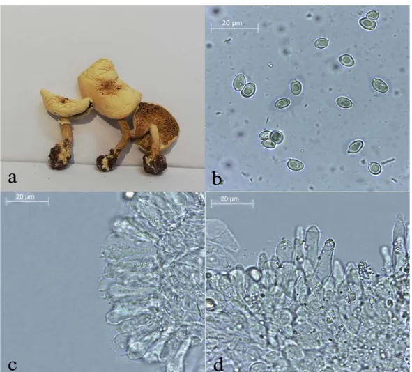
Pileus 50-60 mm, conic when young then flattened with slightly umbo, pale white-cream, surface smooth, silky, robust, radial fibrillose. Lamellae crowded, pale white when young, creamy when mature. Stipe 40-80 × 5-10 mm, whitish grey, silky, cyclindric, expanded towards the base, has universal veil remnants, bulbous base, annulus membranous, persistent, not movable, white. Spores 6-8(9) × 4-5.5 μm, hyaline, smooth, amydaloid. Basidia 18-30 × 6-10 μm, clavate to capitate, with 4 spores. Cheilocystidia 25-52 × 5.5-12 µm, irregular, lageniform to cyclindrical, clavate, usually has crystal shaped at apex. Pleurocystidia absent. Pleipellis cutis made up of 3-15 µm wide, septate, hyaline in KOH, thin-walled hyphae, clamp absent.
Ecology
Under Quercus sp., Tarlabaşı village, Bingöl province, 37° 21'409"K - 44° 30'002"D, 1481 m, 28.10.2018, Acar 1079.
Molecular phylogeny
Sequences of Leucoagaricus generated from the current study and additional sequences retrieved from GenBank were combined and analyzed together to identify our samples correctly and see phylogenetic relationships among species in the phylogenetic tree. The amplified
DNA fragment of the region was approximately 700 bp length encompassing complete ITS1, 5.8S and ITS2 subregions. ITS data matrix comprised a total of 39 sequences including 35 from NCBI. The aligned data included a total of 717 positions, of which 379 were conserved, and 326 were variable (216 variable sites in ITS1 and 110 in ITS2 subregion) nucleotides. Leucoagaricus leucothites (JQ683081) was used as reference sequence.
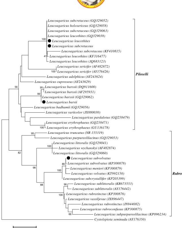
The Maximum Likelihood analysis resulted in similar phylogenetic topologies with Maximum Parsimony, so only one tree (ML) was given to indicate phylogenetic relationships and taxonomic position of studied species. In the ML phylogenetic tree, two Leucoagaricus sections, L. sect. Rubrotincti Singer and L. sect. Piloselli (Kühner ex) Singer, separated from each other and caused two main clades (Figure 3). The taxa that clustered together in a section carried morphological similarities.
L. sect. Rubrotincti is characterized by a reddish-brown pileus, ellipsoid spores, and a negative ammonia reaction (Singer 1986; Vellinga 2001). Section Piloselli is distinguished by basidiomes usually staining orange-red when bruised and turning green with ammonia and spores without germ pore (Singer 1986; Vellinga 2010).
Leucoagaricus subvolvatus grouped with its representative (KP300878) and several retrieved samples in section Rubrotincti with a bootstrap value of 100% (Figure 3). Leucoagaricus barssii, L. leucothites and L. subcretaceus placed in section Piloselli. Leucoagaricus leucothites clustered close to L. subcretaceus. Although these two species are molecularly similar, they can be distinguished from each other based on macroscopic and microscopic characters (Table 2). Leucoagaricus barssii grouped with three representatives from NCBI with 99% bootstrap.
146
Figure 1. Macroscopic and microscopic characters of Leucoagaricus subvolvatus. a. basidiocarps b. spores c. basidia d. cheilocystidia
147
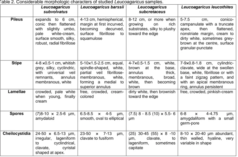
Table 2. Considerablemorphologic characters of studied Leucoagaricus samples. Leucoagaricus
subvolvatus
Leucoagaricus barssii Leucoagaricus subcretaceus
Leucoagaricus leucothites
Pileus expands to 6 cm,
conic then flattened with slightly umbo, pale white-cream, surface smooth, silky, robust, radial fibrillose
4-13 cm, hemispherical, margin at first incurved, becoming decurved, surface fibrillose to squamulose
8-12 cm, or more when growing on rich substrates, silky to plushy toward the edge
5-7.5 cm, conico-campanulate with a truncate apex, then flattened, nonstriate margin, cream to dirty white, sometimes grey-brown at the centre, surface granular-punctate
Stipe 4-8 ×0.5-1 cm, whitish
grey, silky, cyclindric, with universal veil remnants, annulus membranous, white
5-10×1.5-2.5 cm, equal, spindle-shaped, white, partial veil fibrillose-membranous, white, forming a medial to superior annulus
4-7×0.5-1.5 cm, white, brown at the base,
annulus thick, membranous, broad, white, then becoming brown
7-9×0.8-1.8 cm, cylindric-clavate, wide at the swollen base, white, fibrillose or with a faint zigzag pattern, and with an apical membranous ring, annulus persistent
Lamellae crowded, pale white
when young, finally cream
free, crowded, cream-colored
dirty white, then brownish toward the edge
free, crowded, pinkish-cream
Spores (7)8-10 × 2.5-6 μm,
amydaloid
6.5-8.5 × 4-5 µm, smooth, oval to elliptical
(7.5) 8 - 8.5 (10) × 5.5- 6 µm
6-8 × 4-4.75 µm,
amygdaliform with a small germ-pore Cheilocystidia 24-50 × 6.5-13 µm, irregular, lageniform to cyclindrical, clavate, cyristal shaped at apex. 23-50 × 7-13 µm, clavate to fusiform (25) 30-45 (55) × 8 -10 µm, clavate, to lageniform, sometimes capitate 8-10 × 20-40 µm abundant, thin walled, hyaline, very variable in shape
148
Figure 3. Phylogenetic tree of Leucoagaricus species based on ML analysis of the ITS region. Black circle indicates studied specimen. Cystolepiota seminuda was used as outgroup. Bootstrap analysis of ML was based on 1000 replicates
and values higher than 40% were indicated on branches.
Discussion
Section Rubrotincti is characterized a moderately fleshy basidiomata that does not change color when damaged, a red brown, pinkish ochre, olive, or orange pileus with radially arranged hyphae, ellipsoid spores, and a negative ammonia reaction (Singer 1986; Vellinga 2001). Section Piloselli is traditionally distinguished by
basidiomes usually staining orange-red when bruised and turning green with ammonia and spores without germ pore (Singer 1986; Vellinga 2010). These characters were also observed for studied samples and used to identify correctly in section and species levels.
The ITS phylogeny showed that Leucoagaricus subvolvatus is closely related to L. menieri (Sacc.) Singer, Leucoagaricus subcretaceus (GQ329052) Leucoagaricus holosericeus (GQ329058) Leucoagaricus subcretaceus (GQ329063) Leucoagaricus leucothites (GQ329038) Leucoagaricus leucothites Leucoagaricus subcretaceus Leucoagaricus subcretaceus (KF410815) Leucoagaricus leucothites (KF316477) Leucoagaricus leucothites (JQ683123) Leucoagaricus sericifer (AF482872) Leucoagaricus sericifer (AY176426) Leucoagaricus adelphicus (AY243624) Leucoagaricus cupresseus (AY243629)
Leucoagaricus barssii (DQ911600) Leucoagaricus barssii (AF295931) Leucoagaricus barssii (GQ329062) Leucoagaricus barsii Leucoagaricus badhamii (GQ329056) Leucoagaricus variicolor (JX880030) Leucoagaricus pardalotus (GQ258479) Leucoagaricus erythrophaeus (GQ258471)
Leucoagaricus erythrophaeus (GU136178)
Piloselli
Leucoagaricus truncatus (NR 155319)
Leucoagaricus purpureolilacinus (GQ329053) Leucoagaricus littoralis (GQ329041)
Leucoagaricus wychanskyi (AF482874) Leucoagaricus littoralis (GQ329060) Leucoagaricus subvolvatus Leucoagaricus subvolvatus (KP300878) Leucoagaricus menieri (KP300879) Leucoagaricus volvatus (KT992150) Leucoagaricus subcrystallifer (KP205399) Leucoagaricus sublittoralis (KR673553) Leucoagaricus sublittoralis (AY176442) Leucoagaricus rubrotinctus (KP300876) Leucoagaricus vassiljevae (JX896447) Leucoagaricus rubrotinctus (JN944082) Leucoagaricus rubroconfusus (KP300875) Leucoagaricus subpurpureolilacinus (KP096234) Rubrotincti
Cystolepiota seminuda (AY176350)
100 86 100 96 100 100 66 100 88 95 98 95 99 82 56 41 100 0.05
149
L. volvatus Bon & A. Caball. and L. subcrystallifer Ge & Yang. Morphologically these species have white basiomata, similar basidiospores and cheilocystidia with crystals at the apex. However, L. menieri has broad, fragile, shining basidiomata and smaller basidiospores (6–7 × 4.7–5 µm; Candusso and Lanzoni 1990; Bas et al., 1999). Leucoagaricus subcrystallifer has a greenish gray pileus with a dark gray umbo, amygdaliform basidiospores, narrowly fusiform cheilocystidia with crystals at the apex, and filamentous hyphae (Ge et al. 2015). Leucoagaricus subvolvatus and L. volvatus are two white volvate species. L. subvolvatus has fleshy basidiomata with a white to cream pileus, roundish stipe base that is somewhat volvate and narrowly lageniform to cyclindrical cheilocystidia which bear cyristal shaped at apex (Candusso and Lanzoni 1990; Bas et al., 1999). Leucoagaricus volvatus has some olivaceous tinges in the pileus, but also has crystals on the cystidia and a slightly gelatinized pileus. The current name of L. subvolvatus is same name in Mycobank database but Lepiota subvolvata in the Index Fungorum.Leucoagaricus leucothites, L. subcretaceus and L. barssii clustered in section Piloselli. Leucoagaricus leucothites is a widespread species and grows grasslands, lawns, meadows and road verges. Leucoagaricus subcretaceus and L. leucothites are
similar but L. subcretaceus is larger and more coloured (Bon, 1981; Reid and Eicker, 1993). In the constructed tree, these three species were located in the Piloselli clade. Leucoagaricus barssii is one of the few species within the genus to possess pleurocystidia. It has a radially fibrillose and white to greyish pileus (Desjardin et al., 2015; Vellinga 2000; 2001). The ellipsoid spores do not have germ pore, and pleurocystidia are observed close to the edge of lamella (Vellinga 2000; 2001).
Identification of Leucoagaricus samples is difficult because of close relationships with some Lepioid genera such as Leucocoprinus. Therefore, molecular data is needed to support the diagnosis that is completed based on only morphological characters. In this study, four Leucoagaricus species were identified by using both morphological and molecular data and Leucoagaricus subvolvatus was determined as a new record. The number of Leucoagaricus species in Turkey was increased from 10 to 11 with the results of the current study.
Acknowledgements
This study was financially supported by Van Yuzuncu Yil University (Scientific Research Project Foundation, FHD-2018-6858), Van, Turkey.
References
Bas, C., Kuyper, Th. W., Noordeloos, M. E. & Vellinga, E.C. (1999). Flora Agaricina Neerlandica. 4th ed. CRC Press. Rotterdam, Netherlands.
Breitenbach, J., Kränzlin, F. (1991). Fungi of Switzerland. Verlag Mykologia. 3rd ed. Lucerne, Switzerland.
Bon, M. (1981). Clé monographique des Lépiotes d'Europe (Agaricaceae, Tribus Lepioteae et Leucocoprineae),Documents Mycologiques, 11:43.
Candusso M, Lanzoni G. (1990). Fungi Europaei 4. Lepiota s.l. Saronno, Italy: Giovanna Biella. 743 p.
Desjardin, D.E., Wood, M.G. & Stevens, F.A. (2015). California Mushrooms: The Comprehensive Identification Guide. Timber Press: Portland, OR. 560 p.
Doyle, J.J., Doyle, J.L. (1987). A rapid DNA isolation procedure for small quantities of fresh leaf tissue. Phytochemical Bulletin, 19,11-15.
Felsenstein, J. (1985). Confidence limits on phylogenies: An approach using the bootstrap. Evolution, 39,783-791.
Ge, Z.W., Yang, Z.L., Qasim, T., Nawaz, R., Khalid A.N., Vellinga, E.C. (2015). Four new species in Leucoagaricus (Agaricaceae, Basidiomycota) from Asia. Mycologia, 107:5, 1033-1044.
Johnson J. (1999). Phylogenetic relationships within Lepiota sensu lato based on morphological and molecular data. Mycologia, 91: 443–458.
Johnson J., Vilgalys R. (1998). Phylogenetic systematics of Lepiota sensu lato based on nuclear large subunit rDNA evidence. Mycologia, 90: 971–979.
Hussain, S., Jabeen, S., Khalidd, A.N., Ahmade, H., Afshan, N., Sherg, H., Pfister, D.H. (2018). Underexplored regions of Pakistan yield five new species of Leucoagaricus. Mycologia, 110: 2, 387–400.
Kumar, T. K. A., Manimohan, P. (2009). The genera Leucoagaricus and Leucocoprinus (Agaricales, Basidiomycota) in Kerala State, India. Mycotaxon,108: 385–428.
Muñoz, G., Caballero, A., Contu, M., Ercole, E., Vizzini, A. (2014). Leucoagaricus croceobasis (Agaricales, Agaricaceae), a new species of section Piloselli from Spain. Mycol Progress, 13:649–655.
Ortiz, A., Franco-Molano, A.E., JR, M.B. (2008). A new species of Leucoagaricus (Agaricaceae) from Colombia. Mycotaxon, 106: 371-378.
150
Reid, D.A., Eicker, A. (1993). South African fungi. 2. Some species of Leucoagaricus and Leucocoprinus, South African Journal of Botany, 59(1): 85 – 97.
Sesli, E., Denchev, C.M. (2014). Checklists of the myxomycetes, larger ascomycetes, and larger basidiomycetes in Turkey. 6th edn. Mycotaxon Checklists Online. (http://www.mycotaxon.com/resources/checklists/sesli-v106-checklist.pdf): 1-136.
Singer, R. (1986). The Agaricales in modern taxonomy, 4th edn. Koeltz Scientific Books, Koenigstein.
Tamura, K., Nei, M. (1993). Estimation of the number of nucleotide substitutions in the control region of mitochondrial DNA in humans and chimpanzees, Molecular Biology and Evolution, 10: 512-526.
Tamura, K., Stecher, G., Peterson, D., Filipski, A., M., Kumar, S. (2013). MEGA 6: Molecular Evolutionary Genetics Analysis Version 6.0., Molecular Biology Evolution, 30(12), 2725–2729.
Thompson, J.D., Higgins, D.G., Gibson, T.J., (1994). Clustal W improving the sensitivity of progressive multiple sequence alignment through sequence weighting, position-specific gap penalties and weight matrix choice. Nucleic Acids Research, 22, 4673–4680.
Vellinga, E.C. (2000). Notes on Lepiota and Leucoagaricus. Type studies on Lepiota magnispora, Lepiota barssii and Leucogaricus americanus. Mycotaxon, 76:429–438.
Vellinga, E.C. (2001). Noordeloos, M.E., Kuyper, Th.W. & Vellinga, E.C. (eds). Flora agaricina neerlandica 5: 85– 108. A.A. Balkema Publishers Lisse/Abingdon/Exton (PA)/Tokyo. 169 pp.
Vellinga, E.C., de Kok, R.P.J. & Bruns, T.D. (2003) Phylogeny and taxonomy of Macrolepiota (Agaricaceae). Mycologia, 95 (3): 442–456.
Vellinga, E.C. (2004). Ecology and distribution of lepiotaceous Fungi (Agaricaceae), Nova Hedwigia, 78: 273–299. Vellinga E.C., Davis R.M. (2006). Lepiotaceous fungi in California, U.S.A. –1. Leucoagaricus amanitoides sp. nov.
Mycotaxon, 98: 197–204.
Vellinga, E.C.; Contu, M.; Vizzini, A. (2010). Leucoagaricus decipiens and La. erythrophaeus, a new species pair in sect. Piloselli. Mycologia, 102(2):447-454.
Wen, J., Zimmer, E.A. (1996). Phylogeny and Biogeography of Panax L. (the Ginseng Genus, Araliaceae): Inferences from ITS Sequences of Nuclear Ribosomal DNA. Molecular Phylogenetics and Evolution, 6, 167–177.