SYNTHESIS of Ag-DOPED ZnO NANOFIBERS USING ELECTROSPINNING METHOD and THEIR PHOTOCATALYTIC ACTIVITIES
1,2Ozlem ALTINTAS YILDIRIM
1Selçuk University, Faculty of Engineering, Department of Metallurgical and Materials Engineering, Konya,
Turkey
2Konya Technical University, Faculty of Engineering and Natural Sciences, Department of Metallurgical and
Materials Engineering, Konya, Turkey
(Received/Geliş: 08.02.2018; Accepted in Revised Form/Kabul: 07.03.2018)
ABSTRACT: Silver (Ag) doped zinc oxide (ZnO) nanofibers with 1 at.% and 3 at.% Ag content were
prepared using the electrospinning technique and their structural, morphological and photocatalytic properties were investigated. Pure ZnO nanofibers were also prepared with the same procedure for structure and property related comparison purposes. The photocatalytic activity of the Ag doped ZnO nanofibers were determined as a function of Ag content by exploring the degradation behavior of methylene blue under UV light irradiation. It was found that photocatalytic ability of fibers was improved with Ag addition and higher Ag incorporation resulted higher methylene blue degradation rate. For pure ZnO fibers, the degraded amount of dye was 52% of its initial amount after 270 min of UV irradiation time. For the same irridation time, 60% and 67% decomposition ratios of the dye molecules were achieved with the fibers containing 1 at.% Ag and 3 at.% Ag, respectively. The origin of the improvement of photocatalytic activity in Ag doped ZnO nanofibers was attributed to the substitutional incorporation of Ag ions into Zn sites within the ZnO crystal. The substitutional incorporation has been proved with the positional shift of the XRD diffraction lines.
Key Words: Ag doping, Electrospinning method, Photocatalytic property, ZnO nanofibers.
Elektro Eğirme Yöntemi Kullanılarak Ag Aşılanmış ZnO Nano Fiberlerin Sentezi ve Foto Katalitik Aktivitelerinin İncelenmesi
ÖZ: %1 ve %3 gümüş (Ag) içeren Ag aşılanmış çinko oksit (ZnO) nano fiberler elektro eğirme yöntemi
kullanılarak hazırlanmış ve yapısal, şekilsel ve foto katalitik özellikleri incelenmiştir. Saf ZnO nano fiberler de yapı ve özelliklerin karşılaştırılması amacı ile aynı yöntemle sentezlenmişlerdir, Ag aşılanmış ZnO nano fiberlerin foto katalitik özellikleri UV ışığı altında metilen mavisini bozma eğiliminin Ag miktarına bağlı olarak değişiminin bir fonksiyonu olarak belirlenmiştir. Ag aşılama ile fiberlerin foto katalitik aktivitelerinin geliştiği ve yüksek miktarda Ag miktarının eklenmesi ile metilen mavisinin bozunum oranının daha iyi olduğu bulunmuştur. Saf ZnO fiberler ile 270 dakikalık UV ışımasından sonra boyanın bozunum miktarının %52 oranında olduğu tespit edilmiştir. Aynı ışıma suresinde %1 Ag içeren fiberlerde boya moleküllerinin bozunum oranı %60 iken, %3 Ag içeren fiberlerde bu oran %77’dir. Ag aşılama ile ZnO fiberlerin foto katalitik aktivitelerinde gözlemlenen iyileşme ZnO kristal yapısında Zn bölgeleri içine Ag iyonlarının yer alan atom olarak geçmesi ile açıklanabilir. Yer alan atom pozisyonunda yerleşme XRD piklerinin pozisyonlarında meydana gelen kayma ile doğrulanmıştır.
INTRODUCTION
During the last few decades, water and air pollution have created a driving force for the fundamental and applied research in the waste stabilization and environmental remediation areas (Sharma and Lewis 1994). The control of the optical properties of semiconductor materials have increased the importance in the technological applications of the environmental remediation. In the literature, studies related with optical properties of II-VI type semiconductor nanomaterials are mainly focused on the CdSe and CdS nanoparticles containing hazardous elements to the environment (Costi et al., 2008; Yan et al., 2009). However, as an environmentally friendly semiconductor, zinc oxide (ZnO) nanostructures with wide band gap (3.37 eV) and large exciton binding energy (60 meV) show unique optical properties, providing that ZnO nanoparticles are in the foreground material in the technological applications in variety fields such as gas sensors, field emitters, solar cells and photocatalytic applications (Lee et al., 2016).
One of the important application area of ZnO nanostructures is the photocatalytic applications. In this application field, the aim is to destroy unwanted organisms in the air, water and various surfaces with a chemical reaction using a common and inexpensive energy source such as light energy (Serpone and Pelizzetti 1989). The working mechanism of photocatalytic devices is based on the principle of accelerating the photo reaction of a semiconductor material with wide band gap under solar energy. For the photocatalytic reactions, although solar energy is harmless and the most common source of the energy, UV radiation is found to be more effective to degrate dye molecules. The photocatalytic activities of semiconductor materials are classified according to their tendency to form electron-hole pairs under the photoluminescence. When semiconductor nanoparticles are exposed to UV radiation with more energy than their band-energy, they will form hole in the valence band and electrons in the conduction band (O'Neil et al., 1990). The photocatalytic reaction will occur when these electron-hole pairs reach the surface of the material. As a catalyst material, ZnO nanostructures have been observed to be highly effective in visible and near-infrared regions because they require high-energy UV radiation to act as catalysts. In addition, rapid recombination of the resulting electron-hole pairs is another limitting for ZnO. Various methods have been tried to solve these problems, such as semiconductor junction (Chen et al. 2008), transition metal doping (Litter, 1999) and noble metal doping (Li et al. 2011). Among these techniques, it has been observed that the photocatalytic activity can be effectively improved with noble metal addition. It has been observed that the addition of palladium, gold and silver as an advanced metal addition enhances photocatalytic activity (Liqiang et al., 2004; Wu and Tseng 2006; Seery et al., 2007).
The development of the photocatalytic activities of ZnO nanoparticles by Ag doping has been particularly attractive (Yıldırım et al., 2013; Patil et al., 2016). It was reported that the surface modifications in terms of size and morphology of the Ag doped ZnO particles significantly affect the photocatalytic properties of the ZnO nanoparticles (Yıldırım et al., 2013). L. Wang reported that morphologically smooth ZnO nanoparticles exhibit better photocatalytic properties than according to a non-homogeneous morphology (Wang, L. et al., 2008). In addition, Y. Wang observed that the nano flower structure has better photocatalytic activity than rod structure (Wang, Y. et al., 2008). Because of the different working requirements in its applications, the synthesis of Ag doped ZnO nanostructures with well-controlled size and morphology have been also attracted a great attention (Wang et al., 2004). These special morphologies of Ag doped ZnO nanostructures such as nanorods (Hsu and Chang, 2014), nanofilms (Wang et al., 2006), nanoellipsoids (Kumar et al., 2015) and nanoparticles (Yıldırım et al., 2013) have been synthesized using methods ranging from gas phase processes to solution routes.
Although ZnO nanoparticles in a few nanometers size scale have been reported a superior photocatalytic activity due to their large surface to volume ratios, in a practical photocatalytic applications, separation of these particles from the solution could be a problem. Furthermore, agglomeration of the particles in the catalyst solution is another common problem causing to reduce photocatalytic activity. For these aspects, nanofiber catalyst can be proper candidate because of their
unique properties originated from well-defined geometry, ordered structure, flexibility of form, three-dimensional open structure, large specific surface area, easy scale-up, and easily separation from the solution. The electrospining method is favorable to synthesize Ag doped ZnO fibers due to higher efficiency. In this study, it was aimed to synthesize Ag doped ZnO fibers via electrospinning technique and to determine effect of the Ag addition on the photocatalytic property of ZnO fibers. For this purpose, ZnO fibers were synthesized with different Ag content and in the studied dopant content range, the optimum amount of Ag required for the best photocatalytic property was determined.
MATERIALS AND METHOD
Zinc acetate dehydrate (C4H6O4Zn2H2O, %99.5, Fluka), polyvinyl alcohol (PVA, (C2H4O)n, MA
~55000, Aldrich), silver acetate (C2H3AgO2, %99.99, Aldrich), ethonol (C2H6O, %99, Merck) were used as
raw materials for the production of ZnO and Ag doped ZnO fibers. All materials were of analytical grade and used without further purification.
Production of Ag-Doped ZnO Nanofibers
Synthesis of ZnO nanofibers was carried out by dissolving of 1.54 g PVA in 10 ml distilled water and mixing of the solution at 70 ° C for 4 hours using a magnetic stirrer. 1 g of zinc acetate dihydrate was added to PVA solution and mixing was continued for 1 hour to obtain homogeneous solution. The zinc acetate dehydrate solution was then added drop by drop to the PVA solution. At the end of the dripping process, final solution was stirred further 2 hours to achieve complete homogeneity. Then, the solution was inserted into a disposable sterile syringe having a needle pin inner diameter of 0.8 mm and connected to an automatic syringe pump to spray at a 0.3 mL per hour rate. Aluminum foil was used to collect the fibers. During production of the fibers, the distance between the syringe tip and foil was set to 15 cm and applied voltage was set to 15 kV in the electrospining unit.
In order to fabricate Ag doped ZnO nanofibers, silver acetate was added in the PVA-ZnO stock solution. For this purpose, silver salt was added to the stock solution at amounts equivalent to 1 and 3 at.% Ag ion. Subsequently, they were mixed at room temperature with a magnetic stirrer for 1.5 hours in order to homogeneously distribute the ions in the solution. The silver containing solution was added to the main solution and stirred at room temperature for approximately 1 day. After mixing, the stock solution containing Ag was taken into a single-use sterile syringe and spraying was performed using the same parameters as the production of ZnO fibers with an automatic syringe pump.
After the end of the electrospinning process of the all ZnO and Ag doped ZnO fibers, aluminum foils were put into a vacuum dryer at 70 °C for 8 hours. In order to remove PVA and organic pollution from the ZnO structure and to increase the crystallinity of the ZnO fiber structure, the fibers were subjected to a heat treatment at 400 °C for 5 hours.
Characterization
The phase distributions of the nanofibers were determined by X-ray diffraction (XRD). The size and morphology of the nanofibers were investigated by scanning electron microscopy (SEM). For the measurement of the photocatalytic activities of ZnO fibers, the degradation rate of methylene blue (MB) dye under UV irradiation were determined by UV-Vis spectrometry. Photocatalytic measurements were firstly carried out in a dark region. Then, 100 W UV lamp was used to observe degradation of the organic dye under UV light. To investigate the photocatalytic ability, 50 mg of MB was dissolved in 50 mL of deionized water to prepare a stock solution. In order to measure the photocatalytic activity of the ZnO nanofibers, 50 mg fiber was added to the dye solution and the solution was then subjected to continuous stirring in a dark box. In order to measure the degree of degradation in the dark media, the absorption spectrum between 300-600 nm was examined by UV-Vis spectroscopy. After the exposure
with the 100 W of UV light, measurements were periodically carried out each 0.5 h by examining degradation of the 4 mL of the solution. Photocatalytic activities of the 1 and 3. at.% Ag doped ZnO fibers were also investigated with the same procedure.
RESULTS and DISCUSSIONS
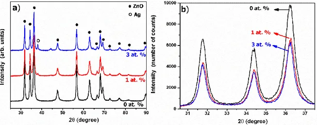
Figure 1 (a) shows x-ray diffractograms of ZnO and Ag-doped ZnO fibers synthesized via electrospinning technique. According to the X-ray diffractograms, fibers are highly crystalline ZnO with the hexagonal wurtzite structure (JCPDS card number: 36-1451). No other crystalline phases were detected from the ZnO fibers. Peak intensities are in agreement with standard peak intensities in the JCPDS card data. For 1 and 3 at.% Ag addition, low-intensity peaks located at 2θ = 38.37° and 46.40° were observed. These diffraction lines corresponds to the metallic silvers (Subhan et al. 2014). The intensity of these metallic Ag peaks increases with an increase in the dopant silver content.
Figure 1. a) XRD diffractogram and b) enlarged XRD diffractogram between 2θ = 30.5-37.5° of ZnO,
1% and 3% Ag-doped ZnO nanofibers
The peak intensities and positions of ZnO diffractions were examined to investigate the effect of the Ag addition on the crystal structure of ZnO fibers. Figure 1 (b) shows the enlarged XRD diagrams between 2θ=31.5-37.5° region. It was observed that 1at.% Ag addition considerably decrease the peak intensity of ZnO but in 3at.% Ag doped ZnO fibers, Ag ions have less effect on the peak intensity. According to peak intensities, Ag addition diminished the crystallinity of ZnO fibers. The crystal structure of ZnO is composed of a zinc rich catalytic active top surface and an oxygen rich catalytic inert bottom surface. These two surfaces are also different in terms of energy. This difference ensures that ZnO has a polar structure. With the effect of this polarity, the particles in the solution medium are willing to enlarge low energy surface areas. The chemical structures present in the synthesis medium allow the particles to grow in the desired direction as a result of interaction with the ZnO core structure (Kolodziejczak-Radzimska and Jesionowski 2014). The formation of a second phase with Ag addition prevented the growth of ZnO crystals through the preferential growth direction. Due to this reason, XRD peak intensity and crystallite of ZnO decrease with Ag addition.
Figure 1 (b) also shows a slight shift in the peak positions of Ag-doped ZnO fibers. The 2θ values as a function of the Ag content are listed in Table 1. For pure ZnO fibers, the highest intense peak of hexagonal wurtzite structure appeared at 2θ = 36.30°, however, the positions change to 2θ = 36.28° and 2θ = 36.25° for 1% and 3% Ag doped ZnO fibers, respectively. This peak shift caused by Ag addition shows some of the Ag ions are substitutionally incorporated into ZnO crystal structure (Yıldırım et al. 2013). Substitutional incorporation of Ag ions can be also proved from the lattice parameters of ZnO. The c-axis lattice constant calculated from the (0002) plane are also listed in the Table I. As observed in
Table I, lattice parameters indicate an enlargement of the lattice from 5.175 to 5.194 Å as the Ag content increased from 0 to 3 at.% because of bigger ionic size of Ag than that of the Zn (Ag+:1.26 Å and Zn2+:
0.74 Å). According to peak shift in the XRD results and lattice parameter change, the Ag ions are substitutionally incorporated into the ZnO crystal by sitting in the Zn2+ sites.
Table 1. (002) peak position and lattice parameters for the undoped ZnO and Ag-doped ZnO fibers
with different Ag contents
Ag:Zn ratio (at.%) (002) Peak Position (degree of 2) Lattice Parameter (c, Å) 0 36.30 5.175 1 34.28 5.178 3 34.25 5.194
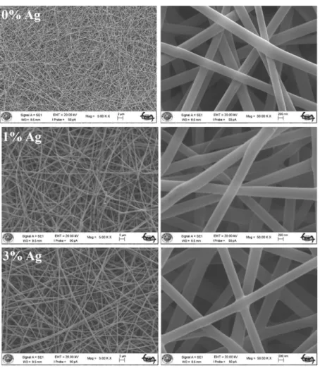
Figure 2 shows low and high magnification SEM micrographs of as-prepared ZnO and 1 at.% and 3 at.% Ag doped ZnO fibers.As can be seen from the images, fibers with a smooth surface are approximately 40 μm in length and 400 nm in diameter. The addition of Ag did not cause a significant change in the morphology of the as-prepared fibers.
3
Figure 2. Low and high magnification SEM images of as-prepared ZnO, 1 at.% and 3 at.% Ag-doped
ZnO fibers.
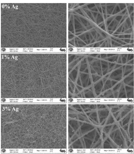
Figure 3 shows low and high magnification SEM images of ZnO, 1% and 3% Ag-doped ZnO nanofibers obtained after heat treatment at 400 °C for 5 hours. With the removal of the polymer by the
effect of heat treatment of the fibers, it can be seen that the diameters of the fibers reduce to nearly 80 nm. After the heat treatment of the fibers, it can be also seen that fibers are formed by the particles which are 30 nm in diameter.
Figure 3. Low and high magnification SEM images of ZnO, 1 at.% and 3 at.% Ag-doped ZnO
nanofibers heat treated at 400 °C for 5 hours.
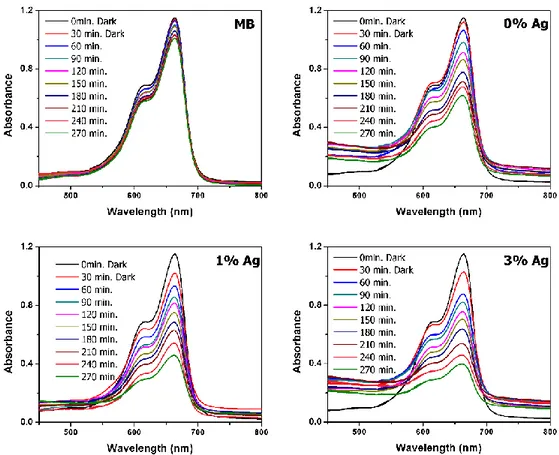
Photocatalytic activities of the nanofiber structures were investigated by the degradation profile of methylene blue under UV light. Figure 4 shows the degradation profile of all ZnO nanofibers. For the comparison purpose, degradation profile of the methylene blue solution without catalyst was also exposed to the UV-light under the same experimental conditions and showed a trace of change with respect to time. A diagram of the reaction of the methylene blue under the influence of light is shown in the Figure 4. It was observed that the methylene blue solution exposed to UV light for 4 hours underwent very little decomposition.
The maximum absorption band at 664 nm was used to degrade the methylene blue. As can be seen from the Figure 4, ZnO began to deteriorate from the 30 minutes and it was observed that the amount of deterioration increased with time due to the effect of UV light. The nanofibers added at 1 and 3 at.% Ag were found to have more efficient for methylene blue dye degradation than that of the bare ZnO fibers. The methylene blue decay according to Figure 4 increases over time in all fiber structures. This degradation in the methylene blue shows the effect of addition of Ag on the photocatalytic properties of ZnO (Chen et al., 2008).
Figure 4. Real-time UV–vis absorption spectra of the photodegradation of methylene blue solutions
containing the undoped and 1 at.% and 3 at.% Ag-doped ZnO nanofibers.
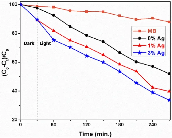
Figure 5 exhibits the UV-light degradation of the dye molecules. After the 270 minutes UV irradiation, approximately 70% decomposition was observed in dye molecules without catalyst addition. For the same irradiation time, ZnO fiber catalyst addition to methylene blue solution resulted in an exponentially decrease with respect to time and 48% decomposition was achieved. According to Figure 5, Ag addition increases the dye decomposition. 60% degradation rate was observed with 1% Ag doped ZnO nanofiber. Furthermore, with an increase in the Ag dopant amount to 3 at.%, degratation rate further increased to 67%. Therefore, it has been observed that Ag addition improves the photocatalytic activities of ZnO nanofibers. This increase can be attributed to the change in the electronic structure of the ZnO by the silver ions sitting into Zn site of the crystal. The photocatalytic activity of semiconductor materials can usually be improved by providing the separation of excited electron–hole pairs (Liu et al. 2011). By employing substitutional incorporation of Ag ions into ZnO crystal structure, the photoexcited electron–hole pairs could be efficiency separated by an extra inner field. Based on the above experimental results, the improved photocatalytic efficiency could be due to substitutionally sitting of Ag ions into ZnO crystal.
Figure 5. Time-dependent degradation profiles of ZnO and 1 at.% and 3 at.% Ag-doped ZnO
nanofibers
CONCLUSIONS
In this study, Ag added ZnO nanofibers with approximately 40 μm in length and 80 nm in diameter were fabricated by the electrospinning technique. The effects of the Ag addition on the structural, morphological and photocatalytic abilities of the ZnO nanofibers were investigated. According to XRD results, the addition of Ag to the ZnO crystal structure led to a shift in diffraction line position and a decrease in the peak intensities of the ZnO peaks. This shift in the peak positions indicates that the added Ag ions are substitutionally incorporated into the ZnO crystal structure. In addition, some of the added Ag ions has formed a separate phase as metallic Ag. The photocatalytic properties of the produced fibers were investigated by examining methylene blue dye degradation under UV light irradiation. It was found that the photocatalytic properties of ZnO nanofibers showing 48% degradation as a result of exposure to 270 minutes ofUV irradiation were improved with 1 and 3 at.% Ag addition to 60% and 67 % degradation rates, respectively. These results show that Ag addition to ZnO fiber structure gives an opportunity to use nanofibers in the large-scale utilization of photocatalysts using UV light to overcome organic and/or water pollution.
ACKNOWLEDGEMENT
This study was carried out within the scope of project number 16401052 supported by Selcuk University Scientific Research Projects (BAP). We thank Selçuk University BAP for their financial support.
REFERENCES
Chen, S., Zhao, W., Liu, W., Zhang, S., 2008, "Preparation, Characterization and Activity Evaluation of p–n Junction Photocatalyst p-ZnO/n-TiO 2", Applied Surface Science, Vol. 255, No. 5, pp. 2478-2484.
Costi, R., Saunders, A. E., Elmalem, E., Salant A., Banin, U., 2008, "Visible Light-induced Charge Retention and Photocatalysis with Hybrid CdSe− Au Nanodumbbells", Nano Letters, Vol. 8, No. 2, pp. 637-641.
Hsu, M.-H., Chang, C.-J., 2014, "Ag-doped ZnO Nanorods Coated Metal Wire Meshes as Hierarchical Photocatalysts with High Visible-Light Driven Photoactivity and Photostability", Journal of
Hazardous Materials, Vol. 278, No. 1, pp. 444-453.
Kolodziejczak-Radzimska, A., Jesionowski, T., 2014, "Zinc Oxide-From Synthesis to Application: A Review", Materials, Vol. 7, No. 4, pp. 2833-2881.
Kumar, R., Rana, D., Umar, A., Sharma, P., Chauhan, S., Chauhan, M. S., 2015, "Ag-doped ZnO Nanoellipsoids: Potential Scaffold for Photocatalytic and Sensing Applications", Talanta, Vol. 137, No. 1, pp. 204-213.
Lee, K., M., Lai, C. W., Ngai, K. S., Juan, J. C., 2016, "Recent Developments of Zinc Oxide based Photocatalyst in Water Treatment Technology: a Review", Water Research, Vol. 88, No. 1, pp. 428-448.
Li, P., Wei Z., Wu, T., Peng, Q., Li, Y., 2011, "Au−ZnO Hybrid Nanopyramids and Their Photocatalytic Properties", Journal of the American Chemical Society, Vol. 133, No. 15, pp. 5660-5663.
Liqiang, J., Baiqi, W., Baifu, X., Shudan, L., Keying, S., Weimin, C., Honggang, F., 2004, "Investigations on the Surface Modification of ZnO Nanoparticle Photocatalyst by Depositing Pd", Journal of
Solid State Chemistry, Vol. 177, No. 11, pp. 4221-4227.
Litter, M. I., 1999, "Heterogeneous Photocatalysis: Transition Metal Ions in Photocatalytic Systems",
Applied Catalysis B: Environmental, Vol. 23, No. 2, pp. 89-114.
Liu, B., Nakata, K., Zhao, X., Ochiai, T., Murakami, T., Fujishima, A., 2011, "Theoretical Kinetic Analysis of Heterogeneous Photocatalysis: the Effects of Surface Trapping and Bulk Recombination Through Defects", The Journal of Physical Chemistry C, Vol. 115, No. 32, pp. 16037-16042.
O'Neil, M., Marohn, J., McLendon, G., 1990, "Dynamics of Electron-Hole Pair Recombination in Semiconductor Clusters", The Journal of Physical Chemistry, Vol. 94, No. 10, pp. 4356-4363. Patil, S. S., Mali, M. G., Tamboli, M. S., Patil, D. R., Kulkarni, M. V., Yoon, H., Kim, H., Al-Deyab, S. S.,
Yoon, S. S., Kolekar, S. S., 2016, "Green Approach for Hierarchical Nanostructured Ag-ZnO and Their Photocatalytic Performance under Sunlight", Catalysis Today, Vol. 260, No. 1, pp. 126-134.
Seery, M. K., George, R., Floris, P., Pillai, S. C., 2007, "Silver Doped Titanium Dioxide Nanomaterials for Enhanced Visible Light Photocatalysis", Journal of Photochemistry and Photobiology A: Chemistry, Vol. 189, No. 2-3, pp. 258-263.
Serpone, N., Pelizzetti, E., 1989, Photocatalysis: fundamentals and applications
Sharma, H. D., Lewis, S. P., 1994, Waste Containment Systems, Waste Stabilization, and Landfills: Design and
Evaluation, John Wiley & Sons.
Subhan, M. A., Awal, M., Ahmed, T., Younus, M., 2014, "Photocatalytic and Antibacterial Activities of Ag/ZnO Nanocomposities Fabricated by Co-Precipitation Method", Acta Metallurgica Sinica
(English Letters), Vol. 27, No. 2, pp. 223-232.
Wang, L., Chang, L., Zhao, B., Yuan, Z., Shao, G., Zheng, W., 2008, "Systematic Investigation on Morphologies, Forming Mechanism, Photocatalytic and Photoluminescent Properties of ZnO Nanostructures Constructed in Ionic Liquids", Inorganic Chemistry, Vol. 47, No. 5, pp. 1443-1452.
Wang, R., Xin, J. H., Yang, Y., Liu, H., Xu, L., Hu, J., 2004, "The Characteristics and Photocatalytic Activities of Silver Doped ZnO Nanocrystallites", Applied Surface Science, Vol. 227, No. 1, pp. 312-317.
Wang, X., Song, C., Geng, K., Zeng, F., Pan, F., 2006, "Luminescence and Raman Scattering Properties of Ag-doped ZnO films", Journal of Physics D: Applied Physics, Vol. 39, No. 23, pp. 4992.
Wang, Y., Li, X., Wang, N., Quan, X., Chen, Y., 2008, "Controllable Synthesis of ZnO Nanoflowers and Their Morphology-Dependent Photocatalytic activities", Separation and Purification Technology, Vol. 62, No. 3, pp. 727-732.
Wu, J.-J., Tseng, C.-H., 2006, "Photocatalytic Properties of nc-Au/ZnO Nanorod Composites", Applied
Catalysis B: Environmental, Vol. 66, No. 1-2, pp. 51-57.
Yan, H., Yang, J., Ma, G., Wu, G., Zong, X., Lei, Z., Shi, J., Li, C., 2009, "Visible-light-driven Hydrogen Production with Extremely High Quantum Efficiency on Pt–PdS/CdS Photocatalyst", Journal of
Catalysis, Vol. 266, No. 2, pp. 165-168.
Yıldırım, O.A., Unalan, H. E., Durucan, C., 2013, "Highly Efficient Room Temperature Synthesis of Silver‐Doped Zinc Oxide (ZnO: Ag) Nanoparticles: Structural, Optical, and Photocatalytic Properties", Journal of the American Ceramic Society, Vol. 96, No. 3, pp. 766-773.