https://doi.org/10.1007/s11033-020-05557-6
ORIGINAL ARTICLE
p60‑katanin: a novel interacting partner for p53
Sirin Korulu1,2 · Aysegul Yildiz3
Received: 8 April 2020 / Accepted: 23 May 2020 / Published online: 28 May 2020 © Springer Nature B.V. 2020
Abstract
Katanin, one of the best-characterized microtubule (MT) severing proteins, is composed of two subunits: catalytic p60-kata-nin, and regulatory p80-katanin. p60-katanin triggers MT reorganization by severing them. MT reorganization is essential for both mitotic cells and post-mitotic neurons in numerous vital processes such as intracellular transport, mitosis, cellular differentiation and apoptosis. Due to the deleterious effect of continuous severing for cells, p60-katanin requires a strategic regulation. However, there are only a few known regulators of p60-katanin. p53 functions in similar cellular processes as katanin such as cell cycle, differentiation, and apoptosis depending on its interacting partners. Considering this similarity, in this study we investigated p53 as a potential regulatory candidate of p60-katanin, and examined their interaction. Co-immunoprecipitation analyses revealed that p60-katanin interacts with p53. We were able to locate a potential interaction site for the two proteins by deleting different candidate regions We showed for the first time that p53 and p60-katanin interact. This interaction appears to occur via p53’s DNA binding domain and p60-katanin’s C-terminal. This study will pave the way for future studies regarding the functional outcomes of this interaction which is vital for understanding the regulation of cellular events such as cell cycle, differentiation, and apoptosis in disease and in health.
Keywords p60-katanin · p53 · Protein–protein interaction · Microtubule related proteins
Introduction
Katanin is a member of the ATPase family, a microtu-bule (MT) severing enzyme [1] which is named after the sword of the Japanese Samurai, Katana. It consists of two subunits: p60-katanin encoded by the KATNA1 gene and p80-katanin encoded by the KATNB1 gene. p60-katanin is responsible for enzymatic activity, whereas p80-katanin is involved in the regulation and localization of p60-katanin
[2–4]. p60-katanin triggers MT reorganization by severing them. The reorganization of MTs is functional for mitotic cells especially in the cell division, while in post-mitotic cells such as neurons, the reorganization of MTs is vital for morphological and functional differentiation of neurons by the formation of axons and dendrites.
In proliferating cells, p60-katanin promotes mitosis by severing the MTs in the mitotic spindle poles and cre-ating a large number of minus MT ends, thereby ensur-ing gamma-tubulin accumulation in mitotic centrosomes which forms the nucleation center for MTs [5]. On the other hand, p60-katanin severs and disassembles MTs at spindle poles to shorten the metaphase spindles [6]. How-ever, in post-mitotic neurons, p60-katanin directs all its power to severe MTs in order to form neuronal extensions such as axons, a process called “neurite growth” which is essential for neuronal network formation [7, 8]. Although MTs are the main cytoskeletal component of the axons, they are too long to be transported into axonal extensions during neurite growth [9]. Thus, p60-katanin severs long MTs into shorter fragments to be easily transported by motor proteins to the axons [10]. Based on all this infor-mation, it is evident that p60-katanin is a critical protein
Electronic supplementary material The online version of this article (https ://doi.org/10.1007/s1103 3-020-05557 -6) contains supplementary material, which is available to authorized users. * Sirin Korulu
1 Department of Molecular Biology and Genetics,
Faculty of Science and Letters, Istanbul Arel University, 34537 Istanbul, Turkey
2 Institute of Natural and Health Sciences, Tallinn University,
10120 Tallinn, Estonia
3 Department of Molecular Biology and Genetics, Faculty
that plays a role in the most vital activities of both mitotic and post-mitotic cells.
Unless its severing activity is controlled, p60-katanin would continuously severe MTs which is undesirable for the cells. Thus, it is evident that katanin requires a strategic regulation within the cell. Despite the fact that MT severing mechanism of p60-katanin has been studied extensively for years, little is known about its regulation. There are almost no known regulators of katanin. There-fore, revealing the regulation partners of p60-katanin is of primary importance for understanding the functional regu-lation of this protein, the MT severing dynamics and also the reciprocal effects of these interactions. In this regard, analyzing the interactions of p60-katanin with its part-ners will provide important insights about the regulatory mechanism of essential cellular events such as cell cycle and cellular differentiation.
Similar to p60-katanin, tumor suppressor p53 plays an essential role in the pendulum between proliferation, apop-tosis and differentiation by binding to different proteins within the cell. p53, also known as a master guardian that controls proliferation in dividing cells, has been shown to be associated with differentiation by affecting neuronal process lengths in non-dividing neurons [11–14].
Furthermore, our previous study that was conducted with primary neurons clearly showed that activating cell cycle by triggering Protein Kinase C caused retraction of neuronal processes with a concomitant increase in p60-katanin levels. Based on this observation, p53 level was then examined and it was indicated that p53 protein level increased by 1.7-fold [15]. This concurrent increase of p53 and p60-katanin proteins suggested that p53 and p60-katanin could affect each other.
Considering the critical role of p60-katanin in MT regulation, especially in mitotic spindle formation for mitotic cells and in the formation of functional structures of neuronal morphology for postmitotic neurons, it is most likely to interact with p53, which has an incontrovertible role in the same processes. To date, p53 has been reported to function in the regulation of proteins responsible for neuronal differentiation such as Koronin1b, Rab13, GAP 43 [14] and it supports the idea that it may interact with another neuronal differentiation regulator, p60-katanin.
Regarding the fact that both p60-katanin and p53’s crit-ical roles in proliferation and differentiation, we aimed to answer the question: “Is it possible for them to inter-act in the cell?”. For this purpose, first, we determined p53–p60-katanin interaction, then identified the possible regions of this interaction. Results of the study showed for the first time that p53 and p60-katanin do interact. The interaction appears to occur via p53’s DNA binding domain and C-terminal of p60-katanin.
Materials and methods
Cell culture
Rat RFL-6 cells were a kind gift from Prof. Arzu Kara-bay (Istanbul Technical University) and were maintained in medium containing F12K (Lonza, Basel, Switzerland), 20% fetal bovine serum (heat inactivated, E.U. 10500064, Thermo Fisher, Gibco), Non-Essential Amino Acids (BE13-114E, Lonza) and 2 mM L-Glutamine (25030081, Thermo Fisher, Gibco). The cells were cultivated in a 5% CO2 incu-bator at 37 °C and plated at a density of 5 × 10⁵ cells/well in
a 6-well plate for Immunoprecipitation and Western blotting experiments.
Cell extraction
Total protein extraction was performed by using Mamma-lian Cell Extraction Kit (K269-500, BioVision, San Fran-cisco Bay, San FranFran-cisco, USA) and specified as Whole Cell Lysate (WCL) through the study. The protein concentration was measured using Bicinchoninic Acid protein assay kit (23235, Pierce, Waltham, MA, USA).
Deletion constructs
All deletion constructs were obtained by cloning differ-ent parts of both Wild Type (WT) p53 (AB082923) and WT p60-katanin (NM_007044) (Fig. 3). p60-katanin and p53-deletion constructs were prepared by sub-cloning dif-ferent parts of WT genes into pcDNA3.1/myc-His and 3XFLAG-CMV™-10 vectors respectively. Prepared con-structs were: Transcription Activation Domain (TAD-p53, p53-b), DNA Binding Domain (DBD-(TAD-p53, p53-c) and Oligomerization-Regulatory Domain (OD-RD-p53, p53-d) constructs for p53 (Fig. 3a); C-terminal containing construct (C-term-p60-katanin, p60-katanin-b) and del201-268 construct (del201-del201-268-p60-katanin, p60-katanin-c) for p60-katanin (Fig. 3b).
Transient transfection
Cells were seeded in 6 well-plates at a density of 5 × 10⁵ and
were cultured overnight in a humidified incubator (5% CO2, 37 °C). Following day, after confluence of the cells reached 70–90%, chemical transfection was performed by using lipo-some-based transfection reagent, Lipofectamine 3000 (L3000-001, Thermo Fisher Scientific, Waltham, MA, USA). In co-immunoprecipitation (Co-IP) Co-IP experiments, for total 2 μg of DNA, different combinations of 1 μg of p53 and 1 μg of p60-katanin deletion constructs were mixed with P3000
reagent in Opti-MEM Medium (31985062, Thermo Fisher, Gibco). These DNAs were then mixed with Lipofectamine 3000 reagent which was diluted in Opti-MEM Medium. The transfection mixture was incubated in room temperature for 15 min and applied onto the cells drop-by-drop. Cells were cultivated for 48 h in 37 °C, 5% CO2 incubator. Finally, total
protein extraction was performed by using BioVision Mam-malian Cell Extraction Kit. A certain amount of total protein was aliquoted as input and the rest of the lysate was used in further Co-IP experiments.
Co‑immunoprecipitation & Western blotting
WCL was mixed with primary mouse anti-p53 antibody (1:500, 2524, Cell Signaling Technology, Danvers, MA, USA) at 4 °C overnight. Next day, antibody–lysate mixture was mixed with Protein G magnetic beads in 4 °C rotator for 1 h. Unbound proteins were washed away for 3 times with PBS and bound proteins were eluted using 50 mM Glycine buffer (pH 2.8). For non-specific IgG and no antibody (beads only) reactions as the negative controls; the same amount of lysate was mixed with either 3 μg normal mouse IgG or the lysate was used without antibody, respectively. Positive con-trol for Co-IP was the total protein lysates aliquoted as input. Eluted proteins, IgG and input samples were separated by 4–12% Bis–Tris SDS-PAGE and transferred onto a nitrocel-lulose membrane. Possible interaction of immunoprecipitated constructs was detected by immunoblotting via rabbit anti-p60-katanin antibody (1:1000, HPA036207, Atlas Antibodies, Bromma, Sweden), mouse anti-p53 antibody (1:500, 2524, Cell Signaling Technology, Danvers, MA, USA). HRP-con-jugated antibodies (goat anti-mouse IgG-HRP: sc-2031; goat anti-rabbit IgG-HRP: sc-2004) were used as the secondary antibody (1:5000, Santa Cruz Biotech., Dallas, TX, USA). Visualization of the protein bands was performed using col-orimetric Amplified Opti-4CN Detection Kit (1708238, Bio-Rad, Hercules, CA, USA).
To reveal the domains that have a role in the interaction, WCL was obtained from the cells that were transfected with deletion constructs. Firstly, 4 μg of mouse monoclonal anti-Octa (FLAG) Antibody (sc-166355, Santa Cruz Biotech., Dallas, TX, USA) was mixed with 500 ng of lysate and incu-bated overnight at 4 °C. The Co-IP analysis was performed as explained previously in detail. Possible interaction of immu-noprecipitated constructs was detected by immunoblotting via rabbit monoclonal anti-His primary antibody (1:1000, 2365S, Cell Signaling Technology, Danvers, MA, USA).
Results
p60‑katanin interacts with p53
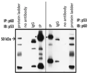
To examine the possible interaction between p60-katanin and p53, we performed co-immunoprecipitation for endog-enous p60-katanin and p53 (Fig. 1 and Supplementary Fig. S1). For this purpose, cell lysates were precipitated with either anti-p60-katanin or anti-p53 katanin and immunob-lotted with anti-p53 and anti-p60-katanin, respectively. As Co-IP data indicates (Fig. 1, IP *) that p60-katanin and p53 interact.
Interaction is provided through C‑terminal of p60‑katanin and DNA binding domain of p53
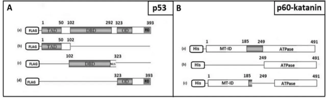
To locate the regions that have a role in this interaction, different FLAG- or His-tagged deletion constructs were pre-pared for both p53 and p60-katanin, respectively (Fig. 2). Besides WT p53 (WT-p53, p53-a), we prepared Transcrip-tion ActivaTranscrip-tion Domain (TAD-p53, p53-b), DNA Binding Domain (DBD-p53, p53-c) and Oligomerization-Regulatory Domain (OD-RD-p53, p53-d) constructs for p53 (Fig. 2a). Similarly, besides WT p60-katanin (WT-p60-katanin, p60-katanin-a), C-terminal containing construct (C-term-p60-katanin, p60-katanin-b) and del201-268 construct (del201-268-p60-katanin, p60-katanin-c) for p60-katanin were prepared (Fig. 2b).
Different combinations of deletion constructs were transferred into the cells by chemical transfection. As
Fig. 1 Western blotting result for co-immunoprecipitation of endog-enous p60-katanin and p53. WCL were immunoprecipitated (IP) with either anti-p60-katanin or anti-p53 antibody and immunoblotted (IB) with either anti-p53 or anti-p60-katanin antibody, respectively. Aster-isk (*) indicates an interaction between p60-katanin and p53. In IgG negative control, there is non-specific antibody during IB, no
anti-illustrated in Fig. 3, first transfection was performed with WT-p53 and C-term-p60-katanin. As Co-IP data indicated (Fig. 3a, 1) analysis was positive in terms of interaction since the signal was in line with input. The C-term-p60-katanin construct was further tested for the interaction with either TAD-p53 (Fig. 3a, 2), DBD-p53 (Fig. 3a, 3) or OD-RD-p53 (Fig. 3a, 4). Last combination that was tested
was for WT-p53 and del201-268-p60-katanin (Fig. 3a, 5). Analyzing whole Co-IP data clearly indicated that C-term-p60-katanin construct interacts only with DBD-p53. Hereby, interaction between p53 and p60-katanin appears to be through the C-terminal of p60-katanin and DNA Binding Domain of p53 (Fig. 4).
Fig. 2 Schematic illustration of deletion constructs. Represents the deletion constructs for either p53 (a) or p60-katanin (b)
Fig. 3 Western blotting image of co-immunoprecipitation analysis. The left part of the figure shows Co-IP analyses results (a) and on the right part there is an illustration of deletion constructs used in Co-IP experiments (b). Input (total protein extraction) is positive control and IgG indicates negative control performed with non-specific IgG.
Lines pointed with either white (in input) or black (in IP) arrowhead show the desired protein signal. Immunoprecipitation was done with anti-FLAG antibody to precipitate FLAG-tagged construct [either WT-p53 (1, 5), TAD-p53 (2), DBD-p53 (3) or OD-RD-p53 (4)]
Discussion
MTs, being essential elements of the cell, are involved in essential functions such as intracellular cargo transport, cell motility, and cell division as well as specialized functions such as formation, growth and maintenance of axons and dendrites in neurons [1, 3]. Besides their inherent dynamic instability, MT severing proteins have also inevitable func-tion in MT reconfigurafunc-tion. Katanin is one of the best-characterized MT severing proteins and is composed of two subunits, regulatory subunit p80-katanin, and catalytic subu-nit p60-katanin [16] of which regulatory partners are still little-known. Similar to p60-katanin, p53 is also important in controlling different cellular mechanisms such as cell cycle arrest, senescence, differentiation and apoptosis by regulat-ing the expression of regulatory proteins [11–14]. Since both p60-katanin and p53 have critical roles in proliferation and differentiation, a reasonable question to ask is: “Is it possible for p60-katanin and p53 to be partners in the cell?”.
To answer this critical question, the first aim of this study was to determine whether p53 and p60-katanin interact in the cell. Our findings revealed that p53 and p60-katanin interact in the cell (Fig. 1). Considering that katanin requires a strict regulation due to its critical MT severing action and p60-katanin and p53 have roles in similar essential cellular events such as proliferation, apoptosis and differentiation, it seems reasonable for these proteins to interact in the cell.
Besides identifying the interaction between p53 and p60-katanin, we also identified the possible regions of this interaction by using different FLAG- or His-tagged
Our results indicated that p53 interacts with p60-katanin through p60-katanin’s C-terminal domain—which also includes ATPase domain (Fig. 4). Through this interaction, it is most likely that p53 may affect the MT severing activ-ity of the p60-katanin which is directed by its C-terminal domain, and thereby indirectly the MT dynamics, depend-ing on intracellular conditions.
The analysis of the interacting regions also revealed that the communication of p60-katanin with p53 takes place via the DNA binding domain of p53. This result indicates that p60-katanin may affect DNA binding function of p53. As with most transcription factors, p53 has various protein–protein interaction sites which are crucial for the fine-tuning of its activity. In concordance with our results, there is a number of protein partners that interact p53 via its DNA-binding domain. Among them, Hypoxia inducible factor-1 alpha (HIF-1a) [17, 18], Heat shock protein 90 (Hsp90) [19], Rad51 [20, 21], Bcl-XL/Bak [22, 23] are the most remarkable. Looking at all these studies, it is evident that these proteins interact with p53 to regulate important cellular activities such as protein homeostasis, DNA dam-age repair, cell proliferation, apoptosis and senescence. However, DNA-binding site of p53 is highly charged to be able to electrostatically interact with DNA. Thus, interac-tion of all these proteins with the DNA-binding site of p53 is strongly dependent on ionic strength as a requirement of electrostatic interaction [18]. For this reason, conditional experiments should be conducted in the future to exam-ine the details of their interaction under different ionic strengths and different pH conditions.
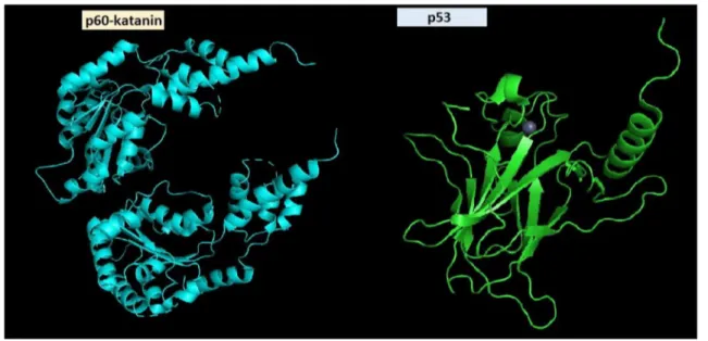
Fig. 4 Schematic illustration of interacting domains. Domain illus-trations are generated via PyMOL and data were taken from https ://www.rcsb.org/ for p60-katanin (5ZQL, crystal structure of human
katanin AAA ATPase domain) and p53 (2FEJ, solution structure of human p53 DNA binding domain)
Considering all these data, it is reasonable to conclude that the interaction between p60-katanin and p53 can have functional results for both proteins, in other words, this inter-action is a mutual interinter-action. The results of this study show that both proteins may have a regulatory effect on each oth-er’s activity in the cell. However, to be able to suggest such a reciprocal regulatory effect, mechanistic studies should be conducted. Identifying their localization of interaction may provide valuable insights about their regulatory effect on each other. Furthermore, revealing their mode of interac-tion, whether direct or via MTs is also crucial to deepen our understanding of functional outcomes of their interaction.
Although there is no reported mutation in p53 of RFL-6 cell line in the literature, and also RFL-6 cell line is not found on the ATTC’s “Validated p53 Hotspot Mutation Cell Line List” [24], it is crucial for our study to have an intrinsic wild type p53 in the cell because it may affect the signifi-cance of the interpretations about the functional outcomes of this cytoplasmic interaction. Thus, further experiments should be conducted in order to make sure whether p53 of RFL-6 cell line is wild type or mutated.
In summary, we showed for the first time that p53 and p60-katanin do interact and this interaction appears to occur via p53’s DNA binding domain and p60-katanin’s C-ter-minal domain (Fig. 4). Further elucidating the functional outcomes of this interaction is vital for better understand-ing of the regulatory mechanism of essential cellular events such as cell cycle, differentiation, and apoptosis in disease and in health.
Acknowledgements We are grateful to Kübra Yener for helping in cloning the deletion constructs and Koray Kırımtay for his help in preliminary Co-IP studies.
Author contributions SK conceived the study, designed and carried out experiments, and contributed to writing the manuscript. AY con-tributed to writing the manuscript. All authors read and approved the final manuscript.
Funding This work was supported by the Scientific and Technological Research Council of Turkey (Grant No. 114Z971 to SK).
Compliance with ethical standards
Conflict of interest The authors declare that they have no conflict of interest.
Ethical approval This article does not contain any studies with human participants or animals performed by any of the authors.
References
1. McNally F, Vale R (1993) Identification of katanin, an ATPase that severs and disassembles stable microtubules. Cell 75:419–429
2. Hartman J, Vale RD (1999) Microtubule disassembly by ATP-dependent oligomerization of the AAA enzyme katanin. Science 286:782–785
3. Quarmby L (2000) Cellular Samurai: katanin and the severing of microtubules. J Cell Sci 113:2821–2827
4. McNally KP, Bazirgan OA, McNally FJ (2000) Two domains of p80 katanin regulate microtubule severing and spindle pole target-ing by p60 katanin. J Cell Sci 113:1623–1633
5. Buster D, McNally K, McNally FJ (2002) Katanin inhibition pre-vents the redistribution of gamma-tubulin at mitosis. J Cell Sci 115:1083–1092
6. McNally K, Audhya A, Oegema K, McNally FJ (2006) Kata-nin controls mitotic and meiotic spindle length. J Cell Biol 175:881–891
7. Poulain FE, Sobel A (2010) The microtubule net work and neu-ronal morphogenesis: dynamic and coordinated orchestration through multi ple players. Mol Cell Neurosci 43:15–32
8. Baas PW, Qiang L (2005) Neuronal microtubules: when the MAP is the roadblock. Trends Cell Biol 15:183–187
9. Baas PW, Vidya Nadar C, Myers KA (2006) Axonal transport of microtubules: the long and short of it. Traffic 7:490–498 10. Yu W, Qiang L, Solowska JM, Karabay A, Korulu S, Baas PW
(2008) The microtubule-severing pro teins spastin and katanin par-ticipate differ ently in the formation of axonal branches. Mol Biol Cell 19:1485–1498
11. Hudson CD, Morris PJ, Latchman DS, Budhram-Mahadeo VS (2005) Brn-3a transcription factor blocks p53-mediated activation of proapoptotic target genes Noxa and Bax in vitro and in vivo to determine cell fate. J Biol Chem 280:11851–11858
12. Ferreira A, Kosik KS (1996) Accelerated neuronal differentiation induced by p53 suppression. J Cell Sci 109:1509–1516
13. Kim J, Lengner CJ, Kirak O, Hanna J, Cassady JP, Lodato MA, Wu S, Faddah DA, Steine EJ, Gao Q, Fu D, Dawlaty M, Jaenisch R (2011) Reprogramming of postnatal neurons into induced pluri-potent stem cells by defined factors. Stem Cells 29(6):992–1000 14. Di Giovanni S, Knights CD, Rao M, Yakovlev A, Beers J, Catania
J, Avantaggiati ML, Faden AI (2006) The tumor suppressor pro-tein p53 is required for neurite outgrowth and axon regeneration. EMBO J 25:4084–4096
15. Korulu S, Yildiz-Unal A, Yuksel M, Karabay A (2013) Protein kinase C activation causes neurite retraction via cyclinD1 and p60-katanin increase in rat hippocampal neurons. Eur J Neurosci 37(10):1610–1619
16. Hartman JJ, Mahr J, McNally K, Okawa K, Iwamatsu A (1998) Katanin, a microtubule-severing protein, is a novel AAA ATPase that targets to the centrosome using a WD40-containing subunit. Cell 93:277–287
17. Hansson LO, Friedler A, Freund S, Rudiger S, Fersht AR (2002) Two sequence motifs from HIF-1α bind to the DNA-binding site of p53. Proc Natl Acad Sci USA 99:10305–10309
18. Sanchez-Puig N, Veprintsev DB, Fersht AR (2005) Binding of natively unfolded HIF-1α ODD domain to p53. Mol Cell 17:11–21 19. Rudiger S, Freund SM, Veprintsev DB, Fersht AR (2002)
CRINEPT-TROSY NMR reveals p53 core domain bound in an unfolded form to the chaperone Hsp90. Proc Natl Acad Sci USA 99:11085–11090
20. Buchhop S, Gibson MK, Wang XW, Wagner P, Sturzbecher HW, Harris CC (1997) Interaction of p53 with the human Rad51 pro-tein. Nucleic Acids Res 25:3868–3874
21. Friedler A, Veprintsev DB, Rutherford T, von Glos KI, Fersht AR (2005) Binding of Rad51 and other peptide sequences to a promiscuous, highly electrostatic binding site in p53. J Biol Chem 280:8051–8059
22. Petros AM, Gunasekera A, Xu N, Olejniczak ET, Fesik SW (2004) Defining the p53 DNA-binding domain/Bcl-x(L)-binding inter-face using NMR. FEBS Lett 559:171–174
23. Sot B, Freund SM, Fersht AR (2007) Comparative biophysical characterization of p53 with the pro-apoptotic BAK and the anti-apoptotic BCL-xL. J Biol Chem 282:29193–29200
24. ATCC (2013) p53 Hotspot mutation cell panels. https ://www. atcc.org/en/Docum ents/Learn ing_Cente r/~/media /5F7B1 CCACF 724E3 398BE 56BFB EE3EF E4.ashx. Accessed 3 Oct 2019
Publisher’s Note Springer Nature remains neutral with regard to jurisdictional claims in published maps and institutional affiliations.