Comparison of platelet-rich plasma and steroid
injection in the treatment of plantar fasciitis
Correspondence: Ferhat SAY, MD, Asst. Prof. Ondokuz Mayıs Üniversitesi Tıp Fakültesi Hastanesi,
Ortopedi ve Travmatoloji Anabilim Dalı, Kurupelit, 55139 Samsun, Turkey. Tel: +90 530 – 566 08 94 e-mail: [email protected]
Submitted: November 22, 2013 Accepted: August 01, 2014 ©2014 Turkish Association of Orthopaedics and Traumatology
Available online at www.aott.org.tr doi: 10.3944/AOTT.2014.13.0142 QR (Quick Response) Code
doi: 10.3944/AOTT.2014.13.0142
Ferhat SAY1, Deniz GÜRLER2, Erkan İNKAYA2, Murat BÜLBÜL3
1Department of Orthopedics and Traumatology, Faculty of Medicine, Ondokuz Mayıs University, Samsun, Turkey; 2Department of Orthopedics and Traumatology, Samsun Training and Research Hospital, Samsun, Turkey;
3Department of Orthopedics and Traumatology, Faculty of Medicine, Medipol University, Istanbul, Turkey
Objective: The aim of this study was to compare the effects of platelet-rich plasma (PRP) and steroid
injections in patients diagnosed with plantar fasciitis.
Methods: A total of 50 patients with chronic plantar fasciitis were included in the study and divided
into 2 groups. In the PRP group (n=25), PRP taken from the patients’ blood was activated using calcium chloride and injected in a single dose. In the steroid group (n=25), a single dose methylpred-nisolone with local anesthetic injection was given. Clinical evaluation was made using the American Foot and Ankle Score (AFAS) and the visual analog scale (VAS).
Results: No complications were seen in any patients. Mean AFAS was 85.5±4.2 at 6 weeks and
90.6±2.6 at 6 months in the PRP group and 75.3±4.8 and 80.3±4.7, respectively, in the steroid group (p<0.001). The difference in the mean VAS between the PRP group (2.4±0.8 and 1±0.8) and the ste-roid group (4±1.1 and 2.6±0.9) at the 6th week and 6th month was statistically significant (p<0.001).
Changes in AFAS and VAS scores were significantly higher in the PRP group (p<0.001).
Conclusion: The application of PRP appears to be more effective than steroid injection in terms of
pain and functional results in the treatment of chronic plantar fasciitis.
Key words: Growth factor; plantar fasciitis; platelet-rich plasma; steroid.
Plantar fasciitis is defined as localized inflammation and degeneration of the plantar aponeuroses. Heel pain is the most common reason for presentation.[1] Approxi-mately 10% of the population will experience heel pain in their life.[2] Reduced ankle dorsiflexion, standing for long periods of time at work, obesity, female gender and advancing age are listed as risk factors.[3,4]
As in other chronic tendinosis, the pathology of plan-tar fasciitis includes degenerative changes in the planplan-tar fascia with fibroblastic proliferation and limited
inflam-matory tissue.[5,6] The pain is generally localized in the medial calcaneal tubercle. In the acute phase, the pain is sharp and typically on the first step of the day or after a period of rest. In the chronic phase, pain is continuous and of a duller nature.[7]
Plantar fasciitis is generally a self-limiting condi-tion. Symptoms in 80 to 90% of cases recover within 10 months.[8] However, this process may be problematic for both the patient and the physician. Due to the natural course of the disease, the majority of treatment
tives are nonsurgical.[1,8] Stretching exercises, splints, or-thoses, non-steroid anti-inflammatory drugs (NSAID), steroid injections, and extracorporeal shockwave therapy (ESWT) are the nonsurgical treatment choices.[1,7] The majority of the nonsurgical treatments have been found to be minimally effective.[9] Today, there is no effective nonsurgical treatment option for plantar fasciitis. Ideal nonsurgical treatment for plantar fasciitis should be as effective as other treatment options with minimal com-plications.
Platelet-rich plasma (PRP) stimulates the natural healing process through growth factors contained in the platelets. PRP applied to the wound area accelerates the physiological healing process, provides support for the connection of cells, reduces pain and has anti-inflamma-tory and anti-bacterial effects.[10]
Studies in the literature have reported the use of PRP in plantar fasciitis and chronic tendinopathy.[11-15] The rationale for using PRP in this degenerative and chronic process is to restart the inflammatory process that com-monly ceases following failed conservative treatment, thus, turning the chronic injury into a new acute injury. [12] The high content of growth factors in PRP increases tissue regenerative abilities and regeneration processes. [14] There are 16 concentration systems which can be used to obtain PRP. Leukocytes and growth factors con-tained in PRP are obcon-tained in different amounts from these systems.[16] Apart from the concentration systems, PRP can be obtained manually from peripheral blood. [17,18] However, many questions remain unanswered re-garding the application of PRP, such as the ideal volume, application frequency, application period and platelet activation.[19]
The aim of this study was to determine the effects on pain and function of PRP obtained manually as a cheap and easy method in the treatment of plantar fasciitis and to compare this data with that of steroid injection which is often used in clinical practice. The hypothesis was that a single dose of manually-prepared PRP would reduce pain associated with plantar fasciitis and increase function and that this effect would be superior to the frequently-used steroid injection.
Patients and methods
Approval for this prospective clinical study was granted by the local ethics committee and informed consent was obtained from all patients participating in the study. Patients who had been diagnosed with plantar fasciitis, monitored for a minimum of 3 months and showed no benefit from conservative treatment starting with stretching exercises and NSAIDs were included in the
study. Diagnosis of plantar fasciitis was made by clinical examination. Direct radiographs were examined to rule out other heel pathologies. Exclusion criteria were sys-temic disease, pregnancy, active tumor or hematological malignant disease, infection, a history of anticoagulant use, use of NSAIDs in the five days prior to the study, Hb values of less than 11 g/dL, thrombocyte count of less than 150,000/mm3, previous steroid injection to the heel area or ESWT therapy, a history of calcaneus frac-ture, or surgery in the heel area.
A total of 50 patients were included in the study. Pa-tients were separated into PRP and steroid groups of 25 subjects each. Patients informed about the treatment op-tions and those who accepted were included in the PRP group (5 males, 20 females; mean age: 47) and the oth-ers in the steroid group (6 males, 19 females; mean age: 48.6).
Platelet-rich plasma was prepared and applied under the same conditions using the method described by Ani-tua et al.[17,18] A total of 30 cc peripheral blood was taken from the antecubital region and mixed with 3.2% sodi-um citrate. Samples were centrifuged at 1800 rpm for 8 minutes at room temperature. From the 3.5 ml PRP obtained, 1 ml was sent to the laboratory for bacterio-logical testing and platelet count. After activation, 2.5 ml of PRP containing 5.5% calcium chloride (Cl2Ca) (50 μl of Cl2Ca in 1 ml of PRP) was administered to the foot from the medial side to maximal tenderness area with palpation under sterile conditions. The patient was kept in the supine position for 20 minutes following admin-istration.
In the steroid group, a mixture of 40 mg/1 ml of methylprednisolone and 1 ml of prilocaine was applied. The peppering injection technique was used in both groups and the fascia was injected in 4 to 5 different lo-cations.
Standard Achilles and plantar fascia stretching and strengthening exercises were applied to all patients. Pa-tients were advised to rest and not stand for the first day after the injection. No NSAID, orthosis or splint was given to any patient.
Clinical evaluation was performed before treatment and at the 6th week and 6th month follow-ups. The Amer-ican Foot and Ankle Score (AFAS)[20] and the visual analog scale (VAS) were used in the clinical evaluation. The AFAS evaluation covered pain, function, maximum walking distance, walking surfaces, gait abnormality, sagittal motion, hindfoot motion, alignment, and ankle-hindfoot stability. Patients were questioned with regard to side effects and subjective satisfaction.
Results were stated as mean±standard deviation (SD). Data were evaluated using the SPSS for Windows v.16.0 (SPSS Inc., Chicago, IL, USA) software. The Mann-Whitney U test was used for the statistical evalu-ation of mean values between groups and for changes over time in the mean clinical scores of the groups. A value of p<0.05 was accepted as statistically significant.
Results
No local or systemic complications were seen in any pa-tient during the application or follow-up.
Both groups were similar in terms of age, gender, side and initial AFAS and VAS scores (Table 1). No statis-tically significant difference was determined between groups in terms of these factors.
Mean platelet count per milliliter in the PRP group was 207,880±51,272 before centrifugation and 818,520±119,236 after (p<0.001).
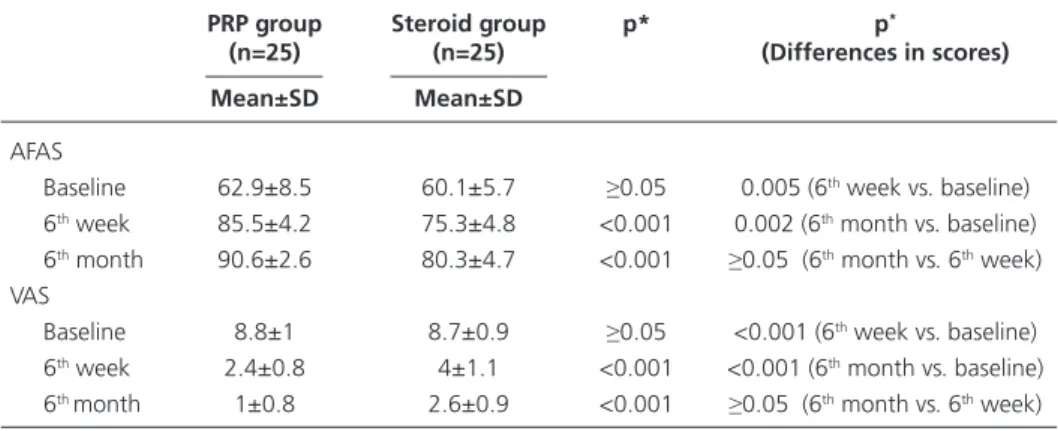
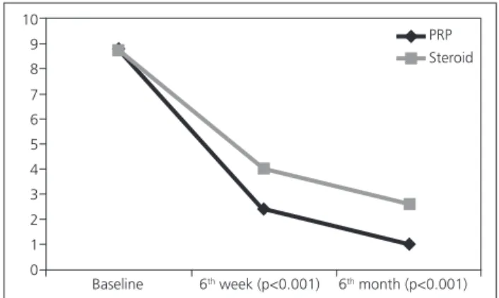
In the PRP group, mean AFAS score was 85.5±4.2 at the 6th week follow-up and 90.6±2.6 at the 6th month follow-up (Fig. 1, Table 2). Mean VAS score was 2.4±0.8 at the 6th week and 1±0.8 at the 6th month
(Fig. 2, Table 2). The differences between the pretreat-ment and follow-up scores were statistically significant (Table 2).
In the steroid group, mean AFAS score was 75.3±4.8 at the 6th week follow-up and 80.3±4.7 at the 6th month follow-up (Fig. 1, Table 2). Mean VAS scores were 4±1.1 and 2.6±0.9 at the 6th week and 6th month follow-ups, respectively (Fig. 2, Table 2). Compared to the pretreat-ment scores, the differences between the 6th week and 6th month scores were statistically significant.
The PRP group had significantly higher mean AFAS and VAS scores at follow-up than the steroid group (p<0.001) (Table 2).
Discussion
Several nonsurgical treatment methods are available for the treatment of plantar fasciitis with various success rates. Ideal treatment for plantar fasciitis has not been determined. The use of PRP in foot and ankle patholo-gies has begun to increase. Our study was designed to compare the effect of PRP and steroid injection in the treatment of chronic plantar fasciitis. A single dose of
Table 1. Comparison of the patients’ characteristics at baseline.
PRP group Steroid group p
(n=25) (n=25)
n Mean±SD n Mean±SD
Age (year) 47±6.8 48.6±6.4 ≥0.05
Male/Female 5/20 6/19 ≥0.05
Affected foot (Right/Left) 10/15 11/14 ≥0.05
AFAS 62.9±8.5 60.1±5.7 ≥0.05
VAS 8.8±1 8.7±0.9 ≥0.05
AFAS: American Foot and Ankle Score; VAS: Visual analog scale; SD: Standard deviation.
Table 2. Comparison of AFAS and VAS scores of the groups at baseline, 6th week and 6th month.
PRP group Steroid group p* p*
(n=25) (n=25) (Differences in scores)
Mean±SD Mean±SD
AFAS
Baseline 62.9±8.5 60.1±5.7 ≥0.05 0.005 (6th week vs. baseline)
6th week 85.5±4.2 75.3±4.8 <0.001 0.002 (6th month vs. baseline)
6th month 90.6±2.6 80.3±4.7 <0.001 ≥0.05 (6th month vs. 6th week)
VAS
Baseline 8.8±1 8.7±0.9 ≥0.05 <0.001 (6th week vs. baseline)
6th week 2.4±0.8 4±1.1 <0.001 <0.001 (6th month vs. baseline)
6th month 1±0.8 2.6±0.9 <0.001 ≥0.05 (6th month vs. 6th week)
*Mann-Whitney U test.
manually obtained PRP was found to be more effective than steroid injection in terms of pain and functional re-sults in the treatment of chronic plantar fasciitis.
The etiology of plantar fasciitis is not fully known. According to the commonly accepted view in the litera-ture, it is an inflammatory response to microtears which form as a result of mechanical loading. Conversely, Lem-ont et al. reported no findings of histological inflamma-tion in histological samples of plantar fasciitis.[6] These paradoxical findings on the etiology of plantar fasciitis have not yet been explained. The use of steroid injections for plantar fasciitis has been reported to be useful in the short-term.[21] In our study, we found a positive effect on pain and functional scores in the steroid group which can be explained by the anti-inflammatory effect. How-ever, steroid injections have been reported to be related to plantar fascia tear,[22] fat pad atrophy, abscess,[23] and osteomyelitis.[24]
Platelet-rich plasma stimulates the proliferation of various cell types in cells and tissue[25] and activates repair cells in the blood circulation.[26] More than 30 bioactive proteins are found within the alpha granules of platelets.[18] Growth factors, such as platelet-derived growth factor, transforming growth factor, vascular en-dothelial growth factor and insulin-like growth factor, and proteins such as fibrin, fibronectin, vitronectin, and thrombospondin, found in PRP, play a role in many stages of tissue healing. These growth factors activate some of the cells that play a function in tissue healing and thus provide soft tissue healing and bone regenera-tion.[27] With its growth factors, PRP stimulates the lo-cal stem cells and activates the repair cells in the circula-tion and bone marrow. Excessive inflammacircula-tion inhibits apoptosis and metalloproteinase activity.[28] Moreover, in tendon recovery, PRP increases tenocyte proliferation in the injured area by providing revascularization by means of the included growth factors and is effective in
increas-ing collagen expression in the tenocytes.[29]
Different methods can be used to obtain PRP; auto-matic machines and commercial kits with double-spin rotation, single-spin rotation and manual PRP separa-tion and selective blood filtrasepara-tion (plateletpheresis).[30] Anitua et al.[18] reported that a platelet count of over 300,000/μl in PRP is effective. In another in vitro study, platelet concentration 2.5 times greater than the basal platelet count was reported to be the most effective.[31] The prepared PRP can be activated by adding bovine or human thrombin or calcium chloride.[32] Growth factors and cytokines are revealed with the formation of plate-let gel from the activated PRP. Some authors used PRP without activation.[11-14] There is no consensus with us-age of PRP in ideal concentration, application frequency or platelet activation.[19] In the current study, PRP was prepared manually as single-spin rotation. In the analy-sis of the prepared PRP, concentration was determined as four times greater than the thrombocyte count in the peripheral blood. The prepared PRP was activated by the addition of calcium chloride.
Platelet-rich plasma was first used in 1987 in heart surgery to prevent excessive blood transfusion.[33] Sev-eral studies have reported the use of PRP in plantar fasciitis and chronic tendinopathy.[11-15] Barrett and Erredge[11] applied PRP together with ultrasonography (USG) to 9 patients and measured the thickness of the plantar fascia on USG. Following treatment, a change in signal intensity and reduced thickness of the plantar fas-cia was determined on USG and after one year, 77.9% of the patients were reported to be symptom-free. Akşahin et al.[12] administered steroids to 30 patients and PRP to 30 patients and reported similar functional and pain scores of both groups at 3 weeks and 6 months. How-ever, when steroid complications were taken into con-sideration, PRP was recommended. Ragab and Othman applied PRP to 25 patients and reported that plantar
Fig. 1. Baseline, 6th week and 6th month AFAS of both groups.
100 95 90 85 80 75 70 65 60
Baseline 6th week (p<0.001) 6th month (p=0.001)
PRP Steroid
Fig. 2. Baseline, 6th week and 6th month VAS scores of both groups.
10 9 8 7 6 5 4 3 2 1 0
Baseline 6th week (p<0.001) 6th month (p<0.001)
PRP Steroid
fascia thickness decreased on USG and 88% of the pa-tients were symptom-free after a mean follow-up of 10 months.[13] Martinelli et al. reported a reduction in pain scores over a 12-month follow-up period in 14 patients treated with PRP.[14] In the evaluation of the results of the current study, the post-treatment 6th week and 6th month functional scores of both groups were signifi-cantly increased compared to the initial values and the pain scores were significantly decreased. The changes in the functional and pain scores of the PRP group were significantly better than those of the steroid group. In the current study, although a clinical recovery was de-termined over a 6 month follow-up, it is not yet known how long this effect will last. Obtaining PRP used in the current study is low-cost. While the cost of automatic devices and kits to obtain PRP is several hundreds of dollars, the cost of the manual method used to prepare PRP was approximately ten dollars.[34]
For PRP obtained from autologous blood, there is no risk of immune reaction or disease transfer. There are no studies in the literature warning of hyperplasia, carci-nogenesis or tumor growth of PRP.[10] No complications were encountered in any patient in the PRP group.
The limitations of this study included its lack of ran-domization, a placebo control group and evaluation of results with functional and pain scores due to lack of radiological and biological results. In addition, the low number of patients and relatively short follow-up period can also be considered limitations.
In conclusion, the administration of PRP in plantar fasciitis treatment appears to be a more effective method than steroid injection for the reduction of pain and pro-vide better functional results at 6th month follow-ups. However, prospective, randomized, placebo-controlled, multicenter studies are required to clarify these results and better understand the effects of PRP.
Acknowledgment: The authors thank Prof. Dr. Nicola
MAFFULLI for his suggestions and also Prof. Dr. Yük-sel BEK for his help with the statistical analysis.
Conflicts of Interest: No conflicts declared.
References
1. League AC. Current concepts review: plantar fasciitis. Foot Ankle Int 2008;29:358-66. CrossRef
2. Crawford F, Thomson C. Interventions for treat-ing plantar heel pain. Cochrane Database Syst Rev 2010;1:CD000416.
3. Riddle DL, Pulisic M, Pidcoe P, Johnson RE. Risk factors for Plantar fasciitis: a matched case-control study. J Bone Joint Surg Am 2003;85-A:872-7.
4. Scher DL, Belmont PJ Jr, Bear R, Mountcastle SB, Orr JD, Owens BD. The incidence of plantar fasciitis in the United States military. J Bone Joint Surg Am 2009;91:2867-72. 5. Jarde O, Diebold P, Havet E, Boulu G, Vernois J.
Degen-erative lesions of the plantar fascia: surgical treatment by fasciectomy and excision of the heel spur. A report on 38 cases. Acta Orthop Belg 2003;69:267-74.
6. Lemont H, Ammirati KM, Usen N. Plantar fasciitis: a de-generative process (fasciosis) without inflammation. J Am Podiatr Med Assoc 2003;93:234-7. CrossRef
7. Healey K, Chen K. Plantar fasciitis: current diagnos-tic modalities and treatments. Clin Podiatr Med Surg 2010;27:369-80. CrossRef
8. Davis PF, Severud E, Baxter DE. Painful heel syn-drome: results of nonoperative treatment. Foot Ankle Int 1994;15:531-5. CrossRef
9. Gill LH, Kiebzak GM. Outcome of nonsurgical treatment for plantar fasciitis. Foot Ankle Int 1996;17:527-32. CrossRef
10. Sánchez M, Anitua E, Orive G, Mujika I, Andia I. Plate-let-rich therapies in the treatment of orthopaedic sport injuries. Sports Med 2009;39:345-54. CrossRef
11. Barrett SL, Erredge SE. Growth factors for chronic plan-tar fasciitis? Podiatry Today 2004;17:36-42.
12. Akşahin E, Doğruyol D, Yüksel HY, Hapa O, Doğan O, Celebi L, et al. The comparison of the effect of cortico-steroids and platelet-rich plasma (PRP) for the treat-ment of plantar fasciitis. Arch Orthop Trauma Surg 2012;132:781-5. CrossRef
13. Ragab EM, Othman AM. Platelets rich plasma for treat-ment of chronic plantar fasciitis. Arch Orthop Trauma Surg 2012;132:1065-70. CrossRef
14. Martinelli N, Marinozzi A, Carnì S, Trovato U, Bianchi A, Denaro V. Platelet-rich plasma injections for chronic plantar fasciitis. Int Orthop 2013;37:839-42. CrossRef
15. de Vos RJ, van Veldhoven PL, Moen MH, Weir A, Tol JL, Maffulli N. Autologous growth factor injections in chronic tendinopathy: a systematic review. Br Med Bull 2010;95:63-77. CrossRef
16. Castillo TN, Pouliot MA, Kim HJ, Dragoo JL. Com-parison of growth factor and platelet concentration from commercial platelet-rich plasma separation systems. Am J Sports Med 2011;39:266-71. CrossRef
17. Anitua E. Plasma rich in growth factors: preliminary re-sults of use in the preparation of future sites for implants. Int J Oral Maxillofac Implants 1999;14:529-35.
18. Anitua E, Andia I, Ardanza B, Nurden P, Nurden AT. Au-tologous platelets as a source of proteins for healing and tissue regeneration. Thromb Haemost 2004;91:4-15. 19. Maffulli N, Del Buono A. Platelet plasma rich products
in musculoskeletal medicine: any evidence? Surgeon 2012;10:148-50. CrossRef
20. Kitaoka HB, Alexander IJ, Adelaar RS, Nunley JA, Myer-son MS, Sanders M. Clinical rating systems for the ankle-hindfoot, midfoot, hallux, and lesser toes. Foot Ankle Int
1994;15:349-53. CrossRef
21. Crawford F, Atkins D, Young P, Edwards J. Steroid injec-tion for heel pain: evidence of short-term effectiveness. A randomized controlled trial. Rheumatology (Oxford) 1999;38:974-7. CrossRef
22. Acevedo JI, Beskin JL. Complications of plantar fascia rupture associated with corticosteroid injection. Foot An-kle Int 1998;19:91-7. CrossRef
23. Buccilli TA Jr, Hall HR, Solmen JD. Sterile abscess forma-tion following a corticosteroid injecforma-tion for the treatment of plantar fasciitis. J Foot Ankle Surg 2005;44:466-8. CrossRef
24. Gidumal R, Evanski P. Calcaneal osteomyelitis following steroid injection: a case report. Foot Ankle 1985;6:44-6. 25. Lucarelli E, Beccheroni A, Donati D, Sangiorgi L,
Cenac-chi A, Del Vento AM, et al. Platelet-derived growth fac-tors enhance proliferation of human stromal stem cells. Biomaterials 2003;24:3095-100. CrossRef
26. Kajikawa Y, Morihara T, Sakamoto H, Matsuda K, Os-hima Y, Yoshida A, et al. Platelet-rich plasma enhances the initial mobilization of circulation-derived cells for tendon healing. J Cell Physiol 2008;215:837-45. CrossRef
27. Alsousou J, Thompson M, Hulley P, Noble A, Willett K. The biology of platelet-rich plasma and its application in trauma and orthopaedic surgery: a review of the literature.
J Bone Joint Surg Br 2009;91:987-96. CrossRef
28. Mishra A, Woodall J Jr, Vieira A. Treatment of tendon and muscle using platelet-rich plasma. Clin Sports Med 2009;28:113-25. CrossRef
29. Baksh N, Hannon CP, Murawski CD, Smyth NA, Ken-nedy JG. Platelet-rich plasma in tendon models: a sys-tematic review of basic science literature. Arthroscopy 2013;29:596-607. CrossRef
30. Andia I, Sánchez M, Maffulli N. Joint pathology and platelet-rich plasma therapies. Expert Opin Biol Ther 2012;12:7-22. CrossRef
31. Graziani F, Ivanovski S, Cei S, Ducci F, Tonetti M, Gabri-ele M. The in vitro effect of different PRP concentrations on osteoblasts and fibroblasts. Clin Oral Implants Res 2006;17:212-9. CrossRef
32. Pietrzak WS, Eppley BL. Platelet rich plasma: biology and new technology. J Craniofac Surg 2005;16:1043-54. 33. Ferrari M, Zia S, Valbonesi M, Henriquet F, Venere G,
Spagnolo S, et al. A new technique for hemodilution, preparation of autologous platelet-rich plasma and intra-operative blood salvage in cardiac surgery. Int J Artif Or-gans 1987;10:47-50.
34. Mei-Dan O, Mann G, Maffulli N. Platelet-rich plasma: any substance into it? Br J Sports Med 2010;44:618-9.
View publication stats View publication stats