See discussions, stats, and author profiles for this publication at: https://www.researchgate.net/publication/256986209
4,5-dianilinophtalimide protects
neuroendocrine cells against serum
deprivation-induced stress and apoptosis
Article in Neuro endocrinology letters · August 2013 CITATION1
READS43
4 authors: Volkan Ergin University of California, Riverside 19 PUBLICATIONS 97 CITATIONS SEE PROFILE Mutlu Erdogan Max Planck Institute of Neurobiology 11 PUBLICATIONS 74 CITATIONS SEE PROFILE Ceren Karasu Gazi University 6 PUBLICATIONS 49 CITATIONS SEE PROFILE Adnan Menevse Gazi University 55 PUBLICATIONS 537 CITATIONS SEE PROFILEAll content following this page was uploaded by Volkan Ergin on 23 May 2014. The user has requested enhancement of the downloaded file.
O
RI
GIN
AL AR
TI
CLE
4,5-dianilinophtalimide protects
neuroendocrine cells against serum
deprivation-induced stress and apoptosis
Volkan Ergin
1*, Mutlu Erdogan
2*, Çimen Karasu
3, Adnan Menevşe
11 Department of Medical Biology & Genetics, Faculty of Medicine, Gazi University, Ankara, Turkey 2 Institute of Materials Science and Nanotechnology, Bilkent University, Ankara, Turkey
3 Cellular Stress Response and Signal Transduction Research Laboratory, Department of Medical
Pharmacology, Faculty of Medicine, Gazi University, Ankara, Turkey
* Authors have equally contributed to this study Correspondence to: Volkan Ergin, MSc.
Department of Medical Biology & Genetics, Faculty of Medicine, Gazi University, 06510 Ankara, Turkey.
tel: +903122024749; fax: +903122124647; e-mail: [email protected]
Submitted: 2013-06-21 Accepted: 2013-06-30 Published online: 2013-08-03 Key words: DAPH; serum deprivation; apoptosis; cellular stress; PC12
Neuroendocrinol Lett 2013; 34(5):359–365 PMID: 23922036 NEL340513A03 © 2013 Neuroendocrinology Letters • www.nel.edu
Abstract
OBJECTIVE: The aim of this study was to reveal the effects of4,5-dianilinoph-thalimide (DAPH), which inhibits amyloid β fibrillization, against serum depriva-tion (SD)-induced apoptosis and the possible mechanisms in differentiated PC12 neuron cells.
METHODS: Firstly, we evaluated whether DAPH protects cell viability exposed to SD by MTT assay. Next, we examined the changes of phospho-p38 MAPK (Thr180/Tyr182), phospho-HSP27 (Ser82), phospho-c-JUN (Ser73) and cleaved-CASP3 (Asp175) profiles by immunoblotting, in PC12 cells exposed to SD. Intra-cellular reactive oxygen species (ROS) level was also measured.
RESULTS: SD induced apoptosis accompanied by up-regulation of phospho-p38 MAPK (Thr180/Tyr182), phospho-HSP27 (Ser82), phospho-c-JUN (Ser73), cleaved-CASP3 (Asp175) and intracellular ROS content. Co-treatment with non-toxic doses of DAPH prevented apoptosis by the attenuation of activated proteins and reduction of ROS level. These results suggest that serum deprivation-induced apoptosis inhibited by DAPH administration.
CONCLUSION: We have provided for the first evidence that DAPH has a neuroprotective effect on SD-caused stress, probably via contributing the re-establishment of redox homeostasis.
Abbrevations:
BCA - Bicinchoninic acid
c-JUN - Proto-oncogene c-JUN
CASP3 - Caspase-3
DAPH - 4,5-Dianilinophthalimide
DCF - 2’,7’-dichlorofluorescein
DCF-DA - 2’,7’-dichlorofluorescein diacetate
GAPDH - Glyceraldehyde-3-phosphate dehydrogenase
HSP27 - Heat shock protein 27
JNK - c-jun N-terminal kinase
IC50 - Half maximal inhibitory concentration
MAPKAPK2 - MAP kinase-activated protein kinase 2
MTT - Thiazoyl blue tetrazolium bromide
NGF - Nerve growth factor
p38 MAPK - p38 mitogen activated protein kinase
PMSF - Phenylmethanesulfonyl fluoride
PVDF - Polyvinylidene difluoride
ROS - Reactive oxygen species
360
Copyright © 2013 Neuroendocrinology Letters ISSN 0172–780X • www.nel.eduVolkan Ergin, Mutlu Erdogan, Çimen Karasu, Adnan Menevşe
INTRODUCTION
Serum is essential for most of the cultured cell lines. Serum constituents such as growth factors, miner-als, lipids and numerous other factors are crucial for cell growth and differentiation (van der Valk et al. 2004). In addition to this, it is well known that the deprivation of serum from medium induces apoptosis in cultured cells. Besides being one of the factors of ischemia (Bialik et al. 1999), removal of serum from culture medium causes massive neuronal cell death (Greene 1978); on the other hand, different stud-ies demonstrated that antioxidant treatment has an inhibitory effect on serum deprivation (SD)-induced apoptosis (Ferrari et al. 1995). While the exact mecha-nism is still unknown, there have been significant amount of reports indicating that SD-induced apop-tosis is correlated with increased ROS, such as super-oxide and hydrogen persuper-oxide (Satoh et al. 1996; King
et al. 2003; Pandey et al. 2003; Zhuge & Cederbaum
2006). In fact, the SD-triggered intracellular signalling pathways are not yet completely clear, however, vari-ous stress-related enzymes, such as caspases and HSPs have apoptotic or protective roles (Stetler et al. 2010; Higuchi et al. 2006).
Neuronal cell death is a characteristic of neuro-degenerative diseases that occurs mainly by necrosis and/or apoptosis (Lipton 1999; Mattson 2000; Yuan
et al. 2003) and neuronal cells deprived of serum go
through apoptotic cell death (LeBlanc et al. 1999; Howard et al. 1993). PC12, a neuroendocrine cell line, which differentiates into a neuronal phenotype when exposed to nerve growth factor, is a useful model system for neuronal differentiation, has been exten-sively used for intracellular signaling studies (Vaudry
et al. 2002). This cell line allows rapid screening of
dif-ferent molecular pathways with minimal preparation time and comprises a convenient model for studying ischemia, neuronal apoptosis and its prevention (Hil-lion et al. 2005; Lee et al. 2012). The cells respond to environmental stresses through various mechanisms ranging from initiation of prosurvival strategies to activation of apoptotic pathways. In this context, there are many stress/apoptosis marker proteins, which are involved in normal signaling and survival pathways, and these marker proteins also interact with eventual cell death pathways. In this present study we assessed the effects of 4,5-dianilinophthalimide (DAPH), a selective inhibitor of formation of Aβ42 fibers and prions associated with various neurodegenerative diseases such as Alzheimer’s disease (Blanchard et al. 2004; Wang et al. 2008), on the expressional profiles of several stress/apoptosis marker proteins in serum deprived PC12 cells. We found that DAPH is signifi-cantly effective on decreasing stress-related and apop-totic conditions triggered by serum deprivation.
MATERIAL & METHODS
Cell culturePC12 cell line, stably overexpressing NGF receptor (PC12 6–15) has been used (Hempstead et al. 1992). This cell line has been kindly provided by Dr. V. Laketa (EMBL, Germany). Cells were maintained in vitro using RPMI medium supplemented with 10% heat-inactivated horse serum, 5% FBS, 2 mM L-glutamine and 1% penicillin-streptomycin (complete medium). Cells were cultured in a humidified atmosphere of 5% CO2 and 95% air at 37 °C. This cell line responds reversibly to NGF (Promega, Madison, WI, USA) and differentiate into neuronal phenotype when plated on Collagen type IV (Sigma-Aldrich, Steinheim, Germany) coated culture flasks in RPMI medium supplemented with 1% heat-inactivated horse serum, 2 mM L-glutamine, 1% penicillin-streptomycin and 100 ng/mL NGF (differentiation medium). All assays are performed on 48 h-differentiated cells in serum deprived (SD) medium.
Cell viability assay
Cell viability is ascertained using Thiazoyl blue tetra-zolium bromide (MTT; Bioworld; Dublin, OH, USA) assay. Briefly, PC12 cells (1 × 10⁴) seeded on
collagen-coated 96-well plates were incubated for 48 h in dif-ferentiation medium. Next, SD medium containing different concentrations of DAPH (CGP52411; Tocris Bioscience, Avonmouth, United Kingdom) were added. At the end of incubation, 10 μL of MTT stock solution (5 mg/mL) was added, and the plates were incubated at 37 °C for 4 h. Culture medium was removed, the resul-tant formazan crystals were dissolved in 100 μL DMSO, and the absorbance values were read on a microplate reader SpectraMax M5e (Molecular Devices, Sunnyvale, CA, USA) at 572 nm wavelength. Cells were assayed in hexaplicate, and three independent experiments were carried out.
Measurement of cellular oxidative stress
The accumulation of intracellular ROS was determined by measuring 2´,7´-dichlorofluorescein (DCF) fluores-cence. ROS cause oxidation of 2,7-dichlorofluorescein diacetate (DCFH-DA) to the fluorescent product DCF in the cell. In brief, cells seeded on collagen-coated 12-well plates and differentiated for 48 h, (1 × 10⁵/well)
then exposed to DAPH for 24 h at 37 °C in serum-deprived medium without NGF. Cell culture plates were washed twice with PBS and incubated with 10 μM DCFH-DA for 30 min (Molecular Probes, Eugene, OR, USA) in PBS. Then DCFH-DA-containing medium was removed; cells were washed twice and DCF fluores-cence was quantified (Ex/Em: 485 nm/535 nm) using a multimode microplate reader SpectraMax M5e (Molec-ular Devices, Sunnyvale, CA).
Western blotting
For protein expression anaylsis, PC12 cells cultured in 60 mm petri-dishes (Sarstedt, Nürnbrecht, Germany) were lysed in 100 μL of lysis buffer (Cell Signaling Tech-nology, Beverly, MA, USA) supplemented with 1 mM PMSF (Roche Diagnostics, Mannheim, Germany). Protein concentrations were determined using the BCA protein assay (Pierce, Rockford, IL, USA). Protein lysates (20 μg) were heated for 5 min at 94 °C in Laem-mli sample buffer containing 5% β-mercaptoethanol and then loaded on 4–15% Tris-glycine SDS-PAGE gels, then transferred electrophoretically onto PVDF membranes. Membranes were blocked with 5% non-fat dry milk for 1 h and incubated overnight at 4 °C with the phospho-HSP27 (Ser82), phospho-c-JUN (Ser73), cleaved CASPASE-3 (Asp175), phospho-p38 MAPK (Thr180/Tyr182) and GAPDH antibodies (Cell Signal-ing Technology, Beverly, MA, USA). Protein bands were detected with horseradish peroxidase-conjugated anti-rabbit secondary antibodies (Cell Signaling Technology, Beverly, MA, USA) and visualized by West-Femto ECL reagents (Pierce, Rockford, IL, USA). Chemilumines-cent signals of immunoblots were documented using Gel Logic 2200 Pro (Carestream Health, Rochester, NY, USA). The net intensity of specific proteins was quanti-fied using Carestream Molecular Image Software.
Statistical analyses
Experiments were performed three times and statistical analysis was conducted using Student’s t-test. Data are expressed as means ± SD and p<0.05 was considered as statistically significant.
RESULTS
Serum withdrawal in the absence and presence of DAPH
Serum starved condition is known to induce cell death in the first 24 h in various cell types, especially in neu-rons (Li et al. 2010). In order to establish the optimal time of SD-induced apoptosis in post-mitotic PC12 cells, a time-dependent study was carried out. We deter-mined that SD induces approx. 40% cell death after 24 h (data not shown). PC12 cells were treated with 0–50 μM DAPH, and cell viability was quantified at 24th h using
MTT assay. The results in Figure 1a showed that DAPH reduced cell viability in a dose-dependent manner. The IC50 value of 20 μM was obtained at 24 h. To test neu-roprotective effect of DAPH on SD-induced cell death, further experiments were performed using non-toxic doses of this chemical lower than value of IC50. Next, we evaluated the neuroprotective effect of DAPH on SD-induced apoptosis in neuronal culture. MTT results confirmed that DAPH has a neuroprotective effect on SD-induced PC12 cells (Figure 1b).
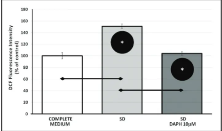
DAPH decreases ROS production in serum deprived cells
A cell membrane-permeable fluorescent dye DCF-DA that is sensitive to oxidation, was used to assess the
levels of intracellular oxidative stress after exposure to both SD and DAPH for 24 h. Differentiated and serum starved PC12 cells treated with 10 μM DAPH for 24 h displayed a decreased fluorescence, about 50% when compared with SD group cells (p<0.05) (Figure 2).
Fig. 1. Cytotoxic levels were assessed by MTT assay. (a) Effect of
DAPH on non-starved PC12 viability. Differentiated PC12 cells were treated with different concentrations of DAPH (0–50 μM) for 24 hrs, Data are means ± SD. *p<0.05 vs. 0 (control), n=6, (b) Effects of non-toxic concentrations of DAPH on serum-deprived (SD) PC12 viability. Data are means ± SD. †p<0.05 vs. 0 (control), *p<0.05 vs. SD, n=6.
Fig. 2. Effect of SD and DAPH treatment on elevating ROS
level of differentiated PC12 cells was determined by spectrophotometrically and showed by DCF fluorescence
362
Copyright © 2013 Neuroendocrinology Letters ISSN 0172–780X • www.nel.eduVolkan Ergin, Mutlu Erdogan, Çimen Karasu, Adnan Menevşe
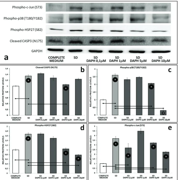
DAPH leads to diminished apoptotis and decreases stress related protein levels
Western blot analysis of PC12 cells exposed to SD revealed expression levels of various proteins, which were markedly downregulated during DAPH treatment (Figure 3a). DAPH co-applied with SD had inhibitory effects on apoptosis compared to SD control. To evalu-ate the protective effect of DAPH on serum-deprived cells, we first tested the caspase-3 status. Caspase-3 is a critical executioner of apoptosis. Activation of caspase-3 requires proteolytic processing of its
inac-tive zymogen into activated fragments (Nicholson et
al. 1995). SD itself induced cleavage of pro-caspase-3,
resulting in the formation of the 19-kDa active form of this enzyme. Western blot analysis showed that DAPH at a concentration of 5 μM, significantly attenuated the SD-induced formation of the active caspase-3 (p<0.05), (Figure 3b). We next examined if p38 MAPK pathway is involved in DAPH protection. p38 is activated by a variety of cellular stresses including inflammatory cyto-kines, LPS and UV (Rouse et al. 1994; Lee et al. 1994). In PC12 cells, to evaluate the expression profile of p38
Fig. 3. (a) Western blot analysis of phospho-c-Jun (S73), phospho-p38 MAPK (T180/Y182), phospho-HSP27 (S82), cleaved CASP3 (N175) and
GAPDH. (b) Bar graphs showing protein levels of cleaved CASP3 (N175), (c) phospho-p38 MAPK (T180/Y182), (d) phospho-HSP27 (S82),
MAPK in SD and DAPH treated conditions, p38 activ-ity was monitored by anti-phospho-p38 (T180/Y182) antibody that specifically binds activated p38. Treat-ment with DAPH at 5 and 10 μM, significantly reduced p38 phosphorylation, the indicator of p38 activation (p<0.05), (Figure 3c). In response to stress, HSP27 expression increases several-fold to create cellular resistance to the adverse environmental stimuli. HSP27 is phosphorylated at Ser15, Ser78, and Ser82 by MAP-KAPK-2 as a result of the activation of the p38 MAPK pathway (Rouse et al. 1994; Landry et al. 1992). Serum deprivation for 24 h caused an increase in activation of HSP27 (Ser82), which was significantly attenuated by addition of DAPH in a dose-dependent manner (p<0.05), (Figure 3d). In addition to this, in vitro exper-iments on neurons have demonstrated that activation of c-JUN by Ser63 and Ser73 phosphorylation can pro-mote apoptosis following serum withdrawal (Ham et al. 2000). Hence, we wanted to test whether suppression of c-JUN by DAPH contributed to the neuroprotec-tive effect of DAPH against serum deprivation, and we demonstrated that DAPH has a significant inhibitory effect on c-JUN phosphorylation at its non-toxic con-centrations (p<0.05), (Figure 3e). Taken together, these results indicate that SD triggers several stress related and proapoptotic pathways in differentiated PC12 cells, on the other hand, DAPH has a neuroprotective effect during serum starvation.
DISCUSSION
SD-induced PC12 cell death was used as an apoptotic model to investigate the therapeutic potential of DAPH as a neuroprotectant in this study. The major finding of this study is that DAPH has a significant antiapoptotic effect on differentiated PC12 cells exposed to SD. The mechanism of this protective action seems to be medi-ated through reducing activation of CASP3, p38 MAPK, HSP27 and c-JUN. We further showed that the protec-tive mechanisms includes reduction of oxidaprotec-tive stress. Here we provide the first evidence for direct neuro-protective effect of DAPH against SD-induced apopto-sis. It is known that PC12 cell apoptosis is induced by various stimuli, including SD and neurotrophic factor withdrawal. In cultured PC12 cells, it has been shown that SD rapidly induces apoptosis, and some studies reported that SD leads to oxidative stress, which is a mediator of neuronal apoptosis. However, the mecha-nism by which serum deprivation causes ROS pro-duction is not clear; our results are consistent with previously reported studies that claim oxidative stress is involved in SD-induced cell death in PC12 cells (Rukenstein et al. 1991; Atabay et al. 1996; Lee et al. 2010). Moreover, we demonstrated that DAPH admin-istration reversed the oxidative stress status.
Mitogen-activated protein kinases (MAPK) path-ways play an important role in cell death and survival. It has been reported that apoptosis induced by
with-drawal of trophic factors is mediated by increased p38 MAPK activity which is activated by inflammatory cytokines, environmental stressors, including UV, heat and hyperosmolarity (Han et al. 1994; Kummer et al. 1997; Kyriakis & Avruch 1996). p38 MAPK has previ-ously been reported to be activated in oxidant-induced apoptosis in cortical neuron model, considered to be a key factor in cell death (Namgung et al. 2000). Also p38 MAPK has been shown to promote neuronal cell death in in vivo experimental models of other neuro-degenerative diseases (Legos et al. 2001; Segura Terros
et al. 2006) and it has been shown to be activated in
patients with Alzheimer’s disease (Zhu et al. 2001). Our data indicate that the oxidative stress and activation of p38 MAPK attenuated by DAPH treatment.
Activation of p38 MAPK then leads to cleavage of pro-CASP3, yielding active cleaved-CASP3, one of the key effectors of apoptosis (Khreiss et al. 2002). It was reported that, upon serum starvation, PC12 cells exhibit increased activation of CASP3, which is considered as an indicator of cell death. (Kim et al. 2000) These results are consistent with ours, in addition to this, we found that DAPH induction resulted in decreased formation of cleaved-CASP3.
Upon stimulation by stress, p38MAPK is phos-phorylated, which then phosphorylates MAPKAPK2 to phosphorylate and activate HSP27 (Rouse et al. 1994). Phosphorylation of HSP27 is observed in response to various stimuli that have either inhibitory (oxida-tive stress, serum starvation) or stimulatory (serum, mitogens) effects on cell proliferation. Other than that, phosphorylation of HSP27 is under the control of intracellular levels of ROS (Mehlen & Arrigo 1994). We observed that HSP27 is phosphorylated during serum starvation, on the other hand, DAPH exposure to SD cells decreased the expression level of HSP27 phosphorylation. This indicates that DAPH contrib-uted to PC12 survival via decreasing lethal effects of SD-induced ROS, which eliminates the requiriment for HSP27 regulated prosurvival pathways.
Several studies reported that JNK is another protein activated by oxidative stress, which is a consequence of SD-induction (Marques et al. 2003) and it is considered as an essential molecule in neurodegeneration (Herd-egen et al. 2001). Our results show that oxidative stress and c-JUN, an important transcription factor that is activated by JNK, are attenuated by DAPH treatment in differentiated PC12 cells. These results suggested that DAPH may act as an anti-oxidant molecule against SD conditions. Taken all together, in the study reported here, we have documented that DAPH has an inhibi-tory effect on SD-induced stress and apoptosis.
In conclusion, we have provided for the first evi-dence that DAPH can attenuate serum withdrawal induced apoptosis in neurons. The down-regulation of activated CASP3, p38 MAPK, HSP27 and c-JUN might be responsible for this protective effect. This indicates that DAPH is at least partially contributes to
mainte-364
Copyright © 2013 Neuroendocrinology Letters ISSN 0172–780X • www.nel.eduVolkan Ergin, Mutlu Erdogan, Çimen Karasu, Adnan Menevşe
nance of cellular homeostasis during serum starvation. Our findings indicate that DAPH has a neuroprotec-tive effect on SD-caused stress, probably via contribut-ing the re-establishment of redox homeostasis. Hence, taken together with the other studies reporting that DAPH has a preventive effect on development of Alzhe-mier’s disease, our results suggest that DAPH could be a potential therapeutic agent for neurodegenerative disorders.
ACKNOWLEDGMENTS
We are grateful to Dr. Vibor Laketa (EMBL, Germany) for providing PC12 (6–15) cell line. Part of this work was presented as a poster at the Molecular Medicine Con-ference, Bangkok, Thailand, December 19–22, 2012.
REFERENCES
1 Atabay C, Cagnoli CM, Kharlamov E, Ikonomovic MD, Manev H (1996). Removal of serum from primary cultures of cerebellar granule neurons induces oxidative stress and DNA fragmenta-tion: protection with antioxidants and glutamate receptoran-tagonists. J Neurosci Res. 43: 465–475.
2 Bialik S, Cryns VL, Drincic A, Miyata S, Wollowick AL, Srinivasan A et al (1999). The mitochondrial apoptotic pathway is activated by serum and glucose deprivation in cardiac myocytes. Circ Res.
85: 403–414.
3 Blanchard BJ, Chen A, Rozeboom LM, Stafford KA, Weigele P, Ingram VM (2004). Efficient reversal of Alzheimer’s disease fibril formation and elimination of neurotoxicity by a small molecule. Proc Natl Acad Sci USA. 101: 14326–14332.
4 Ferrari G, Yan CY, Greene LA (1995). N-acetylcysteine (D- and L-stereoisomers) prevents apoptotic death of neuronal cells. J Neurosci. 15: 2857–2866.
5 Greene LA (1978). Nerve growth factor prevents the death and stimulates the neuronal differentiation of clonal PC12 pheochro-mocytoma cells in serum-free medium. J Cell Biol. 78: 747–755. 6 Ham J, Eilers A, Whitfield J, Neame SJ, Shah B (2000). c-Jun and
the transcriptional control of neuronal apoptosis. Biochem Phar-macol. 60: 1015–1021.
7 Han J, Lee JD, Bibbs L, Ulevitch RJ. A MAP kinase targeted by endotoxin and hyperosmolarity in mammalian cells. Science.
265: 808–811.
8 Hempstead BL, Rabin SJ, Kaplan L, Reid S, Parada LF, Kaplant DR (1992). Overexpression of the trk tyrosine kinase rapidly accel-erates nerve growth factor-induced differentiation. Neuron. 9: 883–896.
9 Herdegen T, Waetzig V (2001). AP-1 proteins in the adult brain: facts and fiction about effectors of neuroprotection and neuro-degeneration. Oncogene. 20: 2424–2437.
10 Higuchi A, Shimmura S, Takeuchi T, Suematsu M, Tsubota K (2006). Elucidation of apoptosis induced by serum deprivation in cultured conjunctival epithelial cells. Br J Ophthalmol. 90: 760–764.
11 Hillion JA, Takahashi K, Maric D, Ruetzler C, Barker JL, Hallenbeck JM (2005). Development of an ischemic tolerance model in a PC12 cell line. J Cereb Blood Flow Metab. 25: 154–162.
12 Howard MK, Burke LC, Mailhos C, Pizzey A, Gilbert CS, Lawson WD et al (1993). Cell cycle arrest of proliferating neuronal cells by serum deprivation can result in either apoptosis or differentia-tion. J neurochem. 60: 1783–1791.
13 Khreiss T, József L, Hossain S, Chan JS, Potempa LA, Filep JG (2002). Loss of pentameric symmetry of C-reactive protein is associated with delayed apoptosis of human neutrophils. J Biol Chem. 277: 40775–40781.
14 Kim HY, Akbar M, Lau A, Edsall L (2000). Inhibition of neuronal apoptosis by docosahexaenoic acid (22:6n-3). Role of phosphati-dylserine in antiapoptotic effect. J Biol Chem. 275: 35215–35223. 15 King AR, Francis SE, Bridgeman CJ, Bird H, Whyte MK, Crossman
DC (2003). A role for caspase-1 in serum withdrawal induced apoptosis of endothelial cells. Lab Invest. 83: 1497–1508. 16 Kummer JL, Rao PK, Heidenreich KA (1997). Apoptosis induced
by withdrawal of trophic factors is mediated by p38 mitogen-activated protein kinase. J Biol Chem. 272: 20490–20494. 17 Kyriakis JM, Avruch J (1996). Protein kinase cascades activated
by stress and inflammatory cytokines. Bioessays. 18: 567–577. 18 Landry J, Lambert H, Zhou M, Lavoie JN, Hickey E, Weber LA et
al (1992). Human HSP27 is phosphorylated at serines 78 and 82
by heat shock and mitogen-activated kinases that recognize the same amino acid motif as S6 kinase II. J Biol Chem. 267: 794–803. 19 LeBlanc A, Liu H, Goodyer C, Bergeron C, Hammond J. (1999).
Caspase-6 role in apoptosis of human neurons, amyloidogenesis, and Alzheimer’s disease. J Biol Chem. 274: 23426–23436. 20 Lee JC, Laydon JT, McDonnell PC, Gallagher TF, Kumar S, Green D
et al (1994). A protein kinase involved in the regulation of
inflam-matory cytokine biosynthesis. Nature. 372: 739–746.
21 Lee SB, Kim JJ, Kim TW, Kim BS, Lee MS, Yoo YD (2010). Serum deprivation-induced reactive oxygen species production is mediated by Romo1. Apoptosis. 15: 204–218.
22 Lee WC, Chen YY, Kan D, Chien CL (2012). A neuronal death model: Overexpression of neuronal intermediate filament pro-tein peripherin in PC12 cells. J biomed sci. 19: 1–13.
23 Legos JJ, Erhardt JA, White RF, Lenhard SC, Chandra S, Parsons AA (2001). SB 239063, a novel p38 inhibitor, attenuates early neuronal injury following ischemia. Brain Res. 892: 70–77. 24 Li J, Li Y, Ogle M, Zhou X, Song M, Yu SP et al (2010).
DL-3-n-butylphthalide prevents neuronal cell death after focal cerebral ischemia in mice via the JNK pathway. Brain Res. 1359: 216–226. 25 Lipton P (1999). Ischemic cell death in brain neurons. Physiol
Rev. 79: 1431–1568.
26 Mattson MP (2000). Apoptosis in neurodegenerative disorders. Nat Rev Mol Cell Biol. 1: 120–129.
27 Marques CA, Keil U, Bonert A, Steiner B, Haass C, Muller WE (2003). Neurotoxic mechanisms caused by the Alzheimer’s disease-linked Swedish amyloid precursor protein mutation: oxi-dative stress, caspases, and the JNK pathway. J Biol Chem. 278: 28294–28302.
28 Mehlen P, Arrigo AP (1994). The serum-induced phosphorylation of mammalian hsp27 correlates with changes in its intracellular localization and levels of oligomerization. Eur J Biochem. 221: 327–334.
29 Namgung U, Xia Z (2000). Arsenite-induced apoptosis in corti-cal neurons is mediated by c-JUN N-terminal protein kinase 3 and p38 mitogen-activated protein kinase. J Neurosci. 20: 6442–6451.
30 Nicholson DW, Ali A, Thornberry NA, Vaillancourt JP, Ding CK, Gallant M et al (1995). Identification and inhibition of the ICE/ CED-3 protease necessary for mammalian apoptosis. Nature.
376: 37–43.
31 Pandey S, Lopez C, Jammu A (2003). Oxidative stress and activa-tion of proteasome protease during serum deprivaactiva-tion-induced apoptosis in rat hepatoma cells; inhibition of cell death by mela-tonin. Apoptosis. 8: 497–508.
32 Rouse J, Cohen P, Trigon S, Morange M, Alonso-Llamazares A, Zamanillo D et al (1994). A novel kinase cascade triggered by stress and heat shock that stimulates MAPKAP kinase-2 and phosphorylation of the small heat shock proteins. Cell. 78: 1027–1037.
33 Rukenstein A, Rydel RE, Greene LA (1991). Multiple agents rescue PC12 cells from serum-free cell death by translation- and tran-scription-independent mechanisms. J Neurosci. 11: 2552–2563. 34 Satoh T, Sakai N, Enokido Y, Uchiyama Y, Hatanaka H (1996).
Survival factor-insensitive generation of reactive oxygen species induced by serum deprivation in neuronal cells. Brain Res. 733: 9–14.
365
Neuroendocrinology Letters Vol. 34 No. 5 2013 • Article available online: http://node.nel.edu
35 Segura Torres JE, Chaparro-Huerta V, Rivera Cervantres MC, Mon-tes-González R, Flores Soto ME, Beas-Zárate C (2006). Neuronal cell death due to glutamate excitotocity is mediated by p38 activation in the rat cerebral cortex. Neurosci Lett. 403: 233–238. 36 Stetler RA, Gan Y, Zhang W, Liou AK, Gao Y, Cao G et al (2010).
Heat shock proteins: cellular and molecular mechanisms in the central nervous system. Prog neurobiol. 92: 184–211.
37 van der Valk J, Mellor D, Brands R et al (2004). The humane col-lection of fetal bovine serum and possibilities for serum-free cell and tissue culture. Toxicol In Vitro. 18: 1–12.
38 Vaudry D, Stork PJS, Lazarovici P, Eiden LE (2002). Signaling path-ways for PC12 cell differentiation: making the right connections. Sci Signal. 296: 1648.
39 Wang H, Duennwald ML, Roberts BE, Rozeboom LM, Zhang YL, Steele AD et al (2008). Direct and selective elimination of specific prions and amyloids by 4, 5-dianilinophthalimide and analogs. Proc Natl Acad Sci USA. 105: 7159–7164.
40 Yuan J, Lipinski M, Degterev A (2003). Diversity in the mecha-nisms of neuronal cell death. Neuron 40: 401–413.
41 Zhu X, Castellani RJ, Takeda A, Nunomura A, Atwood CS, Perry G (2001). Differential activation of neuronal ERK, JNK/SAPK and p38 in Alzheimer disease: the ‘two hit’ hypothesis. Mech Ageing Dev. 123: 39–46.
42 Zhuge J, Cederbaum AI (2006). Serum deprivation-induced HepG2 cell death is potentiated by CYP2E1. Free Radic Biol Med.