http://journals.tubitak.gov.tr/medical/ © TÜBİTAK
doi:10.3906/sag-1505-69
The effect of kisspeptin on spermatogenesis and apoptosis in rats
Nilüfer AYTÜRK1, Tülin FIRAT2, Aysel KÜKNER2,*, Candan ÖZOĞUL3, Fatma TÖRE4, İsmail Engin KANDIRALI5, Bayram YILMAZ6
1Department of Histology and Embryology, Faculty of Medicine, Medipol University, İstanbul, Turkey 2Department of Histology and Embryology, Faculty of Medicine, Abant İzzet Baysal University, Bolu, Turkey
3Department of Histology and Embryology, Faculty of Medicine, Gazi University, Ankara, Turkey 4Department of Physiology, Faculty of Medicine, SANKO University, Gaziantep, Turkey 5Department of Urology Clinics, Bağcılar Education and Research Hospital, İstanbul, Turkey 6Department of Physiology, Faculty of Medicine, İstanbul Yeditepe University, İstanbul, Turkey
1. Introduction
Kisspeptins, the peptide products of the KiSS-1 gene, bind to the G protein-coupled receptor 54 (GPR54), a critical regulator of GnRH secretion. The N-terminally truncated peptide metastin 45-54 exhibits 10-fold higher receptor-binding affinity than full-length metastin; it also shows agonistic KISS-1R activity (1).
Kisspeptins are known to play a role in puberty, cancer metastasis, and vasoconstriction (2,3–7). They are synthesized in the arcuate and anteroventral periventricular hypothalamic neurons and in the perioptic area (8). Kisspeptin expression is variable according to sex in rats; its expression is more marked in female animals (9). Its known derivatives are kisspeptin-10, kisspeptin-13, kisspeptin-14, and kisspeptin-54 (10– 12). All kisspeptins seem to have the same interactions under in vitro conditions. Kiss-10 is well characterized
in mammals, in which it is found in large concentrations (10,11). It has been suggested, however, that Kiss-54 is the most effective form (12). Kisspeptin has been reported to also be synthesized in the testes, ovaries, pancreas, gut, liver, lung, muscle tissue, kidney, nervous system, and most densely in the placenta (13,14). As for its receptor, G protein-coupled receptor-54 (GPR54) is mostly expressed in the hypophysis, placenta, and pancreas (10,15,16). Kisspeptins are highly potent neuropeptides that stimulate the secretion of LH and FSH from the hypophysis, an effect exerted through the release of GnRH (7,17). While this pathway has been satisfactorily defined in several mammal species, the molecular and cellular events at the proencephalic origin of this process are incompletely elucidated. In recent years, studies have indicated that kisspeptin plays a role in the transition to puberty (18,19). Kiss-1 knockout mice showed hypogonadism and low Background/aim: To study the effect of kisspeptin, a gonadotropin release stimulator, on the testicular tissue of the rat.
Materials and methods: Four groups were formed as follows: control, Kiss-10 50 nmol administration for 1 day, Kiss-10 administration
for 13 days, and one last group kept for 7 days following Kiss-10 applied for 13 days. Testicular tissues were stained with hematoxylin-eosin, periodic acid Schiff, Masson trichrome staining, terminal deoxynucleotidyl transferased UTP nick-end labeling, and Ki-67 immune staining. Serum testosterone levels were determined.
Results: Serum testosterone level increased following acute application, while it was reduced by chronic treatment. Spermatogenic
cells as stained by Ki-67 and TUNEL increased in the treated groups compared to the controls. Following a 7-day rest after treatment, a decrease in testosterone levels and Ki-67–stained cell numbers and an increase in TUNEL-stained cells were observed. Leydig cells showed increased vacuolization in the Kiss-1 group. Leydig cell vacuolization continued in the Kiss (13) group and was reduced in the Kiss (13 + 7) group.
Conclusion: Kiss-10 increased spermatogenic cell proliferation, while testosterone level and proliferation decreased and apoptosis
increased during the waiting period.
Key words: Kisspeptin, spermatogenesis, apoptosis, Leydig cells
Received: 15.05.2015 Accepted/Published Online: 21.04.2016 Final Version: 27.02.2017 Research Article
AYTÜRK et al. / Turk J Med Sci levels of circulating gonadotropin (20,21). Kisspeptin
has been shown to control seasonal fertility in sheep (22) and hamsters (23,24). A literature search revealed only a limited number of studies on the effects of kisspeptin.
The aim of the present study was to study the structural and biochemical effects of acute and chronic kisspeptin administration on seminiferous tubular cells, spermatogenesis, apoptosis, and Leydig cells and the reversibility of such effects.
2. Materials and methods 2.1. Experimental animals
All experimental protocols were performed according to the guidelines for the ethical treatment of experimental animals and were approved by the Animal Care and Use Local Ethics Committee. Eight-week-old male Wistar albino rats (200–230 g) were housed at a constant room temperature (21 ± 2 °C) under a 12-h light/dark cycle. They were fed standard rat chow (210 kcal/100 g per day) and drank tap water.
Twenty-four male Wistar rats weighing 200–300 g were used in the study. Four groups were constructed:
Control group (n = 6): Received intraperitoneal normal saline only.
Kiss (1) (n = 6): Kiss-10 [metastin (45-54) amide, M2816-1MG, SIGMA], 50 nmol, intraperitoneally was given once to these animals.
Kiss (13) (n = 6): These animals received Kiss-10, 50 nmol, intraperitoneally daily for 13 days.
Kiss (13 + 7) (n = 6): After receiving Kiss-10, 50 nmol, intraperitoneally daily for 13 days, these animals were kept for 7 days without any treatment. Kisspeptin-10 was used because it is the minimal sequence required for full receptor binding and activation (10,11). The single injection treatment was defined as acute and the 13-day treatment as chronic. The rats were weighed before the study and at its termination. Testes were removed and weighed. Gonadosomatic index percent (GSI %) was determined using the following formula (25):
GSI (%) = Testes weight × 100 Body weight
2.2. Biochemistry
Blood was drawn under anesthesia from the inferior vena cava, centrifuged at 1000 × g for 20 min, and kept at –20 °C. Total testosterone level was measured using an ELISA kit (USCN Life Science Inc., Wuhan, E90458Ra, L101101379, P.R. China). The measurement was performed following the instructions provided with the kit.
2.3. Light microscopy
The left testes were fixated in a 10% solution of neutral formalin. The right testes were frozen at –96 °C to examine with immunofluorescence microscopy. Blocks
were prepared after routine preparation. Sections were stained with hematoxylin-eosin, periodic acid Schiff (PAS), and Masson’s trichrome methods; the diameters of the seminiferous tubules were measured. Staining with Ki-67 was performed (9106S1003B, Thermo Scientific, USA) for marking proliferating cells and with terminal deoxynucleotidyl transferased UTP nick-end labeling (TUNEL) (ApopTag Plus Peroxidase In Situ Apoptosis Detection Kit, S7101, Chemicon International; Lot PSO1736498) for apoptotic cells. The numbers of apoptotic and proliferating cells were determined by counting stained dark-brown nuclei in 10 cross-sectional fields of seminiferous tubules per testis section. Tissues were frozen at –96 °C for immune fluorescence determination of Kiss-10 expression. Sections 15-µm thick obtained by a freezing microtome were stained using a Kiss-1 antibody [(C-20), goat polyclonal IgG, #H1910, Santa Cruz Biotechnology, Inc., Dallas, TX, USA] and examined under an immune fluorescence microscope.
2.4. Electron microscopy
Tissues were taken and fixed in 2.5% glutaraldehyde in 0.1 M sodium phosphate buffer (pH 7.2) for 24 h. After rinsing with phosphate buffer, tissues were postfixed with 2% osmium tetroxide in sodium phosphate buffer. Dehydration was accomplished by gradual ethanol series and embedded in epoxy resin. Semithin sections (1 µm) were stained toluidine blue. Sections were then viewed and photographed with light microscopy.
2.5. Statistical evaluation
The study data were evaluated using GraphPad Prism software (version 3.00, GraphPad Software, La Jolla, CA, USA). The Kruskal–Wallis test was used for between-group comparison of apoptotic and Ki-67(+) cell counts and Dunn’s multiple comparison test for within-group evaluations. Subject weights, testosterone levels, testis weights, gonadosomatic index (GSI %), and tubular diameters were evaluated by the Mann–Whitney U test. Biochemical measurements were compared using one-way ANOVA, while within-group comparisons used Tukey’s test. P-values smaller than 0.05 were accepted as significant.
3. Results
There was no difference in body weight, testicular weight, GSI %, or diameter of seminiferous tubules among the groups. Kiss-1 immune positive cells were not detected in any group (data not shown).
3.1. Serum testosterone levels
While serum testosterone levels were significantly elevated after a single administration of kisspeptin (P < 0.03) they decreased in the Kiss (13) and Kiss (13 + 7) groups, although with no significant difference from the controls (Figure 1).
3.2. Proliferating cell count
Ki-67 immune staining was applied to determine the number of proliferative cells in the seminiferous tubules. The Ki-67–stained spermatogenic cells were counted for all groups. Ki-67–stained cell counts were higher in the Kiss (13) group compared to both Kiss (1) and the controls (P < 0.001). As for the (13 + 7) group, after a 7-day rest following 13 consecutive days of treatment, the stained cell count was higher than that in both the controls and Kiss (1) (P < 0.001 and 0.01, respectively) but lower than that of Kiss (13). No statistical significance was detected between the Kiss (13) and Kiss (13 + 7) groups (Figure 2).
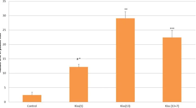
3.3. Apoptotic cell count
TUNEL staining was applied to mark apoptotic cells in the seminiferous tubules. TUNEL (+)-stained spermatogenic cells were counted in all groups (Figure 3).
A lower number of TUNEL (+) cells were detected in the Kiss (1) group, but the difference was not significant. Apoptotic TUNEL (+) cell counts were significantly higher in the Kiss (13) and Kiss (13 + 7) groups compared to the controls (P < 0.05) and Kiss (1) (P < 0.001).
3.4. Light microscopy findings
Kisspeptin expression was not observed in either the control or the treatment groups
(data not shown). Seminiferous tubules in treated animals were largely similar to those of the control group (Figures 4a–4d). Cell shedding was present but not widespread in the lumens of some tubules in the treatment groups. Tissues from all groups were stained with PAS. No disruption or thickening of the basal membrane was seen with PAS staining in the treatment groups. Staining intensity was comparable throughout all animal groups. Connective tissue proliferation among seminiferous
tubules was not observed following Masson trichrome staining (data not shown).
The appearance of Sertoli and Leydig cells, spermatogonia, primary spermatocytes, spermatids, developing acrosomes, and spermatozoa was by definition normal for the control group (Figure 5a). The seminiferous tubule basal membrane was markedly corrugated in the Kiss (1) group, where vacuolization in the Sertoli cells and spermatocytes, increased intercellular intervals, and numerous vacuoles in Leydig cells were observed (Figure 5b). Leydig cell vacuolization continued in the Kiss (13) group (Figure 5c). In the Kiss (13 + 7) group, such vacuoles in the Leydig cells were fewer; excessive intercellular distances remained (Figure 5d).
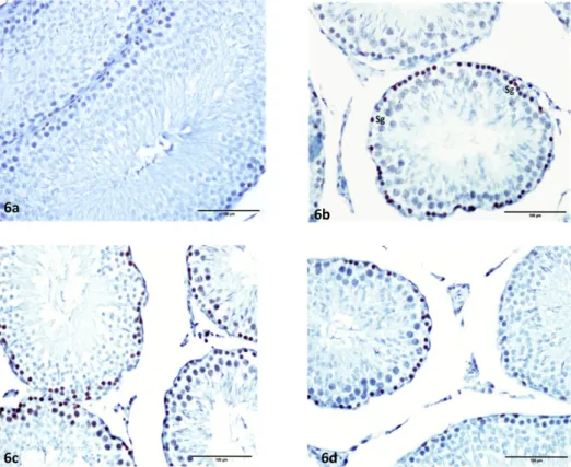
The cells that stained positive with Ki-67 were generally identified as spermatogonia. No immune staining was found in Leydig or Sertoli cells. Mitotic activity was least in the controls and highest in the Kiss (13) group (Figures 6a–6d).
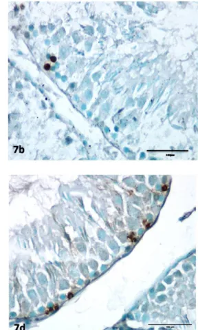
TUNEL (+)-stained cells, the spermatogonia, were very few in the controls and more abundant in the treatment groups. TUNEL (+) immune staining was not observed in the Sertoli or Leydig cells (Figures 7a–7d).
4. Discussion
Since 2003, research has revealed the important role of kisspeptins in initiation and timing of puberty and in regulating fertility in adulthood. Continuous administration of kisspeptins downregulates the hypothalamic–gonadal–pituitary (HPG) axis (26). Kisspeptin is a potent peptide that initiates luteinizing hormone (LH) release in male and female mice (27), in both prepubertal and adult rats (28) and in prepubertal donkeys (29). Plasma LH and testosterone levels were reported to Figure 1. Plasma testosterone levels. *P < 0.03 compared to other groups.
AYTÜRK et al. / Turk J Med Sci
Figure 2. Number of Ki-67(+) cells. Ki-67 staining cells in the acute treatment, Kiss (1) (*), chronic treatment Kiss (13)
(**) and additional waiting period
Kiss (13 + 7) (***) groups were statistically significantly higher than in the control group (P < 0.001) *Control vs. Kiss (1) group, P < 0.001
**Control vs. Kiss (13) group, P < 0.001 ***Control vs. Kiss (13 + 7) group, P <0.001 #Kiss (1) vs. Kiss (13) group, P < 0.001 ##Control vs. Kiss (13 + 7) group, P <0.01
Figure 3. Apoptotic TUNEL (+) cell counts were significantly higher in the Kiss (13) and Kiss (13 + 7) groups
compared to the controls (*P < 0.05) and Kiss (1) (**P < 0.001).
*Control vs. Kiss (13) group, P < 0.05
***Control vs. Kiss (13 + 7) group, P < 0.05 **Kiss (1) vs. Kiss (13) group, P < 0.001 **Kiss (1) vs. Kiss (13 + 7) group, P < 0.001
be increased on the day following the first administration of Kisspeptin-54 (50 nmol/day), an effect that disappeared after the second day of treatment, when hormone levels returned to normal. This result was interpreted as being due to desensitization of the HPG axis by the continuous use of kisspeptin (12). A single dose of Kiss-54 was reported to cause ovulation in rats (30) and to significantly increase sex steroid levels in prepubertal fish (31). Gonadotropin increases were registered following a single IV or IM dose of Kiss-10 in prepubertal cats (32), sheep (33), and goats (34). It was reported that Kiss-10 given at varying doses reduces testosterone levels, a reduction that was attributed to testicular degeneration rather than a fall in LH secretion (25). However, another published study indicated that different doses of Kiss-10 were all followed by a testosterone increase within 20 min of administration (35). We observed that Kiss-10 significantly increased total testosterone plasma levels in the acute phase while
chronic administration was associated with a decrease, which continued in the additional waiting period without reaching the control levels. The increased vacuolization in the Leydig cells was interpreted as a possible enlargement of endoplasmic reticulum, consistent with active secretion. Kisspeptin mRNA, characterized in human testicular tissue (11), was not identified in rat testes (36). Wang et al. reported that Kiss-10 expression is strong in Leydig cells of 5-week- and 15-week-old mice, while it is weak in 2-week-old mice (37). Salehi et al., while observing Kisspeptin expression in Leydig cells, found no development in germ cells or Sertoli cells at any stage (38). We did not detect any positive immune staining for Kiss-1 in any group in our study.
Kisspeptins were reported to have a predominately autocrine and paracrine action in the testes (39,40). Our results are similar to those reported by Thompson et al. in that testosterone levels decreased over a 13-day Figure 4. Light microscopic view of seminiferous tubules in experimental groups. The seminiferous tubules structure can
be seen to resemble each other. Control (4a), Kiss (1) (4b), Kiss (13) (4c), and Kiss (13 + 7) (4d) groups. Hematoxylin and eosin stain.
AYTÜRK et al. / Turk J Med Sci
Figure 5. Semithin section view of experimental groups. Seen intact spermatogenic and Sertoli cells in the control group (5a). More
folding basement membrane, many vacuoles in the Sertoli and Leydig cell cytoplasm are seen in the Kiss (1) group (5b). Increased vacuoles in the Leydig cells cytoplasm of the Kiss (13) group (5c) and decreased vacuoles in the Leydig cells cytoplasm of the Kiss (13 + 7) group (5d) are seen. Sp: Spermatid, Ps: Primary spermatocytes, SC: Sertoli cells, LC: Leydig cells. Toluidine blue stain.
Figure 6. Ki-67 immunohistochemistry stained view of groups. Control (6a), Kiss (1) (6b), Kiss (13) (6c), and Kiss (13 + 7) (6d) groups.
Ki-67-positive–stained spermatogenic cells of Kiss (1) and Kiss (13) groups are increased compared to the control and Kiss (13 + 7) groups.
treatment period and adult male rats were used (12,39). The increase seen in Ki-67–stained spermatogonia within the seminiferous tubules in our study was interpreted as related to the increase in testosterone levels; their proliferation increased with both acute and chronic treatment and decreased in the additional waiting period without, however, reaching control levels. This finding was attributed to the reversibility of Kiss-10 action.
A twice-weekly high-dose (250 ng/g) Kiss-10 treatment for 7 weeks increased the GSI of prepubertal white bass, while no significant change could be detected in striped bass (41). Another report indicated that Kiss-15 increases GSI in prepubertal fish (31). Reports vary according to subject type and physiological development stages. Varying stimuli have been determined for different modes of administration (central or systemic) and different physiological stages (42,43). Kisspeptin-54, 50 nmol daily for 13 days, led to testicular degeneration in adult rats (12,39). Application of Kiss-10 led to seminiferous tubule degeneration in prepubertal rats; cellular degeneration was seen to increase along with increasing dose. Cytoplasmic vacuolization was demonstrated by light and electron
microscopy in Sertoli cells and spermatogonia (25). In our study, no obvious degeneration was observed in seminiferous tubule cells even though the dose administered amounted to 50 nmol; in contrast, the increased testosterone level increased spermatogenesis. While apoptotic spermatogonia were identified in our study, no apoptosis was seen in the Sertoli or Leydig cells. Semithin sections showed a large degree of cytoplasmic vacuolization in the Sertoli cells and spermatogonia. Vacuolization decreased in the cells of animals in the groups subjected to chronic treatment and in those with the additional waiting period. Apoptotic cell counts were low in the acute treatment animals and increased in the two other treatment groups. The increase in Ki-67 staining cells in acute treatment animals paralleled the increase in testosterone level. However, proliferation increased in chronic treatment animals despite the testosterone decrease. The increased apoptotic cell count was possibly offsetting the increase in total cell count that occurred over the same period. Testosterone levels continued to decrease, apoptosis counts increased, and proliferation of spermatogenic cells decreased over the 7-day waiting period following chronic administration. Figure 7. TUNEL-positive staining in spermatogenic cells in the Kiss (13 + 7) group is higher than the other groups.
AYTÜRK et al. / Turk J Med Sci To summarize, acute or chronic administration of
50 nmol/kg kisspeptin-10 was not observed to cause degeneration in the rat seminiferous tubules, while it increased cellular proliferation; the interruption of kisspeptin was associated with a decrease in proliferation and testosterone levels and with an increase in apoptosis. Kiss-10 was seen to exert its effects through testosterone
secretion; these effects were reversible. The increased proliferation in spermatogenic cells may lead to new developments in the treatment of infertility.
Acknowledgment
This study was supported by Abant İzzet Baysal University Scientific Research Foundation (2010.08.01.348).
References
1. Asami T, Nishizawa N, Matsui H, Nishibori K, Ishibashi Y, Horikoshi Y, Nakayama M, Matsumoto S, Tarui N, Yamaguchi M et al. Design, synthesis, and biological evaluation of novel investigational nonapeptide KISS1R agonists with testosterone-suppressive activity. J Med Chem 2013; 56: 8298-8307. 2. Lee JH, Miele ME, Hicks DJ, Phillips KK, Trent JM, Weissman
BE, Welch DR. KiSS-1, a novel human malignant melanoma metastasis suppressor gene. J Natl Cancer Inst 1996; 88: 1731-1737.
3. Lee JH, Welch DR. Identification of highly expressed gene in metastasis-suppressed chromosome 6/human malignant melanoma hybrid cells using subtractive hybridization and differential display. Int J Cancer 1997; 71: 1035-1041.
4. Lee DK, Nguyen T, O’Neill GP, Cheng R, Liu Y, Howard AD, Coulombe N, Tan CP, Tang-Nguyen AT, George SR et al. Discovery of a receptor related to the galanin receptors. FEBS Lett 1999; 446: 103-107.
5. Mead EJ, Maguire JJ, Kuc RE, Davenport AP. Kisspeptins: a multifunctional peptide system with a role in reproduction, cancer and the cardiovascular system. Br J Pharmacol 2007; 151: 1143-1153.
6. Popa SM, Clifton DK, Steiner RA. The role of kisspeptins and GPR54 in the neuroendocrine regulation of reproduction. Annu Rev Physiol 2008; 7: 213-238.
7. Oakley AE, Clifton DK, Steiner RA. Kisspeptin signaling in the brain. Endocr Rev 2009; 30: 713-743.
8. Decourt C, Caraty A, Briant C, Guillaume D, Lomet D, Chesneau D, Lardic L, Duchamp G, Reigner F, Monget P et al. Acute injection and chronic perfusion of kisspeptin elicit gonadotropins release but fail to trigger ovulation in the mare. Biology of Reproduction 2014; 90: 1-12.
9. Overgaard A, Tena-Sempere M, Franceschin I, Desroziers E, Simonneaux V, Mikkelsen JD. Comparative analysis of kisspeptin-immunoreactivity reveals genuine differences in the hypothalamic Kiss1 systems between rats and mice. Peptides 2013; 45: 85-90.
10. Kotani M, Detheux M, Vandenbogaerde A, Communi D, Vanderwinden JM, Le Poul E, Brézillon S, Tyldesley R, Suarez-Huerta N, Vandeput F et al. The metastasis suppressor gene KiSS-1 encodes kisspeptins, the natural ligands of the orphan G protein-coupled receptor GPR54. J Biol Chem 2001; 276: 34631-34636.
11. Ohtaki T, Shintani Y, Honda S, Matsumoto H, Hori A, Kanehashi K, Terao Y, Kumano S, Takatsu Y, Masuda Y et al. Metastasis suppressor gene KiSS-1 encodes peptide ligand of a G-protein-coupled receptor. Nature 2001; 411: 613-617. 12. Thompson EL, Murphy KG, Patterson M, Bewick GA, Stamp
GW, Curtis AE, Cooke JH, Jethwa PH, Todd JF, Ghatei MA et al. Chronic subcutaneous administration of kisspeptin-54 causes testicular degeneration in adult male rats. Am J Physiol Endocrinol Metab 2006; 291: E1074-E1082.
13. Kuohung W, Kaiser UB. GPR54 and KiSS-1: role in the regulation of puberty and reproduction. Rev Endocr Metab Disor 2006; 7: 257-263.
14. Mark PJ, Jones ML, Lewis JL, Waddell BJ, Smith JT. Kiss1 and Kiss1 rmRNA expression in the rat placenta: changes with gestational age and regulation by glucocorticoids. Placenta 2013; 34: 657-662.
15. Shahed A, Young KA. Differential ovarian expression of KiSS-1 and GPR-54 during the estrous cycle and photoperiod induced recrudescence in Siberian hamsters (Phodopus sungorus). Mol Reprod Dev 2009; 76: 444-452.
16. Muir AI, Chamberlain L, Elshourbagy NA, Michalovich D, Moore DJ, Calamari A, Szekeres PG, Sarau HM, Chambers JK, Murdock P et al. AXOR12, a novel human G protein-coupled receptor, activated by the peptide KiSS-1. J Biol Chem 2001; 276: 28969-28975.
17. Mikkelsen JD, Bentsen AH, Ansel L, Simonneaux V, Juul A. Comparison of the effects of peripherally administered kisspeptins. Regul Pept 2009; 152: 95-100.
18. Tena-Sempere M. GPR54 and kisspeptin in reproduction. Hum Reprod Update 2006; 12: 631-639.
19. Tena-Sempere M. Kisspeptin signaling in the brain: recent developments and future challenges. Mol Cell Endocrinol 2010; 314: 164-169.
20. Funes S, Hedrick JA, Vassileva G, Markowitz L, Abbondanzo S, Golovko A, Yang S, Monsma FJ, Gustafson EL. The KiSS-1 receptor GPR54 is essential for the development of the murine reproductive system. Biochem Biophys Res Commun 2003; 312: 1357-1363.
21. Seminara SB, Messager S, Chatzidaki EE, Thresher RR, Acierno JS Jr, Shagoury JK, Bo-Abbas Y, Kuohung W, Schwinof KM, Hendrick AG et al. The GPR54 gene as a regulator of puberty. N Engl J Med 2003; 349:1614-1627.
22. Smith JT, Coolen LM, Kriegsfeld LJ, Sari IP, Jaafarzadehshirazi MR, Maltby M, Bateman K, Goodman RL, Tilbrook AJ, Ubuka T et al. Variation in kisspeptin and RF amide-related peptide (RFRP) expression and terminal connections to gonadotropin-releasing hormone neurons in the brain: a novel medium for seasonal breeding in the sheep. Endocrinology 2008; 149: 5770-5782.
23. Revel FG, Saboureau M, Masson-Pevet M, Pevet P, Mikkelsen JD, Simonneaux V. Kisspeptin mediates the photoperiodic control of reproduction in hamsters. Curr Biol 2006; 16: 1730-1735.
24. Simonneaux V, Bur I, Ancel C, Ansel L, Klosen P. A kiss for daily and seasonal reproduction. Prog Brain Res 2012; 199: 423-437.
25. Ramzan F, Qureshi IZ. Intraperitoneal kisspeptin-10 administration induces dose-dependent degenerative changes in maturing rat testes. Life Sciences 2011; 88: 246-256. 26. Dedes I. Kisspeptins and the control of gonadotrophin
secretion. Syst Biol Reprod Med 2012; 58: 121-128.
27. Gottsch ML, Cunningham MJ, Smith JT, Popa SM, Acohido BV, Crowley WF, Seminara S, Clifton DK, Steiner RA. A role for kisspeptins in the regulation of gonadotropin secretion in the mouse. Endocrinology 2004; 145: 4073-4077.
28. Navarro VM, Castellano JM, Fernandez-Fernandez R, Barreiro ML, Roa J, Sanchez-Criado JE, Aguilar E, Dieguez C, Pinilla L et al. Developmental and hormonally regulated messenger ribonucleic acid expression of KiSS-1 and its putative receptor, GPR54, in rat hypothalamus and potent luteinizing hormone-releasing activity of KiSS-1 peptide. Endocrinology 2004; 145: 4565-4574.
29. Shahab M, Mastronardi C, Seminara SB, Crowley WF, Ojeda SR, Plant TM. Increased hypothalamic GPR54 signaling: a potential mechanism for initiation of puberty in primates. Proc Natl Acad Sci USA 2005; 102: 2129-2134.
30. Matsui H, Takatsu Y, Kumano S, Matsumoto H, Ohtaki T. Peripheral administration of metastin induces marked gonadotropin release and ovulation in the rat. Biochem Biophys Res Commun 2004; 320: 383-388.
31. Selvaraj S, Ohga H, Nyuji M, Kitano H, Nagano N, Yamaguchi A, Matsuyama M. Subcutaneous administration of Kiss1 pentadecapeptide accelerates spermatogenesis in prepubertal male chub mackerel (Scomber japonicas). Comp Biochem Phys A 2013; 166: 228-236.
32. Ezzat AA, Saito H, Sawada T, Yaegashi T, Yamashita T, Hirata T, Sawai K, Hashizume T. Characteristics of the stimulatory effect of kisspeptin-10 on the secretion of luteinizing hormone, follicle-stimulating hormone and growth hormone in prepubertal male and female cattle. J Reprod Dev 2009; 55: 650-654.
33. Redmond JS, Macedo GG, Velez IC, Caraty A, Williams GL, Amstalden M. Kisspeptin activates the hypothalamic-adenohypophyseal-gonadal axis in prepubertal ewe lambs. Reproduction 2011; 141: 541-548.
34. Saito H, Sawada T, Yaegashi T, Goto Y, Jin J, Sawai K, Hashizume T. Kisspeptin-10 stimulates the release of luteinizing hormone and testosterone in pre- and postpubertal male goats. Anim Sci J 2012; 83: 487-492.
35. Curtis AE, Cooke JH, Baxter JE, Parkinson JRC, Bataveljic A, Ghatei MA, Bloom SR, Murphy KG. A kisspeptin-10 analog with greater in vivo bioactivity than kisspeptin-10. Am J Physiol Endocrinol Metab 2010; 298: E296-E303.
36. Terao Y, Kumano S, Takatsu Y, Hattori M, Nishimura A, Ohtaki T, Shintani Y. Expression of KiSS-1, a metastasis suppressor gene, in trophoblast giant cells of the rat placenta. Biochim Biophys Acta 2004; 1678: 102-110.
37. Wang JY, Hsu MC, Tseng TH, Wu LS, Yang KT, Chiu CH. Kisspeptin expression in mouse Leydig cells correlates with age. Journal of the Chinese Medical Association. http://dx.doi. org/10.1016/j.jcma.2015.01.004.
38. Salehi S, Adeshina I, Chen H, Zirkin BR, Hussain MA, Wondisford F, Wolfe A, Radovick S. Developmental and endocrine regulation of kisspeptin expression in mouse Leydig cells. Endocrinology 2015; 156: 1514-1522.
39. Ohga H, Fujinaga Y, Selvaraj S, Kitano H, Nyuji M, Yamaguchi A, Matsuyama M. Identification, characterization, and expression profiles of two subtypes of kisspeptin receptors in a scombroid fish (chub mackerel). Gen Comp Endocr 2013; 193: 130-140.
40. Thompson EL, Amber V, Stamp GW, Patterson M, Curtis AE, Cooke JH, Appleby GF, Dhillo WS, Ghatei MA, Bloom SR et al. Kisspeptin-54 at high doses acutely induces testicular degeneration in adult male rats via central mechanisms. Br J Pharmacol 2009; 156: 609-625.
41. Beck BH, Fuller SA, Peatman E, McEntire ME, Darwish A, Freeman DW. Chronic exogenous kisspeptin administration accelerates gonadal development in basses of the genus Morone. Comp Biochem Phys A 2012; 162: 265-273.
42. Caraty A, Decourt C, Briant C, Beltramo M. Kisspeptins and the reproductive axis: potential applications to manage reproduction in farm animals. Domest Anim Endocrinol 2012; 43: 95-102.
43. Okamura H, Tsukamura H, Ohkura S, Uenoyama Y, Wakabayashi Y, Maeda K. Kisspeptin and GnRH pulse generation. Adv Exp Med Biol 2013; 784: 297-323.