Relationship between serum DHEAS and
oxidative stress levels of body mass index
in healthy postmenopausal women
Burhan Goy
1, Murat Atmaca
2, Mehmet Aslan
1,R
ıfkı Ucler
2, Murat Alay
2,
Ismet Seven
1, Halit Demir
3, Mustafa Ozturk
41
Department of Internal Medicine, Medical Faculty, Yuzuncu Yil University, Van, Turkey,2Department of Endocrinology and Metabolism, Medical Faculty, Yuzuncu Yil University, Van, Turkey,3Department of Chemistry, Division of Biochemistry, Faculty of Science, Yuzuncu Yil University, Van, Turkey,4Department of Endocrinology and Metabolism, Medical Faculty, Medipol University, Istanbul, Turkey
Objectives:Menopause is a natural step in the process of aging. Postmenopausal women have decreased levels of antioxidants and increased oxidative stress, the latter of which plays an important role in atherogenesis. The aim of the present study was to evaluate the relationship of the body mass index (BMI) with serum catalase activity, malondialdehyde (MDA), and dehydroepiandrosterone sulfate (DHEAS) levels in healthy postmenopausal women and estimate whether the MDA/DHEAS ratio is a possible marker of oxidative stress for determining cardiovascular risk in these women.
Methods:We investigated serum catalase activity, MDA, and DHEAS levels, parity history, age, and BMI in 96 healthy postmenopausal women aged 50–82 years. The serum MDA levels and catalase activity were measured spectrophotometrically. The serum DHEAS levels were measured using an enzyme-linked immunosorbent assay. The ratio percentage of the serum DHEAS levels to serum MDA levels was designated as a biomarker for oxidative stress.
Results:The mean BMI of the patients was 31.72± 6.16 kg/m2(range= 20.5–47.94). The MDA/DHEAS ratio was significantly decreased in patients with a BMI over 30 compared to that of patients with a BMI between 25 and 30 (P = 0.025). Moreover, BMI was positively correlated with serum DHEAS levels (r = 0.285, P < 0.01) and negatively correlated with the MDA/DHEAS ratio (r = −0.241, P < 0.05) in postmenopausal women. Furthermore, BMI was observed to be a potential predictor of the MDA/DHEAS ratio based on covariance analysis (P = 0.039).
Conclusions: Our results indicate that healthy, obese, postmenopausal women have a decreased MDA/ DHEAS ratio. Additionally, BMI was observed to be a potential predictor of the MDA/DHEAS ratio.
Keywords: Menopause, Number of pregnancies, Catalase, Oxidative stress, Body mass index, Dehydroepiandrosterone sulfate
Introduction
Dehydroepiandrosterone sulfate (DHEAS), which is secreted by the adrenal glands, comprises the largest quantity of the sex steroids in the human body.1,2 The physiological significance of DHEAS is poorly understood. DHEAS levels decline markedly as a person ages, and the level of DHEAS at age 65 is less than one-fifth of the level measured at age 20.3–5 The decline in DHEAS levels with age has led one to contemplate that DHEAS itself might play a role in the human lifespan. Low DHEAS levels in elderly men have been associated with increased mortality due to cardiovascular issues as well as other
causes.6–9 However, this association has not been clearly studied in either healthy women or women with a comorbidity.6,8–12
DHEAS has been shown to exert an antioxidant effect. Animal and in vitro tissue studies have shown that treatment with a DHEAS replacement lowers the oxidative stress levels.13,14 Recent claims suggest that oxidative stress is associated with aging and that the degree of oxidative damage controls the rate of aging.15
Obesity is a chronic disease characterized by low-degree inflammation induced by oxidative stress and is a significant health problem in Western countries. Moreover, obesity is a critical risk factor for athero-sclerotic cardiovascular disease.16 Some studies have reported elevated levels of malondialdehyde (MDA),
Correspondence to: Murat Atmaca, Medical Faculty, Department of Endocrinology and Metabolism, Yuzuncu Yil University, Van, Turkey. Email: [email protected]
a marker of oxidative stress, in healthy obese sub-jects.17,18 Although there are several reports of weight gain in menopausal women,19,20some studies have shown that weight gain due to menopause is associated with a decrease in estrogen levels and a reduced basal metabolism.21,22
In the present study, we evaluated the effect of the body mass index (BMI) on serum catalase activity and MDA and DHEAS levels in postmenopausal healthy women and estimated whether the MDA/ DHEAS ratio could serve as a novel marker of oxi-dative stress for determining cardiovascular risk.
Materials and methods Subjects
This study was performed between May and June 2014 at the Departments of Endocrinology and Metabolism and Internal Medicine of Yuzuncu Yil University, Faculty of Medicine. A total of 96 subjects older than 50 years of age were randomly selected from voluntary attendees during a routine checkup. Their current ages, age at menopause, and number of preg-nancies and abortions were recorded.
Subjects who had diabetes mellitus, hyperlipidemia, chronic renal failure, congestive heart failure, postme-nopausal bleeding, chronic liver disease, or rheumato-logic disease were excluded from the study. Also smokers and patients who were treated with either hormone replacement therapy or steroid were excluded from the study. None of the patients were receiving regular antioxidant vitamin supplements, such as vita-mins E and C.
All of the patients’ basic physical parameters (e.g. weight and height) were measured, and the BMI values were calculated as follows: weight (kg)/height (m2).
The study protocol was carried out in accordance with the Helsinki Declaration as revised in 2000. The study protocol was accepted by the Ethical Committee of Yuzuncu Yil University (Van, Turkey), and informed consent was obtained from each subject.
Blood collection
Following a 12-hour fasting period, blood samples were collected at 8:00 and 11:00 a.m. after an over-night fasting period. The samples were collected into empty tubes and immediately stored on ice at 4°C. The serum was then separated from the cells by cen-trifugation at 1409g for 10 minutes. The resulting serum samples were stored at−20°C until they were used to measure catalase activity and the DHEAS and MDA levels.
Measurement of serum DHEAS levels
The serum DHEAS levels were measured using the microparticle chemiluminescence method on an
ARCHITECT i2000SR device. The results are expressed asμg/dl.
Measurement of serum MDA levels
To determine the amount of lipid peroxidation in the serum, the MDA levels were analyzed spectrophoto-metrically using the modified thiobarbituric acid-reac-tive substance method as described by Yagi.23 The results are expressed as nmol/ml.
Measurement of serum catalase activity
Catalase activity was measured using H2O2as the
sub-strate. The change in the H2O2levels was followed at
240 nm. The enzyme activity is expressed as units per liter of serum (U/l) at 25°C.24
Statistical analysis
The study results were analyzed using the SPSS® for Windows computing program (version 16; SPSS, Inc., Chicago, IL, USA). Comparisons of three groups were performed with the analysis of variance test Variables potentially affecting the MDA/ DHEAS ratio were analyzed using covariance analy-sis. The correlation between the variables was evalu-ated with the Pearson correlation. The results are expressed as the mean± standard deviation (SD), and statistical significance was set at P< 0.05.
Results
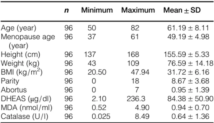
A total of 96 postmenopausal women older than 50 years of age were included in the study. The mean age was 61± 8 years, and the mean BMI was 31.72± 6.16 kg/m2. The average number of pregnan-cies and abortions were 8.67± 3.68 and 0.95 ± 1.39, respectively. The mean values of the demographic characteristics and laboratory tests are shown in Table 1.
The number of pregnancies in 28 of the subjects was less than 6, between 6 and 10 in 35 of the subjects, and greater than 10 in 33 of the subjects. Additionally, 41 of the 96 cases had a history of abortions. There was no statistically significant difference between the
Table 1 Description of the cases included in this study n Minimum Maximum Mean± SD Age (year) 96 50 82 61.19± 8.11 Menopause age (year) 96 37 61 49.19± 4.98 Height (cm) 96 137 168 155.59± 5.33 Weight (kg) 96 43 109 76.59± 14.18 BMI (kg/m2) 96 20.50 47.94 31.72± 6.16 Parity 96 0 18 8.67± 3.68 Abortus 96 0 7 0.95± 1.39 DHEAS (μg/dl) 96 2.10 236.3 84.38± 50.90 MDA (nmol/ml) 96 0.52 4.90 0.94± 0.70 Catalase (U/l) 96 0.025 8.49 0.64± 1.36 BMI, body mass index; DHEAS, dehydroepiandrosterone sulfate; MDA, malondialdehyde.
number of pregnancies experienced and abortion history with regard to demographic characteristics and laboratory tests (P> 0.05).
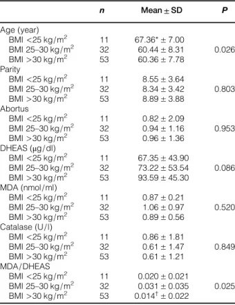
The BMI was less than 25 kg/m2in 11 of the sub-jects, between 25 and 30 kg/m2in 32 of the subjects, and greater 30 kg/m2 in 53 of the subjects. Comparative statistics of demographic characteristics
and laboratory tests according to BMI groups are shown in Table 2. The mean age was significantly higher in women with BMI <25 kg/m2 than in either women with a BMI between 25 and 30 kg/m2 or women with a BMI>30 kg/m2groups (P= 0.026). The MDA/DHEAS ratio was significantly lower in women with BMI>30 kg/m2than in women with a BMI between 25 and 30 kg/m2 (P= 0.025). There were no statistically significant differences for the other parameters based on the BMI groups (P> 0.05) (Table 2).
The Pearson correlation analysis showed significant positive correlations between number of pregnancies and abortions (r= 0.352, P < 0.01). Catalase activity was positively correlated with the number of abortions (r= 0.232, P < 0.05), whereas the serum DHEAS levels were positively correlated with BMI (r= 0.285, P < 0.01). However, the MDA/DHEAS ratio was negatively correlated with the BMI (r= −0.241, P < 0.05) (Table 3).
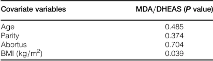
Factors (e.g. age, number of pregnancies, number of abortions, and BMI) that might affect the MDA/ DHEAS ratio were analyzed in a covariance analysis. BMI was found to be a potential predictor of the MDA/DHEAS ratio (P = 0.039) (Table 4).
Discussion
In the present study, we observed that the MDA/ DHEAS ratio was significantly decreased in healthy, obese, postmenopausal women. To the best of our knowledge, this is the first study to investigate the MDA/DHEAS ratio in healthy, obese, postmenopau-sal women.
There are claims suggesting that oxidative stress is associated with aging and that the degree of oxidative damage controls the rate of aging.15Menopause nor-mally manifests in women between ages 45 and 55 as Table 2 Comparison of the BMI groups with regard to the
demographic and laboratory data of the cases
n Mean± SD P Age (year) BMI<25 kg/m2 11 67.36*± 7.00 BMI 25–30 kg/m2 32 60.44± 8.31 0.026 BMI>30 kg/m2 53 60.36 ± 7.78 Parity BMI<25 kg/m2 11 8.55± 3.64 BMI 25–30 kg/m2 32 8.34± 3.42 0.803 BMI>30 kg/m2 53 8.89 ± 3.88 Abortus BMI<25 kg/m2 11 0.82± 2.09 BMI 25–30 kg/m2 32 0.94± 1.16 0.953 BMI>30 kg/m2 53 0.96 ± 1.36 DHEAS (μg/dl) BMI<25 kg/m2 11 67.35± 43.90 BMI 25–30 kg/m2 32 73.22± 53.54 0.086 BMI>30 kg/m2 53 93.59 ± 45.30 MDA (nmol/ml) BMI<25 kg/m2 11 0.87± 0.21 BMI 25–30 kg/m2 32 1.06± 0.97 0.520 BMI>30 kg/m2 53 0.89 ± 0.56 Catalase (U/l) BMI<25 kg/m2 11 0.86± 1.81 BMI 25–30 kg/m2 32 0.61± 1.47 0.849 BMI>30 kg/m2 53 0.61 ± 1.21 MDA/DHEAS BMI<25 kg/m2 11 0.020± 0.021 BMI 25–30 kg/m2 32 0.031± 0.035 0.025 BMI>30 kg/m2 53 0.014†± 0.022
*Comparison of BMI<25 kg/m2group to BMI between 25 and 30 and BMI>30 kg/m2group.
†Comparison of BMI>30 to BMI between 25 and 30.
BMI, body mass index; DHEAS, dehydroepiandrosterone sulfate; MDA, malondialdehyde.
Table 3 Correlation analysis results of the cases
Age Menopause age Menopause duration BMI (kg/m2) Parity Abortus DHEAS (μg/dl) MDA (U/l) Catalase (nmol/ml) MDA/ DHEAS Age 1 Menopause age 0.172 1 Menopause duration 0.828** −0.409** 1 BMI (kg/m2) −0.184 −0.172 −0.073 1 Parity 0.184 0.159 0.080 0.071 1 Abortus −0.051 0.113 −0.112 0.046 0.352** 1 DHEAS (μg/dl) −0.171 −0.072 −0.118 0.285** −0.132 0.010 1 MDA (U/l) −0.053 −0.063 −0.013 −0.103 −0.057 −0.122 −0.122 1 Catalase (nmol/ml) 0.192 0.088 0.128 0.029 0.024 0.232* 0.003 −0.064 1 MDA/ DHEAS 0.006 0.0082 −0.041 −0.241* 0.085 −0.064 −0.589** 0.653** −0.093 1 *P < 0.05. **P < 0.01.
a natural event in the process of aging. The estradiol level rapidly declines in women with the start of meno-pause. In vitro studies have shown that estrogens exert an antioxidant effect on membrane phospholipid per-oxidation.22,25Compared with the decrease in estrogen levels during menopause, the decrease in androgen levels is slower and less pronounced.26
DHEAS comprises the largest quantity of the sex steroids in the human body.2DHEAS levels decline markedly as a person ages, and the level of DHEAS at age 65 is less than one-fifth of its measured level at age 20.4,5The results of in vivo and in vitro studies have shown that DHEAS limits lipid peroxi-dation.27–29 DHEAS has also been shown to play a role in various physiological and pathological pro-cesses such as age-related cancer,30 atherosclerosis,30 obesity,31 infections,32 insulin sensitivity,33 and aging.34 The antioxidant characteristic of DHEAS can explain these effects.
In our study, we did not observe a statistically sig-nificant correlation between age and the DHEAS levels, probably due to the limited number of cases available to us as well as the intensity of the menopau-sal transition of some of our cases. Recently, it has been shown that during the menopausal transition, there is a modest increase in the production of DHEAS.35
MDA is one of the toxic end-products of non-enzy-matic hydrolysis of oxidative lipid peroxides.36 Recently, it has been shown that serum MDA levels increase with age in the 40–50 age group but remain constant after age 50.37 In the present study, we found no correlation between age and MDA levels. All of our subjects were healthy women over age 50. Thus, the lack of an increase in the MDA levels after age 50 is consistent with data from the literature. Victorino et al.38observed no difference between pre-menopausal and postpre-menopausal women with regard to serum MDA levels. However, Sánchez-Rodríguez et al.39
reported higher levels of MDA in postmeno-pausal women than in perimenopostmeno-pausal women. The effect of the aging process on serum MDA and dehy-droepiandrosterone (DHEA) levels has also been assessed. Sreeramulu et al.37 reported that serum MDA and DHEA levels in women are lower than those in men. They also observed a negative
correlation between age and serum DHEA levels but a positive correlation between age and MDA levels.
In our study, we report lower levels of oxidative stress in our obese subjects. There was a negative cor-relation between the MDA/DHEAS ratio as an oxidative stress index and BMI in healthy postmeno-pausal women. However, there was no association between serum MDA levels and BMI.
When the BMI groups were compared, there was no significant difference between the groups with regard to the MDA and DHEAS levels, but the MDA/ DHEAS ratio was significantly lower in women with a BMI over 30 than in women with a BMI between 25 and 30. The covariance analysis of the factors that can affect MDA/DHEAS ratio revealed that BMI was the only potential predictor.
In contrast to our study, Mittal and Kant40 ident-ified a positive correlation between MDA levels and body weight in healthy postmenopausal women. Though some studies have reported weight gain during menopause,19,20several studies have attributed weight gain during menopause to decreased estrogen levels and a lowered basal metabolism.21,22 An increase in oxidative stress in conjunction with an increase in body weight can result in the development of metabolic syndrome, diabetes mellitus, hyperten-sion, dyslipidemia, and atherosclerosis in obese sub-jects.41 However, several authors have reported increased MDA levels (a marker of oxidative stress) in healthy obese subjects.17,18
Atherosclerosis is the major cause of morbidity and death in Western countries and involves complicated interactions between arterial cells, blood cells, and plasma lipoproteins.42Oxidative stress (i.e. an imbal-ance between the amount of reactive oxygen species and antioxidant defense mechanisms) plays an impor-tant role in atherogenesis.43 Obesity is a chronic disease characterized by low-degree inflammation induced by oxidative stress. This oxidative stress plays a central role in the pathogenesis of obesity-associated disorders.44 A Turkish study on the DHEAS levels in obese patients reported a positive correlation between the DHEAS levels and BMI in both women and men.45 Likewise, Saruc et al. 46 reported that DHEA and DHEAS levels are positively correlated with the BMI and waist/hip ratio in Turkish postmenopausal women. Cao et al.47 assessed the association between DHEAS and BMI in early and late postmenopausal women and observed higher levels of DHEAS in women with a BMI≥24 kg/m2 than in women with a normal body weight. Consistent with data published in the literature, we observed a positive correlation between the serum DHEAS levels and BMI.
Catalase is an antioxidant enzyme that contributes to the maintenance of oxidative balance by converting Table 4 Covariance analysis results for MDA/DHEAS
Covariate variables MDA/DHEAS (P value)
Age 0.485
Parity 0.374
Abortus 0.704
BMI (kg/m2) 0.039
BMI, body mass index; DHEAS, dehydroepiandrosterone sulfate; MDA, malondialdehyde.
hydrogen peroxide, a by-product of oxidative stress, into water and molecular oxygen.48 Vaishali et al.49 found that postmenopausal women had significantly higher levels of catalase than premenopausal women. Similarly, Arora et al.50reported higher levels of cat-alase in postmenopausal women than in premenopau-sal women. In our study, we observed no association between age and catalase levels because all of our cases were postmenopausal women. We also found no association between BMI and catalase levels. In contrast, Mittal and Kant40reported a positive corre-lation between catalase and body weight in healthy postmenopausal women.
In the present study, we measured the weight and height of each subject to calculate the BMI. We did not measure the waist/hip ratio because we were unaware of that type of obesity measurement for our subjects. Additionally, visceral fat is known to be associated with atherosclerosis, and perhaps our sub-jects have subcutaneous adipose tissue. Furthermore, the paradoxical results may be because none of the enrolled patients have a metabolic comorbidity such as hypertension, diabetes mellitus, or hypertriglyceridemia. In conclusion, our study indicates that healthy, obese, postmenopausal women have a decreased MDA/DHEAS ratio. BMI was found to be a poten-tial predictor of the MDA/DHEAS ratio, which can be used as a marker of oxidative stress in postmeno-pausal women.
Acknowledgments
The authors thank staff of Faculty of Science, Department of Chemistry at Yuzuncu Yil University for their generous friendly assistance in every step of this study.
Disclaimer statements
Contributors B.G., M.A., M.A., M.O., and I.S.: con-ception and design; M.A., M.A., H.D., and M.O.: analysis and interpretation of the data; B.G., M.A., M.A., and R.U.: critical revision of the article for important intellectual content; B.G., M.A., M.A., and M.A.: final approval of the article; and B.G., I.S., and M.A.: collection and assembly of data. Funding None.
Conflicts of interest None. Ethics approval Yes.
References
1 Longcope C. Metabolism of dehydroepiandrosterone. Ann N Y Acad Sci 1995;774:143–8.
2 Labrie F. Adrenal androgens and intracrinology. Semin Reprod Med 2004;22:299–309.
3 Yen SS. Dehydroepiandrosterone sulfate and longevity: new clues for an old friend. Proc Natl Acad Sci U S A 2001;98:8167–9.
4 Orentreich N, Brind JL, Rizer RL, Vogelman JH. Age changes and sex differences in serum dehydroepiandrosterone sulfate con-centrations throughout adulthood. J Clin Endocrinol Metab 1984;59:551–5.
5 Nafziger AN, Bowlin SJ, Jenkins PL, Pearson TA. Longitudinal changes in dehydroepiandrosterone concentrations in men and women. J Lab Clin Med 1998;131:316–23.
6 Trivedi DP, Khaw KT. Dehydroepiandrosterone sulfate and mortality in elderly men and women. J Clin Endocrinol Metab 2001;86:4171–7.
7 Barrett-Connor E, Khaw KT, Yen SS. A prospective study of dehydroepiandrosterone sulfate, mortality, and cardiovascular disease. N Engl J Med 1986;315:1519–24.
8 Berr C, Lafont S, Debuire B, Dartigues JF, Baulieu EE. Relationships of dehydroepiandrosterone sulfate in the elderly with functional, psychological, and mental status, and short-term mortality: a French community-based study. Proc Natl Acad Sci U S A 1996;93:13410–5.
9 Mazat L, Lafont S, Berr C, Debuire B, Tessier JF, Dartigues JF, et al. Prospective measurements of dehydroepiandrosterone sulfate in a cohort of elderly subjects: relationship to gender, sub-jective health, smoking habits, and 10-year mortality. Proc Natl Acad Sci U S A 2001;98:8145–50.
10 Barrett-Connor E, Khaw KT. Absence of an inverse relation of dehydroepiandrosterone sulfate with cardiovascular mor-tality in post-menopausal women. N Engl J Med 1987;317: 711.
11 Barrett-Connor E, Edelstein SL. A prospective study of dehy-droepiandrosterone sulfate and cognitive function in an older population: the Rancho Bernardo Study. J Am Geriatr Soc 1994;42:420–3.
12 Tilvis RS, Kahonen M, Harkonen M. Dehydroepiandrosterone sulfate, diseases and mortality in a general aged population. Aging (Milano) 1999;11:30–4.
13 Camporez JP, Akamine EH, Davel AP, Franci CR, Rossoni LV, Carvalho CR. Dehydroepiandrosterone protects against oxi-dative stress-induced endothelial dysfunction in ovariectomized rats. J Physiol 2011;589:2585–96.
14 Aragno M, Tamagno E, Gatto V, Brignardello E, Parola S, Danni O, et al. Dehydroepiandrosterone protects tissues of streptozotocin-treated rats against oxidative stress. Free Radic Biol Med 1999;26:1467–74.
15 Peppa M, Uribarri J, Vlassara H. Aging and glycoxidant stress. Hormones (Athens) 2008;7(2):123–32.
16 Aslan M, Horoz M, Sabuncu T, Celik H, Selek S. Serum paraox-onase enzyme activity and oxidative stress in obese subjects. Pol Arch Med Wewn 2011;121(6):181–6.
17 Amirkhizi F, Siassi F, Minaie S, Djalali M, Rahimi A, Chamari M. Is obesity associated with increased plasma lipid peroxidation and oxidative stress in women? ARYA Atheroscler J 2007;2(4): 189–92.
18 Khan NI, Naz L, Yasmeen G. Obesity: an independent risk factor for systemic oxidative stress. Pak J Pharm Sci 2006; 19(1):62–5.
19 Heymsfield SB, Gallagher D, Poehlman ET, Wolper C, Nonas K, Nelson D, et al. Menopausal changes in body composition and energy expenditure. Exp Gerontol 1994;29(3–4):377–89. 20 Simkin-Silverman LR, Wing RR. Weight gain during
meno-pause: is it inevitable or can it be prevented? Postgrad Med 2000;108(3):47–50, 53–6.
21 Yagi K. Female hormones act as natural antioxidants– a survey of our research. Acta Biochim Pol 1997;44:701–9.
22 Ruiz-Larrea MB, Martín C, Martínez R, Navarro R, Lacort M, Miller NJ. Antioxidant activities of estrogens against aqueous and lipophillic radicals: differences between phenol and catechol estrogens. Chem Phys Lipids 2000;105:179–88.
23 Yagi K. Assay for blood plasma or serum. Methods Enzymol 1984;105:328–33.
24 Aebi H. Catalase in vitro. Methods Enzymol 1984;105:121–6. 25 Sugioka K, Shimosegawa Y, Nakano M: Estrogens as natural
antioxidants of membrane phospholipid peroxidation. FEBS Lett 1987;210:37–9.
26 Hurt LS, Ronsmans C, Thomas SL. The effect of number of births on women’s mortality: a systematic review of the evidence for women who have completed their childbearing. Popul Stud (Camb) 2006;60:55–71.
27 Boccuzzi G, Aragno M, Seccia M, Brignardello E, Tamagno E, Albano E, et al. Protective effect of dehydroepiandrosterone against copper-induced lipid peroxidation in the rat. Free Radic Biol Med 1997;22:1289–94.
28 Aragno M, Brignardello E, Tamagno E, Gatto V, Danni O, Boccuzzi G. Dehydroepiandrosterone administration prevents the oxidative damage induced by acute hyperglycemia in rats. J Endocrinol 1997;155:233–40.
29 Khalil A, Lehoux JG, Wagner RJ, Lesur O, et al. Dehydroepiandrosterone protects low density lipoproteins against peroxidation by free radicals produced by gamma-radiolysis of ethanol–water mixtures. Atherosclerosis 1998;136:99–107. 30 Rao M, Subbarao A, Reddy J. Inhibition of spontaneous
testicu-lar Leydig cell tumor development in F-344 rats by dehydroe-piandrosterone. Cancer Lett 1992;65:61–6.
31 Nestler JE, Barlascini CO, Clore JN, Blackard WG. Dehydroepiandrosterone reduces serum low density lipoprotein levels and body fat, but does not alter insulin sensitivity in normal man. J Clin Endocrinol Metab 1988;66:57–71. 32 Loria R, Inge T, Cook S, Szakol A, Regelson W. Protection
against acute lethal viral infections with the native steroid dehy-droepiandrosterone (DHEA). J Virol 1988;26:301–14.
33 Buffington CK, Givens JR, Kitabchi AE. Opposing actions of dehydroepiandrosterone and testosterone on insulin sensitivity. Diabetes 1991;40:693–700.
34 Morales AJ, Nolan JJ, Nelson JC, Yen SSC. Effects of replace-ment dose of dehydroepiandrosterone in men and women of advancing age. J Clin Endocrinol Metab 1994;78:1360–7. 35 Lasley BL, Crawford SL, McConnell DS. Ovarian adrenal
inter-actions during the menopausal transition. Minerva Ginecol 2013;65(6):641–51.
36 Nielsen F, Mikkelsen BB, Nielsen JB, Andersen HR, Grandjean P. Plasma malondialdehyde as biomarker for oxidative stress: reference interval and effects of life-style factors. Clin Chem 1997;43:1209–14.
37 Sreeramulu D, Ramalakshmi BA, Balakrishna N, Raghuramulu N. Serum dehydroepiandrosterone and lipid peroxides in human volunteers of different age groups. Indian J Clin Biochem 2004; 19(1):79–82.
38 Victorino VJ, Panis C, Campos FC, Cayres RC, Colado-Simão AN, Oliveira SR, et al. Decreased oxidant profile and increased antioxidant capacity in naturally postmenopausal women. Age (Dordr) 2013;35(4):1411–21.
39 Sánchez-Rodríguez MA, Zacarías-Flores M, Arronte-Rosales A, Correa-Muñoz E, Mendoza-Núñez VM. Menopause as risk factor for oxidative stress. Menopause 2012;19(3):361–7. 40 Mittal PC, Kant R. Correlation of increased oxidative stress to
body weight in disease-free post menopausal women. Clin Biochem 2009;42:1007–11.
41 Matsuda M, Shimomura I. Increased oxidative stress in obesity: implications for metabolic syndrome, diabetes, hypertension, dyslipidemia, atherosclerosis, and cancer. Obes Res Clin Pract 2013;7(5):330–41.
42 Ross R. Atherosclerosis– an inflammatory disease. N Engl J Med 1999;340:115–26.
43 Harrison D, Griendling KK, Landmesser U, Hornig B, Drexler H. Role of oxidative stres in atherosclerosis. Am J Cardiol 2003; 91:7–11.
44 Vincent HK, Taylor AG. Biomarkers and potential mechanisms of obesity induced oxidant stress in humans. Int J Obes (Lond) 2006;30:400–18.
45 Topsakal S, Akin F, Yerlikaya E, Erurker T, Dogu H. Dehydroepiandrosterone sulfate levels in Turkish obese patients. Eat Weight Disord 2014;19(2):261–5.
46 Saruc M, Yuceyar H, Ayhan S, Turkel N, Tuzcuoglu I, Can M. The association of dehydroepiandrosterone, obesity, waist–hip ratio and insulin resistance with fatty liver in postmenopausal women– a hyperinsulinemic euglycemic insulin clamp study. Hepatogastroenterology 2003;50(51):771–4.
47 Cao Y, Zhang S, Zou S, Xia X. The relationship between endogenous androgens and body fat distribution in early and late postmenopausal women. PLos ONE 2013;8(3):58448–53. 48 Begenik H, Soyoral YU, Erkoc R, Emre H, Taskın A, Tasdemir
M. et al. Serum malondialdehyde levels, myeloperoxidase and catalase activities in patients with nephrotic syndrome. Redox Rep 2013;18(3):107–12.
49 Vaishali S, Sanjeev S, Neelima S, Shaila S. Status of antioxidant enzymes and trace metals in postmenopausal women. J Obstet Gynecol India 2005;55(1):64–6.
50 Arora KS, Gupta N, Sıngh RA, Nagpal S, Arora D. Role of free radicals in menopausal dıstress. J Clin Diagn Res 2009;3: 1900–2.