Original Article / Özgün Makale
Chitosan Polysaccharide Suppress Toll Like Receptor Dependent
Immune Response
Çitosan Polisakkaridi Toll Benzeri Reseptöre Bağlı Bağışıklık Yanıtını Baskılar
Gizem Tincer,1 Banu Bayyurt,1 Yakup M. Arıca,2 İhsan Gürsel1
ABSTRACT
Objectives: Chitosan is a widely used vaccine or anti-cancer delivery vehicle. In this study, we
investigated the immunomodulatory effect of chitosan/pIC nanocomplexes on mouse immune cells.
Materials and methods: Proliferative and cytotoxic features of chitosan were tested via CCK-8 assay
on RAW 264.7. IL-1b production was assessed via ELISA from PEC supernatants. TNF-a, and NO induction from chitosan treated RAW cells detected by ELISA and Griess assay, respectively. mRNA message levels of TLRs and cytokines on macrophages in response to chitosan/pIC nanocomplex treatments were evaluated by RT-PCR.
Results: Results revealed that chitosan is non-toxic to cells, however, proliferative capacities of
macrophages were reduced by chitosan administration. Mouse PECs treated with chitosan, led to NLRP3 dependent inflammasome activation as evidenced by dose-dependent IL-1b secretion. Chitosan/pIC nanocomplexes did not improve immunostimulatory action of pIC on RAW cells, since TNF-a and NO productions remained unaltered. Expression levels of several TLRs, CXCL-16 and IFN-a messages from mouse splenocytes were down regulated in response to chitosan/pIC nanocomplex treatment.
Conclusion: Our results revealed that chitosan is an anti-proliferative and inflammasome triggering
macromolecule on immune cells. Utilization of chitosan as a carrier system is of concern for immunotherapeutic applications.
Keywords: Biomaterial; chitosan; double-stranded ribonucleic acid; innate immunity; polysaccharide; Toll like
receptor 3. 1Department of Molecular Biology
and Genetics, Thorlab, Therapeutic Oligonucleotide Research Laboratory, Bilkent University, Ankara, Turkey. 2Gazi University, Biochemical Processing and Biomaterial Research Laboratory, Ankara, Turkey
Correspondence:
İhsan Gürsel, MD.
Bilkent Üniversitesi, Thorlab, Moleküler Biyoloji ve Genetik Bölümü, Fen Fakültesi, 06800 Bilkent, Ankara, Türkiye
Tel: +90 312 - 290 24 08 e-mail: [email protected] ©2015 Turkish Journal of Immunology. All rights reserved.
doi: 10.5606/tji.2015.329
Received: January 29, 2015 Accepted: March 18, 2015
ÖZET
Amaç: Bu çalışmada çitosan/pIC nanokomplekslerinin fare immün hücreleri üzerinde immün
düzenleyici etkisi araştırıldı.
Gereç ve yöntemler: Çitosanın proliferatif ve sitotoksik özellikleri RAW 264.7’de cell counting
kit-8 ile test edildi. İnterlökin-1b üretimi ELISA ile peritoneal eksüda hücre süpernatantlarından değerlendirildi. Çitosan ile tedavi edilen RAW hücrelerinden tümör nekroz faktör-a ve nitrik oksit indüksiyonu sırasıyla ELISA ve Griess testleriyle tespit edildi. Toll benzeri reseptörlerin ribonükleik asit mesaj düzeyleri ve çitosan/pIC nanokompleks tedavilerine yanıt olarak makrofajlardaki sitokinler ters transkripsiyon polimeraz zincir reaksiyonu ile değerlendirildi.
Bulgular: Bulgular çitosanların hücrelere toksik olmadığını gösterdi; öte yandan, makrofajların
proliferatif kapasiteleri çitosan uygulaması ile azaltıldı. Çitosan ile tedavi edilen fare peritoneal eksüda hücreleri doz bağımlı interlökin-1b sekresyonu ile gösterildiği üzere nükleotid bağlayıcı oligomerizasyon domain benzeri reseptör protein 3 bağımlı inflamazom aktivasyonuna yol açtı. Tümör nekroz faktör-a ve nitrik oksit üretimleri değişmediğinden, çitosan/pIC nanokompleksleri pIC’nin RAW hücreleri üzerindeki immünstimülatör aksiyonunu iyileştirmedi. Toll benzeri reseptörlerin, kemokin (C-X-C motif) ligand 16’nın ve interferon-a mesajlarının fare splenositlerinden ifade düzeyleri çitosan/pIC nanokompleks tedavilerine yanıt olarak azaldı.
Sonuç: Bulgularımız çitosanın immün hücreler üzerinde antiproliferatif ve inflamazom tetikleyici
bir makromolekül olduğunu gösterdi. Çitosanın taşıyıcı bir sistem olarak kullanımı immünterapötik uygulamaların konusudur.
Anahtar sözcükler: Biyomalzeme; çitosan; çift sarmal ribonükleik asit; doğal bağışıklık; polisakkarit; Toll benzeri
Chitosan polysaccharides and their derivatives are used as adjuvants for mucosal immunity, drug carriers and anti-cancer agents.[1] They are widely studied natural
polysaccharides for the controlled delivery of a drug or an adjuvant, as well as a vaccine delivery system. Inclusion of chitosan improves the stability of drugs, genes or proteins when formulated as micro/nanocarriers.[1-3]
Lately these biomaterials were used as delivery systems for labile Toll-like receptor agonists (TLR) in order to improve their immunostimulatory capabilities. When the derivatives of chitosan biopolymers were co-administered with Toll-like receptor 7 (TLR7) ligand; imiquimod or with TLR9 ligand CpG oligodeoxynucleotides (CpG ODN), the nanocapsules containing TLR agonists elicited a protective immune response against hepatitis B.[4]
Moreover, when chitosan derivatives were complexed with CpG ODN, the uptake of CpG ODNs and the cytokine production were improved. As an immune adjuvant, chitosan nanoparticles containing CpG ODN provoked strong humoral as well as Th1 type cellular immune responses in mucosal immunity.[5,6]
Being a valuable biomaterial and an immunotherapeutic agent that might be harnessed as a novel drug delivery system, the immunomodulatory effects of standalone chitosan is still elusive. Chitosan and its derivatives are recognized by various types of innate immune receptors, Toll-like receptor (TLR) 2, C-type lectin Dectin-1 and the mannose receptor, have been implicated in mediating a variety of immune responses such as TNF-α, IL-10 and IL-17 cytokine production.[7] Moreover, chitosan was
shown to activate the NLRP3 inflammasome, leading to robust IL-1β responses by a phagocytosis-dependent mechanism.[8]
In the present study, chitosan derivatives were studied to assess their immunostimulatory potential. For this, we first checked their cytotoxicity and proliferative effect on macrophages. Next, we investigated the inflammasome inductive capacity of these biopolymers in the context of their IL-1β production potentials from peritoneal exudate cells. Furthermore, in order to evaluate the immunostimulatory potential of chitosan as a delivery system, we nano-complexed chitosan with a TLR3 ligand; polyinosinic:polycytidylic acid (pIC) and evaluated the TNF-α cytokine and anti-bacterial nitric oxide (NO) inductive capacity , along with mRNA message levels of different TLRs and type I IFN triggering in mouse macrophages.
MATERIALS AND METHODS
All cell culture media components were from Lonza (Switzerland) and Hyclone (USA). Cell Counting Kit-8 (CCK-8) was provided from Dojindo Molecular Devices
(Japan). Chitosan biopolymer was purchased from Sigma Aldrich. TLR3 ligand polyinosinic:polycytidylic acid (pIC) was purchased from GE Healthcare Life Sciences. Alum was obtained from Thermo Scientific (USA). Chitosan complexes with pIC (1:1 w/w, chitosan:ligand ratio) were prepared overnight incubation at 4 °C.
Cytotoxicity and proliferation assays
Evaluation of the cytotoxicity and proliferation was performed by cell counting kit-8 (CCK-8) assay. RAW 264.7 cells (mouse macrophage cell line) were distributed into 96-well plates as 5000 cell/ml and 105 cell/ml for
cytotoxicity and proliferation assays, respectively. Cells were either incubated with 3 weeks chitosan treated (conditioned) media (CM) or with chitosan polymers for 24h, 48h and 72h at 37 °C in culture. A total of 100 μl of media from each sample was incubated with 10 µl CCK-8 in 96-well plate and incubated at 37 °C for 3h. Cytotoxicity of chitosan and cell proliferation rate were measured as the absorbance at 450 nm using a microplate reader (BioTek, µQuant, USA).
Stimulation assays
Peritoneal macrophages from BALB/c mice were collected into cold PBS, 4d after intraperitoneal (i.p.) injection of 2.5 ml, sterile 4% thioglycollate broth. Cells were washed at 1500 rpm for 5 mins and resuspended in 10% FBS regular RPMI 1640 media. Cells were counted with BD AccuriTM C6 (BD, USA) and seeded into plates
as 1.25x10⁶/ml. Mouse cells were primed with 100 ng/ml ultrapure lipopolysaccharide (LPS) for 4h (control cells were left unprimed), followed by incubation with the positive control alum and chitosan at 20 µg/ml, 100 µg/ml and 500 µg/ml doses for 20h. Supernatants were collected for IL-1β and TNF-α ELISA assays.
ELISA and NO assays
Immulon 2 HB microtitre plates (Thermo Scientific, USA) were coated with anti IL-1β and anti TNF-α antibodies (Biolegend, USA) for overnight at 4 °C and then blocked with PBS-BSA for 2h @ RT). Serially diluted recombinant proteins and culture supernatants were transfered into plates for overnight at 4°C. IL-1β and TNF-α cytokines were detected using 50 µl biotinylated anti-cytokine Ab for 2h (B122: IL-1β and 6B8: TNF-α clones, Biolegend, USA) followed by 50 µl phosphatase-streptavidin (Perbio Pierce, USA) addition for 1h and 50 µl PNPP substrate addition at RT to develop in the wells. The cytokine content was read at 405 nm with ELISA plate reader (BioTek, µQuant, USA). Nitric oxide detection from RAW 264.7 cells (105/ml) was carried out by Griess method, according to manufacturers protocol (Fluka, USA). Cells were incubated with the stimulants
for 24h and supernatants were read at 540 nm using a plate reader.
TLR and cytokine RT-PCR
Total RNA was isolated from 2x10⁶ RAW 264.7 cells, which were stimulated either with (i) chitosan,
(ii) pIC and (iii) chitosan/pIC nanocomplexes for 2h
via TRIzol (Invitrogen, USA) extraction method. RNA samples were reverse-transcribed and amplified to obtain single stranded cDNA (Finnzymes, Finland). cDNA synthesis was performed using 12.5 µl of master mix (Finnzymes PCR Master Mix), 1 µl cDNA, 10 pmol/µl sense and antisense primers specific for each target genes. PCR conditions for β-actin, TLR 1, 2, 3, 4, 6, 7, 9 and CXCL-16 were as follows; 94 °C for 30 sec, 55 °C for 30 sec, 72 °C, 1 min, 35 cycles, for IFN-α; initial denaturation at 94 °C for 2 mins, 94 °C for 30 sec, 64.3 °C for 30 sec, 72 °C for 1 min, 40 cycles and final extension at 72 °C for 10 mins.
Statistical analyses
Statistical analyses were performed using Sigma STAT4 software. Student’s t-test was used to evaluate the statistical differences between stimulation groups. P values < 0.05 were considered as significant.
RESULTS
Non-toxic chitosan polymer inhibited macrophage proliferation
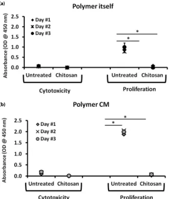
Initial experiments were performed to delineate the cytotoxic effects of biopolymer chitosan. For this, RAW 264.7 cells were stimulated with, (i) 3 weeks polymer incubated media or (ii) polymer itself for 1 day, 2 and 3 days. As seen in the Figure 1a, CCK-8 assay revealed that, chitosan did not induce any toxic effect on the cells, as evidenced by low absorbance values at 450 nm. However, chitosan strongly affected the proliferation of macrophages even after the first day of incubation. When mouse macrophages were incubated with biopolymer conditioned media, similar findings were observed, as presented in the Figure 1b. Three weeks old chitosan conditioned media negatively affected the proliferation capacities of macrophages. These data indicated that, chitosan polymer or its by-product leached into culture media, were non-toxic, however, they reduced the proliferative capacities of mouse macrophages.
Chitosan biopolymer induced inflammasome dependent IL-1β production
As shown above, chitosan failed to contribute to proliferation rate of mouse macrophages. Whether they suppressed TLR dependent immune activation via
triggering inflammasome activation was not explored. Next, we aimed to investigate these possibilities. Thioglycollate treated peritoneal macrophages were isolated from BALB/c mice. Cells were primed with LPS to initiate pro-IL-1β accumulation and provide a substrate for subsequent processing via caspase-1 leading to mature IL-1β secretion via the assembly of inflammasome multiprotein complexes. After 4h of priming, cells were incubated with NLRP3 inflammasome ligand alum and chitosan in various doses for additional 20h. Results showed that, chitosan treated peritoneal macrophages released IL-1β in a dose dependent manner. The IL-1β levels were comparable to alum, a gold standard NLRP3 inflammasome ligand (Figure 2). Our data, pointed that chitosan could modulate caspase-dependent inflammasome activation. These findings collectively (Figures 1 and 2), may explain the low proliferation rate induced by chitosan on mouse macrophages, since inflammasome caspase pathway induces pyroptosis or necrosis.
(a)
(b)
Figure 1. Effects of chitosan biopolymers on mouse macrophage proliferation. (a) RAW 264.7 cells were incubated with 20 µg/ml chitosan or (b) with 3 weeks chitosan conditioned media (Polymer CM) for 24h-72h. Cytotoxic property of biopolymers and proliferation status of macrophage cells were detected with CCK-8 assay. The results represent mean values from three independent experiments ± SD. Student’s t-test revealed that * p<0.05.
Chitosan/pIC nanocomplexes suppressed TLR specific message transcripts but did not influence cytokine production
Recent reports implicated that when either TLR7 or TLR9 ligands encapsulated within chitosan particles immune stimulatory potentials exceeded with respect to their free counterparts.[4-6] Next,
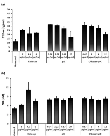
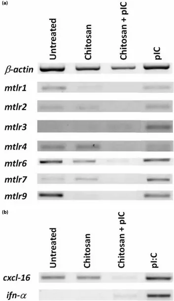
we aimed to test whether nanocomplexes of TLR3 ligand, pIC, with chitosan reproduces this improved effect on immune cells. We prepared and tested chitosan/pIC nanocomplexes (1:1, w/w) and compared immunostimulatory activity to that of free pIC on immune cells. When RAW 264.7 cells incubated with pIC, chitosan alone or their complexed forms for 24h in culture, ELISA results demonstrated that chitosan/pIC complexes had no substantial positive or negative effect on TNF-α secretion compared to pIC alone (Figure 3a). Similar to TNF-α cytokine production, complexes did not improve NO production compared to pIC treated cells in dose dependent manner (Figure 3b). Strikingly, as presented in Figure 4a chitosan/pIC complexes significantly downregulated TLR 2,3,6,7 and 9 message transcipt expressions compared to pIC or chitosan alone treated groups. Moreover TLR1 and TLR4 mRNA levels in pIC alone treatments were slightly decreased compared to chitosan/pIC complexes. Furthermore, chitosan downregulated the chemokine CXCL-16 and IFN-α gene expressions when complexed with pIC in contrast to pIC alone after 2h stimulation (Figure 4b). Other cytokines and chemokines such as; IP-10, MIP3-α and TNF-α were also studied but there were no differences among above mentioned treatment groups (data not shown).
DISCUSSION
Immunomodulatory activities of chitosan and its derivatives have been studied for their potential applications against allergy, infectious diseases or cancer.[9-11] Previous findings suggested that chitin and
chitosan induce various pro-inflammatory (IL-1β) or inflammatory cytokines (TNF-α, IL-10, IL-17) upon incubating them with macrophages or keratinocytes.[12-15]
Present study, established that an analog of dsRNA, pIC, that triggers TLR3 mediated signaling cascade failed to contribute to improved activity when complexed with chitosan and tested on RAW 264.7 cells. While we clearly indentified that cytotoxic effect of chitosan on RAW 264.7 cells were absent, our findings pointed out that in vitro proliferation rate of mouse macrophages was negatively influenced by chitosan treatment. This reduction is not only mediated by chitosan but also regulated by the degradation by-products of this biopolymer, since macrophages incubated with three weeks old chitosan conditioned media significantly diminished proliferation of the cells.
Figure 2. Inflammasome activation by chitosan were provided from mouse PECs. Quantification of IL-1β production with 4h LPS (100 ng/ml) priming prior to stimulation of BALB/c PECs with chitosan (20 µg/ml, 100 µg/ml, 500 µg/ml) and as positive control with alum (20 µg/ml, 100 µg/ml, 500 µg/ml) for 18h. Representative plots of three independent experiments are shown. p<0.01 between untreated and chitosan (except 20 µg/ml) treated groups.
Figure 3. Dose dependent TNF-α and NO production were provided from mouse macrophages with pIC, chitosan and chitosan+pIC complexes (a) TNF-α and (b) NO levels were detected by ELISA and Griess assay respectively from from RAW cell supernatants after 24h stimulation. Result represents combination of at least two independent experiments.
(a)
Natural biomaterials like chitosan have been demonstrated to induce NLRP3 inflammasome dependent IL-1β production in bone marrow derived macrophages.[14] Regarding to the anti-proliferative effect
of chitosan, we aimed to investigate the inflammasome dependent IL-1β production in peritoneal mouse macrophages. LPS primed macrophages induced more IL-1β production upon chitosan stimulation. This production was parallel to alum, one of the well-established NLRP3 ligands (Figure 2). High levels of IL-1β release from macrophages could be attributed to the cell death and anti-proliferative effect of chitosan.
The most extensively studied pathogen recognition receptors (PRRs) are TLRs. They recognize specific pathogen associated molecular patterns (PAMPs).[16]
A subclass of TLRs are also known as, endosome associated TLRs. These are TLR3, 7/8 and 9. They are specialized to sense pathogenic nucleic acid moieties.[17] TLR3 detects
double stranded RNA, or synthetic poly (inosinic: cytidylic acid; pIC) and used as immunotherapeutic agent.[18] When these ligands are given in vivo, they are
rapidly cleared by nucleases, decreasing their therapeutic effects.[19] Various carrier systems such as liposomes,
biodegradable carriers and soluble macromolecules were widely studied for labile nucleic acids attempting to increase their in vivo performances.[20-22]
Although numerous studies investigated chitosan as a delivery and drug carrier systems for proteins plasmids and peptides, there are only few studies in the literature aiming to develop chitosan as delivery system for labile nucleic acid based TLR ligands. Co-delivery of TLR7 agonist, imiquimod and the recombinant hepatitis B surface antigen with chitosan resulted improved protective immune response and immunological memory.[4] A recent study demonstrated
that, chitosan-CpG ODN carrier system synergistically promoted the secretion of Th1 and Th17 dependent cell polarizing cytokines from mouse dendritic
cells.[23] Chitosan-silica-CpG ODN nanohybrids
enhanced cellular uptake and induction of IL-6 to a greater degree than chitosan/CpG ODN nanocomplexes in human peripheral blood mononuclear cells.[5]
In light of the recently accumulated data, our goal was to develop a chitosan/pIC nano delivery system and expand its beneficial immunotherapeutic applications. When complexes were assayed, we found that, nano-formulations did not improve TNF-α production in a dose-dependent manner compared to pIC or chitosan alone treated cells (Figure 3a). We further assessed the production of macrophage mediated anti-bacterial cytotoxic effect by checking NO production and found that there was no improvement in NO secretion (Figure 3b). Surprisingly, chitosan/pIC complexes,
downregulated the mRNA levels of TLR 1, 2, 3, 4, 6, 7 and 9 compared to the pIC alone treated RAW 264.7 mouse macrophages. Moreover, gene expression levels of scavenger receptor CXCL-16 and anti-viral Type I interferon alpha were also down-regulated (Figure 4). Contrary to the previous findings reported with TLR7/8 and TLR9 ligands, chitosan/pIC complexes failed to develop a Th1 based inflammatory response. However, parallel studies of chitosan + TLR7/8 agonist; R848 or gardiquimod, TLR9 agonist; CpG ODN complexes along with pIC (TLR3 ligand) should be performed in order to prove that TLR7/8 and TLR9 ligands synergize with chitosan, while TLR3 failed to reproduce similar effects.
Figure 4. TLR and cytokine gene expression profile of chitosan and pIC. mRNA levels of (a) TLRs, (b) CXCL-16 and IFN-α were assessed by reverse transcriptase PCR from chitosan (20 µg/ml), pIC (20 µg/ml) and chitosan/pIC (20 µg/ml:20 µg/ml) treated RAW 264.7 cells after 2h in vitro incubation.
(a)
In conclusion, the non-toxic, anti-proliferative chitosan showed inflammasome dependent inflammatory response, yet did not improve immunostimulatory effect of associated pIC.
Declaration of conflicting interests
The authors declared no conflicts of interest with respect to the authorship and/or publication of this article.
Funding
The authors received no financial support for the research and/or authorship of this article.
REFERENCES
1. Arca HC, Günbeyaz M, Senel S. Chitosan-based systems for the delivery of vaccine antigens. Expert Rev Vaccines 2009;8:937-53.
2. Jabbal-Gill I, Watts P, Smith A. Chitosan-based delivery systems for mucosal vaccines. Expert Opin Drug Deliv 2012;9:1051-67. 3. Li X, Min M, Du N, Gu Y, Hode T, Naylor M, et al. Chitin,
chitosan, and glycated chitosan regulate immune responses: the novel adjuvants for cancer vaccine. Clin Dev Immunol 2013;2013:387023.
4. Vicente S, Peleteiro M, Díaz-Freitas B, Sanchez A, González-Fernández Á, Alonso MJ. Co-delivery of viral proteins and a TLR7 agonist from polysaccharide nanocapsules: a needle-free vaccination strategy. J Control Release 2013;172:773-81. 5. Chen S, Zhang H, Chinnathambi S, Hanagata N. Synthesis of
novel chitosan-silica/CpG oligodeoxynucleotide nanohybrids with enhanced delivery efficiency. Mater Sci Eng C Mater Biol Appl 2013;33:3382-8.
6. Zhang H, Chen S, Zhi C, Yamazaki T, Hanagata N. Chitosan-coated boron nitride nanospheres enhance delivery of CpG oligodeoxynucleotides and induction of cytokines. Int J Nanomedicine 2013;8:1783-93.
7. Bueter CL, Specht CA, Levitz SM. Innate sensing of chitin and chitosan. PLoS Pathog 2013;9:1003080.
8. Bueter CL, Lee CK, Wang JP, Ostroff GR, Specht CA, Levitz SM. Spectrum and mechanisms of inflammasome activation by chitosan. J Immunol 2014;192:5943-51.
9. Chen YL, Wang CY, Yang FY, Wang BS, Chen JY, Lin LT, et al. Synergistic effects of glycated chitosan with high-intensity focused ultrasound on suppression of metastases in a syngeneic
breast tumor model. Cell Death Dis 2014;5:1178.
10. Bowman K, Leong KW. Chitosan nanoparticles for oral drug and gene delivery. Int J Nanomedicine 2006;1:117-28.
11. Zaharoff DA, Rogers CJ, Hance KW, Schlom J, Greiner JW. Chitosan solution enhances both humoral and cell-mediated immune responses to subcutaneous vaccination. Vaccine 2007;25:2085-94.
12. Da Silva CA, Hartl D, Liu W, Lee CG, Elias JA. TLR-2 and IL-17A in chitin-induced macrophage activation and acute inflammation. J Immunol 2008;181:4279-86.
13. Da Silva CA, Chalouni C, Williams A, Hartl D, Lee CG, Elias JA. Chitin is a size-dependent regulator of macrophage TNF and IL-10 production. J Immunol 2009;182:3573-82.
14. Bueter CL, Lee CK, Rathinam VA, Healy GJ, Taron CH, Specht CA, et al. Chitosan but not chitin activates the inflammasome by a mechanism dependent upon phagocytosis. J Biol Chem 2011;286:35447-55.
15. Koller B, Müller-Wiefel AS, Rupec R, Korting HC, Ruzicka T. Chitin modulates innate immune responses of keratinocytes. PLoS One 2011;6:16594.
16. Kawai T, Akira S. The role of pattern-recognition receptors in innate immunity: update on Toll-like receptors. Nat Immunol 2010;11:373-84.
17. Ishii KJ, Koyama S, Nakagawa A, Coban C, Akira S. Host innate immune receptors and beyond: making sense of microbial infections. Cell Host Microbe 2008;3:352-63.
18. Zhang SY, Herman M, Ciancanelli MJ, Pérez de Diego R, Sancho-Shimizu V, Abel L, et al. TLR3 immunity to infection in mice and humans. Curr Opin Immunol 2013;25:19-33. 19. Barry ME, Pinto-González D, Orson FM, McKenzie GJ, Petry
GR, Barry MA. Role of endogenous endonucleases and tissue site in transfection and CpG-mediated immune activation after naked DNA injection. Hum Gene Ther 1999;10:2461-80. 20. Tincer G, Yerlikaya S, Yagci FC, Kahraman T, Atanur OM,
Erbatur O, et al. Immunostimulatory activity of polysaccharide-poly(I:C) nanoparticles. Biomaterials 2011;32:4275-82. 21. Dow S. Liposome-nucleic acid immunotherapeutics. Expert
Opin Drug Deliv 2008;5:11-24.
22. Hafner AM, Corthésy B, Merkle HP. Particulate formulations for the delivery of poly(I:C) as vaccine adjuvant. Adv Drug Deliv Rev 2013;65:1386-99
23. Mori A, Oleszycka E, Sharp FA, Coleman M, Ozasa Y, Singh M, et al. The vaccine adjuvant alum inhibits IL-12 by promoting PI3 kinase signaling while chitosan does not inhibit IL-12 and enhances Th1 and Th17 responses. Eur J Immunol 2012;42:2709-19.