ARTICLE IN PRESS
www.icvts.org doi:10.1510/icvts.2007.162669
Interactive CardioVascular and Thoracic Surgery 6 (2007) 744–747
䊚 2007 Published by European Association for Cardio-Thoracic Surgery
Institutional report - Cardiac general
Mid-term results of peripheric cannulation after port-access surgery
夞
Ertan Sagbas , Baris Caynak *, Cihan Duran , Onur Sen , Birol Kabakci , Ilhan Sanisoglu , Belhhan Akpinar
a a, b a a a aDepartment of Cardiac Surgery, Istanbul Science University, Group Florence Nightingale Hospitals, Abidei Hurriyet Caddesi 290, 34381 Sisli, Istanbul, Turkey a
Department of Radiology, Istanbul Science University, Group Florence Nightingale Hospitals, Abidei Hurriyet Caddesi 290, 34381 Sisli, Istanbul, Turkey b
Received 9 July 2007; received in revised form 24 August 2007; accepted 24 August 2007
Abstract
Several minimally invasive approaches, avoiding median sternotomy, have been described within the last few years for cardiac surgery. Femoral arterial and venous cannulation for extracorporeal perfusion are required for many of these operations. The aim of this report is to assess the long-term outcomes of femoral cannulations in patients who underwent minimally invasive procedures. One hundred and sixty patients underwent operations by the port-access method between January 2002 and October 2006. Cardiopulmonary bypass was established by femoral artery-vein cannulation, and a transthoracic clamp was used for the aortic occlusion. One hundred and twenty-one patients were under follow-up in the outpatient clinic and 85 patients underwent Doppler ultrasonography (US) for femoral arterial and venous stenosis. The mean follow-up was 27.9 months (range 1–57 months). There were three hospital mortalities (1.86%), and five late mortalities in this series. The mean follow-up for the Doppler examination was 20.54 months (range 1–56 months). There were two seromas and three wound complications (2.48%), all of which healed after outpatient treatment. All of the flow patterns of the common femoral arteries (CFA) were triphasic except in three of the patients. Three patients (2.48%) were found to have arterial stenosis. One patient with intermittant claudication underwent percutaneous dilatation and stenting of the CFA. Doppler US detected luminal narrowing in two patients who had been having no symptoms, and they are being followed in the outpatient clinic without any complaints. We found a chronic recanalized thrombotic change in the common femoral vein (CFV) in one patient (0.63%). Our study demonstrates vessel patency andyor stenosis in patients without complaints. In conclusion, femoral artery and vein cannulation for port-access surgery with transthoracic clamping can be performed successfully with excellent results in the mid-term.
䊚 2007 Published by European Association for Cardio-Thoracic Surgery. All rights reserved.
Keywords: Minimally invasive surgery; Complications; Ultrasound; Vascular disease
1. Introduction
Median sternotomy with cannulation of the ascending aorta and the right atriumyvena cava for cardiopulmonary bypass (CPB) has been the standard approach for most cardiac surgery procedures. Several minimally invasive approaches, avoiding median sternotomy, have been described within the last few years w1–3x. Femoral arterial and venous cannulation for extracorporeal perfusion are required for many of these operations. We have been performing port-access approaches since 2002. CPB was established by femoro-femoral cannulation in these operations.
The aim of this report is to assess the mid-term outcomes of femoral cannulations in patients who underwent mini-mally invasive procedures.
2. Materials and methods
The ethical commission of the hospital approved the study protocol. During the follow-up period, data were collected
夞 Dr B. Akpinar discloses that he has a financial relationship with Medtronic, Inc.
*Corresponding author. Tel.: q90 532 5641103; fax: q90 212 2398791.
E-mail address: [email protected] (B. Caynak).
during outpatient visits, and Doppler ultrasonography (US) was performed. One hundred and sixty consecutive port-access operations were performed between January 2002 and October 2006. Patients with severe chest wall de-formities, significant coronary artery disease, aortic valve insufficiency, lung adhesions, or iliac artery disease were excluded. In addition to routine angiography and echocar-diography, each patient underwent a detailed evaluation preoperatively including a computerized axial tomography (CAT) scan andyor magnetic resonance imaging (MRI), as well as transesophageal echocardiography (TEE) and Dopp-ler US andyor MR angiography for identification of such risk factors and proper patient selection. Patient characteristics are shown in Table 1. Most of these patients had mitral valve diseases. The surgical operations and the number of patients are shown in Table 2.
2.1. Surgical procedure
After standard induction of anesthesia, each patient underwent double lumen intubation for single lung venti-lation. Following the administration of 2 mgykg of heparin, a 17 F arterial cannula (DLP, Inc, Grand Rapids, MI) was introduced through the right internal jugular vein percu-taneously to assist venous drainage during CPB. TEE
moni-ARTICLE IN PRESS
745
E. Sagbas et al. / Interactive CardioVascular and Thoracic Surgery 6 (2007) 744–747
Table 1
Patient demographics
Variable n
Patients (n) 160
Patient age (years) 52"8
Maleyfemale 62y98
Isolated mitral valve insufficiency 31
Combined mitral valve disease 66
ASD sinus venosusysecundum 5y58
Tricuspid valve disease 14
Mixoma 1
Atrial fibrillation 75
Reoperation 6
LVEF (%) 55.2"6.3
ASD, Atrial septal defect; LVEF, Left ventricular ejection fraction;
n, Number.
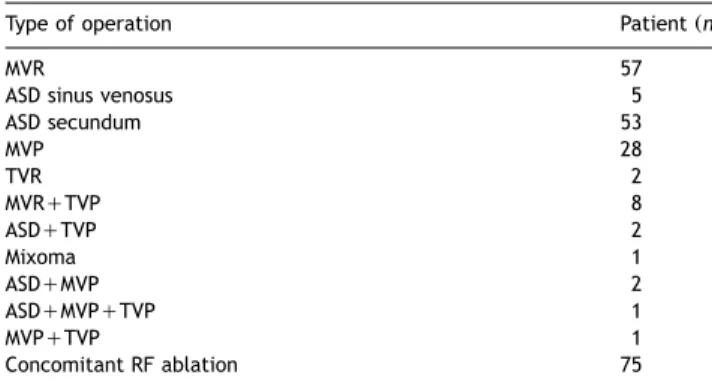
Table 2 Type of operation
Type of operation Patient (n)
MVR 57
ASD sinus venosus 5
ASD secundum 53 MVP 28 TVR 2 MVRqTVP 8 ASDqTVP 2 Mixoma 1 ASDqMVP 2 ASDqMVPqTVP 1 MVPqTVP 1 Concomitant RF ablation 75
MVR, Mitral valve replacement; ASD, Atrial septal defect; MVP, Mitral valve plasty; TVR, Tricuspid valve replacement; TVP, Tricuspid valve plasty; RF, Radiofrequency.
torization was used routinely to evaluate the results of valve repair and detect evacuation of air. Patients were positioned supine with the right shoulder elevated, and external defibrillation pads were placed. A right lateral minithoracotomy (4–6 cm) in the fourth intercostal space was performed. A soft tissue retractor (Heartport Inc, Redwood City, USA) was used for the exposure of the surgical field, avoiding the division or traction of any rib. A 5-mm camera port (Storz, Karl Storz GmbH and Co, Tuttingen, Germany) was introduced through the fourth intercostal space front axillary line. A second port was introduced through the sixth intercostal space mid axillary line for left atrial venting and carbon dioxide insufflation, which began immediately after collapsing the right lung.
Simultaneously, the right femoral artery and vein were prepared by means of a 4-cm oblique incision in the groin. CPB was established by femoro-femoral cannulation. Venous cannulation was performed first. Although the sequence of cannulation can vary between clinics, it is our experience that maneuvering the femoral venous cannula can be more difficult in the presence of a 20 Fr arterial cannula in the femoral artery. The femoral vein was can-nulated through two concentric pursestrings of 5-0 polypro-pylene suture to avoid occlusion of venous return from the right leg. A venotomy was made within the pursestring, and the 24–29 F femoral cannula (DLP, Inc, Grand Rapids, MI) was inserted. The cannula was advanced to the junction of
the superior vena cava and right atrium over a flexible J wire using TEE for guidance. Once properly positioned, the venous cannula was secured and arterial cannulation was performed.
The common femoral artery was clamped proximally and distally. A transverse arteriotomy was made, leaving the posterior one-third of the artery intact. An 18–20 F arterial cannula (DLP, Inc, Grand Rapids, MI) was inserted proxi-mally and secured with a Rummell tourniquet. Cardiopul-monary bypass was then initiated with the femoral arterial-venous cannula and the 17–19 F arterial cannula previously inserted in the right internal jugular vein, thus allowing adequate venous drainage. After weaning from bypass, the venous cannula was removed and the purse-string suture was tied. The arterial cannula was removed, and the transverse arteriotomy was closed using continuous 6-0 polypropylene sutures. In most patients arterial can-nulation was uneventful. The use of open arteriotomy technique enhanced the cannulation. However, in patients with small femoral arteries, we could still manage with 18 Fr cannulas. Special care was given during the closure of the arteriotomy using a 6.0 polypropylene and single suture technique. With most of the patients being mitral valve and ASD closures, atherosclerotic vessels were uncommon.
The pericardium was opened 2 cm above and parallel to the phrenic nerve. Exposure was optimized with several pericardial stay sutures. Patients were cooled down to 28 8C. Both vena cavae were encircled with tapes for a drier operative field. A transthoracic clamp (Chitwood, Scanlan, St Paul, MN, USA) was introduced from the second intercostal space, front axillary line percutaneously. After cross-clamping of the aorta, a blood cardioplegia cannula was inserted in the ascending aorta. The left atrium was opened parallel to the interatrial groove. The Heart Port atrial retractor system (Heartport Inc, Redwood City, CA) was used for the exposure of the atrium.
2.2. Doppler examination
The femoral arteries and veins were evaluated by ultra-sonography (US). US examination was performed by Sie-mens Sonoline Anteras, Germany. A 7-3 MHz multi hertz probe was used. Arteries were evaluated for flow rate and flow pattern. Normal arteries show a triphasic flow pattern in Doppler US. In stenotic arteries, Doppler flow patterns and flow rates change in prestenotic, stenotic, and post-stenotic regions. Proximal to the lesion, the flow pattern is normal, while at the stenosis the peak velocity increases in proportion to the degree of stenosis. Focal areas of doubling of the measured peak systolic velocity have been shown to correspond to lesions of 50% narrowing in the luminal diameter of the artery. Distal to the stenosis, the peak systolic velocity returns to values equal to or lower than those proximal to the stenosis, and in color Doppler imaging there is turbulant flow.
For venous examination, the venous caliber and the exis-tence of the turbulant flow were evaluated.
3. Results
One hundred and sixty patients underwent operations by the port-access method. Thirty-one patients were lost
ARTICLE IN PRESS
746 E. Sagbas et al. / Interactive CardioVascular and Thoracic Surgery 6 (2007) 744–747
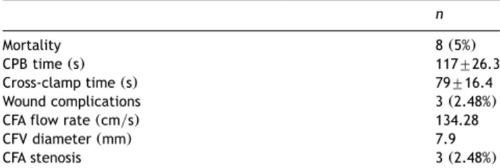
Table 3 Complications n Mortality 8 (5%) CPB time (s) 117"26.3 Cross-clamp time (s) 79"16.4 Wound complications 3 (2.48%)
CFA flow rate (cmys) 134.28
CFV diameter (mm) 7.9
CFA stenosis 3 (2.48%)
CPB, Cardiopulmonary bypass; CFA, Common femoral artery; CFV, Common femoral vein.
during the follow-up period, 121 patients were under follow-up in the outpatient clinic. The mean follow-up was 27.9 months (range 1–57 months). There were three hos-pital mortalities (1.86%), and five late mortalities in this series (Table 3).
Eighty-five patients underwent Doppler US for femoral arterial and venous stenosis. The mean follow-up for the Doppler examination was 20.54 months (range 1–56 months).
There were three wound complications in this series (2.48%), all of which healed after outpatient treatment. There were two seromas; one was healed after needle aspiration and compressed medical dressing, and the other became infected and needed wound treatment in the out-patient clinic (this case was included in the number of wound complications).
All of the flow patterns of the CFA were triphasic except in three of the patients. The flow rate was calculated to be between 60–212 cmys (mean flow rate was 134.28 cmys). The diameter of the CFV was calculated to be between 5.5–12.5 mm (mean diameter of the CFV was 7.9 mm).
In our series, three patients (2.48%) were found to have arterial stenosis. Of those, one patient (a 52-year-old woman) complained of claudication two months following the operation; Doppler US showed 70% narrowing in the lumen of the CFA. This patient underwent percutaneous dilatation and stenting of the CFA. The patient currently has no complaints and a control Doppler US detected no stenosis in the CFA lumen in outpatient examinations. Doppler US detected luminal narrowing in two patients who had been having no symptoms. The flow pattern of the CFA was monophasic and the flow rate was lower than normal (32 cmys in one and 35 cmys in the other). Those patients are being followed in the outpatient clinic without any complaints.
We found chronic recanalized thrombotic changes in CFV in one patient (0.63%). The lumen of the vein had narrowed and the wall was thicker than normal and there was flow in the venous lumen and no turbulance was detected.
4. Discussion
Port-access surgery reduces blood loss, hospital stay, and recovery time w4, 5x. These approaches require femoral vessel cannulation for cardiopulmonary bypass, which intro-duces a risk of femoral arterial andyor venous injury. Studies have shown that the risk of aorto-iliac problems
was high in patients who underwent port-access surgery with endoaortic baloon occlusion. Besides these complica-tions, this method is more expensive than the transthoracic clamp technique w6x. We preferred femoral artery and venous cannulation with a transthoracic clamp as described by Chitwood and colleagues for the occlusion of the ascend-ing aorta to avoid these complications and to decrease the price of the surgery w7x. Therefore, we had no dissection of the aorta and none of the major aortic problems intraoperatively.
In general, minimally invasive procedures can be per-formed with bypass and clamp times that are not signifi-cantly longer than those during conventional techniques. Studies have shown that the prolonged period of limb ischemia in patients undergoing complex minimally invasive operations has the potential to cause ischemic complica-tions in the cannulated extremity w8, 9x. In our group of patients, cardiopulmonary bypass and ischemic times can be compared with the conventional procedures and are found to be reasonable. Therefore, we did not experience any ischemic complications in the cannulated extremity which may have required a fasciotomy in the early post-operative period.
Muhs and friends published a study of arterial injuries from femoral artery cannulation in 739 consecutive patients who had port-access minimally invasive cardiac surgery w6x. They identified patients with new arterial insufficiency from the cannulation site. There were four patients having postoperative claudication, and of those, three of them had iliofemoral arterial occlusion or localized iliofemoral dissection and were treated with iliofemoral bypass, and one patient had localized FA stenosis treated by angioplasty. In our series of 160 patients, there was only one patient who complained of claudication. Doppler US and DSA showed a significant stenosis in this patient, and she underwent a percutaneous dilatation and stenting of the CFA. We examined all of the patients, except for those lost in the follow-up, for femoral cannulation problems in the outpatient clinic, and we performed Doppler US. We detected two patients having CFA stenosis without any symptoms, and treatment has not yet been necessary for these stenoses. Doppler US of the CFV showed chronic recanalized thrombotic changes in CFV in one patient having no symptoms.
Our study demonstrates vessel patency andyor stenosis in patients without complaints. Other studies in the literature have evaluated femoral cannulation problems retrospec-tively in patients with complaints.
In conclusion, femoral artery and vein cannulation for port-access surgery with transthoracic clamping can be performed successfully with excellent results in the mid-term.
5. Study limitations
5.1. This study has some limitations
Mean follow-up is not very long. There was a considerable number of patients who were lost to follow-up and as a consequence the Doppler follow-up was not complete in these patients. This was mainly due to geographical and
ARTICLE IN PRESS
747
E. Sagbas et al. / Interactive CardioVascular and Thoracic Surgery 6 (2007) 744–747
economical constraints, which prevented the recruiting of some of the patients from rural areas during follow-up.
Another limitation is that a Doppler analysis was not performed immediatelly after the operation in all patients; and the comparison between initial status of femoral ves-sels and the data obtained during follow-up were not possible.
References
w1x Galloway AC, Shemin RJ, Glower DD, Boyer JH, Groh MA, Kuntz RE, Burdon TA, Bibakove GH, Reitz BA, Colvin SB. First report of the port access international registry. Ann Thorac Surg 1999;67:51–58. w2x Casselman FP, Van Slycke S, Wellens F, De Geest R, Degrieck I, Van
Praet F, Vermeulen Y, Vanermen H. Mitral valve surgery can now routinely be performed endoscopically. Circulation 2003;108:II48–54. w3x Dogan S, Aybek T, Risteski PS, Detho F, Rapp A, Wimmer-Greinecker G,
Moritz A. Minimally invasive port access vs. conventional mitral valve surgery: prospective randomized study. Ann Thorac Surg 2005;79:492– 498.
w4x Glower DD, Siegel LC, Frischmeyer KJ, Galloway AC, Ribakove GH, Grossi EA, Robinson NB, Ryan WH, Colvin SB. Predictors of outcome in a multicenter port-access valve registry. Ann Thorac Surg 2000;70: 1054–1059.
w5x Aybek T, Dogan S, Risteski PS, Zierer A, Wittlinger T, Wimmer-Grein-ecker G, Moritz A. Two hundred and forty minimally invasive mitral operations through right minithoracotomy. Ann Thorac Surg 2006; 81:1618–1624.
w6x Muhs BE, Galloway AC, Lombino M, Silberstein M, Grossi EA, Colvin SB, Lamparello P, Jacobowitz G, Adelman MA, Rockman C, Gagne PJ. Arterial injuries from femoral artery cannulation with port access cardiac surgery. Vasc Endovascular Surg 2005;39:153–158.
w7x Chitwood WR, Wixon CL, Elbeery JR, Moran JF, Chapman WH, Lust RM. Video-assisted minimally invasive mitral valve surgery. J Thorac Cardio-vasc Surg 1997;114:773–782.
w8x Gates JD, Bichell DP, Rizzo RJ, Couper GS, Donaldson MC. Thigh ischemia complicating femoral venous cannulation for cardiopulmonary bypass. Ann Thorac Surg 1996;61:730–733.
w9x Hendrickson SC, Glower DD. A method for perfusion of the leg during cardiopulmonary bypass via femoral cannulation. Ann Thorac Surg 1998; 65:1807–1808.