Journal of Neurological Sciences [Turkish] 33:(2)# 48; 378-382, 2016 http://www.jns.dergisi.org/text.php3?id=967
Case Report
Temporary Paraplegia Resulting From Venous Air Embolism Following Lumbar Disc Surgery
Kadir KOTİL
Istanbul Arel University, Neurosurgery, İstanbul, Turkey Abstract
A spinal cord ischemic injury defined by acute transient paraplegia, following lumbar disk surgery was not reported in the literature. A lumbar MRI was performed to a 60 years old obese and short female patient due to low back and leg pain. An acute disk extrusion was found at the L4-5 absolute spinal stenosis background. The patient had lumbar disc surgery at the prone position. The patient woke up with paralysis in both extremities under T10 level postoperatively. The MRI showed air bubbles in the spinal canal, originating from the giant venous dural lacs. After 6-8 hours with medical treatment, the patient completely regained the sensory first, then the motor function. Since the fast recovery in the early period and the air seen on MRI, the other reasons such as fibro cartilaginous embolism at the spinal cord level were excluded, and the etiology was thought to be the venous air embolism. This is the first case in the literature with paraplegia due to acute spinal ischemia, which is thought to happen due to venous air embolism during the lumbar decompression surgery.
Keywords: Temporary paraplegia, venous air embolism, lumbar disc surgery
Lomber Disk Cerrahisi Sonucu Gelişen Hava Embolisine Bağlı Geçici Parapleji Olgusu Özet
Lomber disk cerrahisine bağlı bir çok komplikasyon tanımlanmasına rağmen geçici spinal kord iskemisi yaratan bir parapleji olgusu bugune kadar literatürde tanımlanmamıştır. 60 yaşında obez ve kısa boylu bir kadın hastanın bel ve bacak ağrıları nedeniyle lomber MRG çekilmiştir. L4-5 mutlak spinal stenosis zemininde akut disk ekstrüzyonu tespit edilmiştir. Hastaya prone pozisyonunda lomber disk cerrahisi pozisyonunda opere edildi. Postop uyanma esmasında her iki ekstremite TH 10 altı paralizi ile uyandı. Acil çekilen magnetic rozenans çekiminde spinal kanal içinde dev dural venöz laklardan giren hava kabarcıkları tespit edilmiştir. Medikal tedaviye alınan hastanın 6-8 saat sonra önce duyu sonra motor defisiti tam olarak düzeldi. Hem erken dönemde hızla düzelmesi ve MRG de de hava saptanması üzerine fibrokartilogenaz emboli gibi spinal kord seviyesinde görülen diğer nedenler ekarte edilerek etyolojinin venöz hava embolisine bağlı olabileceği düşünülmüştür. Literatürde venöz hava embolisine bağlı olduğu düşünülen lomber dekompresyon cerrahisi esnasında oluşan transient akut spinal iskemi sonucu gelişen paraplejili ilk olgudur.
INTRODUCTION
Intraoperative venous air embolism can be explained as entry of the air into the systemic or local circulation, when the pressure in the vein is lower than the atmospheric pressure in the surgery region. It occurs as the aspiration of the air outside with the opening of a vein with a negative pressure. This situation may occur, especially the neurosurgical operations to the patients at sitting position(3,11), and spinal and pelvic approaches where the intra-abdominal pressure changes(7).
Lumbar disc surgery is the most common intervention in spinal surgery. Many complications may occur after disk surgery, as in other conventional neurosurgical interventions. Venous air embolism is seen usually after posterior fossa surgery in neurosurgical practice, and rare cases are reported in spinal surgery(6,7,11,13,14), but its course is mortal
because of the frequent entry of the air embolism into the systemic circulation, or in rare cases, it is morbid. Another embolism that may cause mortality in spinal surgery is the fibro cartilaginous embolism (FCE), and about 34 cases have been reported up till now(9). Whole of the reported FCE cases died after paraplegia. The FCE occurs with the retrograde embolism of the ventral disk material and blocking the central artery, causing a permanent infarct of the spinal cord, and it is usually irreversible.
The sensory and motor paraplegia following surgery is usually the neural injury or the epidural hemorrhage under the level of the lesion, which we call the cauda equina syndrome. However this case is the first case we met in the literature according to our intensive researches, which caused transient paraplegia due to venous air embolism at a very higher level of operated L4-5 (T10).
CASE PRESENTATION
Sixty years old obese and short (155 cm) female patient and increased waist
circumference (140 cm) presented with long term low back pain and neurogenic claudication. The examinations revealed multilevel spinal stenosis and acute disc hernia at the ground of L4-5, and surgery was planned.
The anamnesis and the physical examination were nonspecific except the intermediate obesity. Cardiac, pulmonary and the abdominal examinations were normal.
Her body temperature was 36.6°C, and the heart beat was 73 bpm/rhythmic. The respiratory rhythm was 20 and the blood pressure was 145/90 mm/Hg. The neurological examination revealed a neurogenic claudication in 30 meters, a severe pain in her left knee and absence of patellar reflex. The muscle strength of the dorsal flexion was 3/5, and there was hypoesthesia in the L5 dermatome.
The blood chemistry screen revealed leukocyte count: 9900, Htc: 39%, platelet count: 390.000, erythrocyte sedimentation rate: 23mm/h, C-reactive protein: 18, prothrombin (PT) and partial thromboplastin time (PTT):11.8 and 29.0 sec.
Chest X-ray showed no specific feature.
Lumbar micro discectomy or decompression surgery was planned with the detection of acute central disc extrusion with a narrow canal at the L4-5 level in lumbar MRI. The patient was prepared to surgery in prone position as her abdomen being in the opening, because of the patient is obese and has an increased waist circumference. The surgical area was opened from left side unilaterally at L4-5 level and sequestrectomy and bilateral decompression of the central canal were performed. Following the extraction of the sequestered disc particles and the spinal stenosis was revealed, the massive venous bleeding was controlled with the pads. The total bleeding volume was 120 cc from the dense and thick varicose veins in the surgical area. The operation lasted for 50
minutes, and the patient was extubated and neurological examination was performed. The patient was free to move her hands, however, both lower extremities didn't respond even to the stimulation with pain from the level of the hips. There was no response to pain from the level of T10. Anal sensory and motor function was lost. The MRI showed dense air particles in the spinal canal at the level of L 2. The patient's vital signs, lung sounds and even the chest x-ray were normal. She was normotensive (120-130/85 mm/Hg), her oxygen saturation level (SpO2) was 99 to 95%, and the heart beat rate was between 56-72/bpm preoperatively. All the blood count levels were within normal range
including the D-dimer value. Spinal cord venous air embolism or fibro cartilaginous embolism were suspected and low molecular weight heparin (LMWH) and steroid (methylprednisolone 500 mg/IV) were started. The blood pressure was tried to stay high (Rheomacrodex 500cc/6 hours, 4x1). After 4 hours, first the sensory deficit, then in 7 hours, the motor deficit started to recover in proximal to distal direction. The anal sensory and motor functions recovered totally. At the third day, the neurological examination was fully recovered and the patient was mobilized. The patient was discharged with physical therapy consultation.
DISCUSSION
The first case of air embolism in the brain surgery literature was reported by Shenkin and Godfeder in 1969(11). Air embolism usually occurs in the posterior fossa surgery during the occipitocervical fixation at the seated position in spinal surgery and
the course of the air in the systemic circulation is usually fatal(11,12). Acute paraplegia is a rare complication of lumbar disc surgery and it usually occurs below the surgical level as a result of the dural tear or iatrogenic nerve injury, presented as cauda equina syndrome(6,13). Transient
Figure 1: A migrated disc with multilevel spinal
stenosis is distinguished in the L4-5 level at the lumbar mild narrow canal base in the lumbar sagittal MRI.
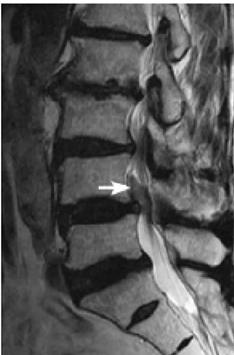
Figure 2: The bubbles are distinguished supporting
the intracanal air embolism in the postoperative early stage sagittal MRI.
paraplegia due to a far neighbour spinal cord segment ischemia, which is far above the surgical decompression area does not exist in the literature. There may be many positional complications in the prone position spinal surgery(13). In this position, intra-abdominal and intracaval pressure rise and increase the intraspinal pressure and a tendency to spinal venous bleeding occurs. Air may enter from these open venous lacs. The air may enter the systemic circulation and may lead to acute cardiovascular arrest or as in our one and only case, it may enter to the spinal circulation and cause spinal cord ischemia and total paralysis. The venous bleeding in our case might also have triggered this; because the venous circulation is from distal to proximal, with its suction power , the air particles in the spinal cord venous system might have made a pressure effect on the large epidural veins.
The acute motor paralyses in the literature are usually in the form of anterior spinal cord syndrome and their origin is fibro cartilaginous embolism which is usually fatal. A major part of these cases are seen above the T11 level, and reported as thoracic paraplegia(13). The number of these cases is 34 in total(2,15), and the surgical decompression is at the upper lumbar level in only 2 cases and only in 1 case it occurred after the sacral region surgery. These FCE cases also rarely occurred after lumbar decompression in the literature(9). In their autopsies and radiological examinations, spinal cord infarct findings were found on the pyramidal tract. As in our case, the spinal cord ischemia was not transient in the total of these FCE cases; it was permanent paraplegia and it occurred at the level of the lesion(6,13,15).
Nevertheless, both sensory and motor involvement, the surgical area being at the lower lumbar region and the neurological injury ending with recovery in 4 hours, not with death, and air found in the spinal canal on spinal MRI, led us to think of
transient spinal cord ischemia due to venous air embolism.
The total of 3 cases with venous air embolism in the Albin et al(1). These cases didn't respond to resuscitation and lasted with death due to the air to enter into the systemic circulation and the cardiac arrest. It was said that the abdominal compression may be a reason of diminishing the spinal cord perfusion and cause venous pooling(2). The treatment of such complications is initially to protect the patient from the risk factors. The precautions are to use anti-embolism lower extremity compression stockings, to free the abdomen at the prone position, to lower the vena cava pressure and to reduce the epidural venous bleeding, to prevent the lack of fluid in the vascular bed, to keep the lower extremities high to increase the venous turn, thus, to increase the venous pressure. Hence, the possibility of embolism is reduced and during the operation, it is crucial to have the equipment to control and follow if the patient has any air in his or her systemic circulation. Because the air in the spinal canal cannot be taken out as the air in the systemic circulation, it didn't cause a pressure on the spinal cord, that's why conservative treatment was performed. 500- 1000 mg of plus methylprednisolone and adjusting the blood pressure to higher levels may obtain the suction of the embolism or relieve of the circulation partly, as in our case(4,10).
As a result, the mentioned precautions should be taken to prevent the venous air embolism in spinal surgery in the prone position, especially in patients with wide abdomen and we can protect our patients from this potentially heavy neurological morbidity(5,7).
In conclusion, if the venous embolism could be detected in the perioperatively by precordial doppler ultrasonography, venous air embolism could be detected into the venous circulation, but we detected only a few air mid-lumbar region after the 2 hours surgery. We didn't detect any large
air at level of T10 or T12. Early MRI investigation could be showed to us giant bubble at this level, but we couldn't it. Yes, small venous air embolism may go clinically unrecognized. Venous embolism very rarely reported in the posterior spine surgery, also no any report in the microdiscectomy. The etiopathogenesis is not clear in our case. This is first documented case of venous air embolism during microdiscectomy, and we need more supportive new documentation.
Correspondence to:
Kadir Kotil
E-mail: [email protected]
Received by: 27 November 2015 Revised by: 22 December 2015 Accepted: 05 January 2016
The Online Journal of Neurological Sciences (Turkish) 1984-2016
This e-journal is run by Ege University Faculty of Medicine,
Dept. of Neurological Surgery, Bornova, Izmir-35100TR
as part of the Ege Neurological Surgery World Wide Web service.
Comments and feedback: E-mail: [email protected] URL: http://www.jns.dergisi.org
Journal of Neurological Sciences (Turkish) Abbr: J. Neurol. Sci.[Turk]
ISSNe 1302-1664
REFERENCES
1. Albin MS, Caroll RG, Maroon JC. Clinical considerations concerning detection of venous air embolism. Neurosurgery 1978,3:380-4
2. Albin MS, Ritter RR, Pruett CE, Kalff K. Venous air embolism during lumbar laminectomy in the prone
position: report of three cases. Anesth Analg 1991,73:346-9
3. Basaldella L, Ortolani V, Corbanese U, Sorbara C, Longatti P. Massive venous air embolism in the semi-sitting position during surgery for a cervical spinal cord tumor: anatomic and surgical pitfalls. J Clin Neurosci 2009,16(7):972-5
4. Harrison EA, Mackersie A, McEvan A, Facer E. The sitting position for neurosurgery in children: A review of 16 years experience. Br J Anaesth 2002,88:12-16
5. Frankel AS, Holzman RS. Air embolism during posterior spinal fusion. Can J Anaesth 1998,35:511 6. Han JJ, Massagli TL, Jaffe KM. Fibrocartilaginous
Embolism—An Uncommon Cause of SpinalCord Infarction: A Case Report and Review. Arch Phys Med Rehabil. 2004, 85(1):153-7. Review
7. Iwabuchi T, Sobata E, Ebina K, Tsubakisaka H, Takiguchi M. Dural sinus pressure: Various aspects in human brain surgery in children and adults. Am J Physiol 1986,250:389-96
8. Langmayr JJ, Ortler M, Obwegeser A, Felber S. Quadriplegia after lumbar disc surgery. A case report [see comment]. Spine 1996,21:1932-5 9. McCarthy RE. Lonstein JE, Mertz JD, Kuslich SD.
Air embolism in spinal surgery. J Spinal Disord 1990;3:1-5
10. McDouall SF, Shlugman D. Fatal venous air embolism during lumbar surgery: the tip of an iceberg? Eur J Anaesthesiol 2007,24:803-16 11. Shenkin HN, Goldfedder P. Air embolism from
exposure of posterior cranial fossa in prone position. JAMA. 1969; 27;210(4):726
12. Toro G, Roman GC, Navarro-Roman L, Cantillo J, Serrano B, Vergara I. Natural history of spinal cord infarction caused by nucleus pulposus embolism. Spine 1994;1;19(3):360-6.
13. Tosi L, Rigoli G, Beltramello A. Fibro cartilaginous embolism of the spinal cord: a clinical and pathogenetic reconsideration. J Neurol Neurosurg Psychiatry. 1996;60(1):55-60
14. Turker RJ, Slack C, Regan Q.Thoracic paraplegia after lumbar spinal surgery. J Spinal Disord 1995;8:195-200
15. Valls PL, Naul LG, Kanter SL. Paraplegia after a routine lumbar laminectomy: report of a rare complication and successful management. Neurosurgery 1990;27:638-40.