© TÜBİTAK
doi:10.3906/kim-1810-26 h t t p : / / j o u r n a l s . t u b i t a k . g o v . t r / c h e m /
Research Article
An exploration of new avenues regarding deep tissue penetration and higher
singlet oxygen efficiencies: novel near-IR photosensitizers for photodynamic
therapy
Nisa YEŞİLGÜL1,, Bilal KILIÇ2,∗,
1National Nanotechnology Research Center (UNAM), Graduate School of Engineering and Science,
Bilkent University, Ankara, Turkey
2Professional Flight Program, Faculty of Aviation and Aeronautical Science, Özyeğin University, İstanbul, Turkey
Received: 13.10.2018 • Accepted/Published Online: 07.01.2019 • Final Version: 03.04.2019
Abstract: A series of novel BODIPY-bearing electron-withdrawing groups at the meso position are reported here.
According to the optical measurements, it may be clearly seen that the introduction of electron-donating groups into 3,5-positions and the presence of electron-withdrawing groups at the meso position of the BODIPY core resulted in spectacular bathochromic shifts (up to ~ 304 nm), and the projected photosensitizers had absorption bands in the therapeutic window of the electromagnetic spectrum (600–900 nm). The absorption maxima of compounds 4, 5, 6, and
7 were at 886 nm, 890 nm, 760 nm, and 761 nm, respectively. The singlet oxygen generation experiments revealed that
compounds 6 and 7, with high singlet oxygen quantum yields (0.52 and 0.93, respectively), were excellent and promising candidates for photodynamic therapy. The singlet oxygen quantum yield of 0.93 was the highest reported value so far for BODIPY-based photosensitizers.
Key words: BODIPY, photosensitizer, photodynamic therapy, singlet oxygen, near-IR photosensitizer
1. Introduction
Cancer is a challenging and terrifying disease and the most widely used treatment modalities are chemotherapy, radiotherapy, and surgery.1−3 These treatment modalities frighten patients due to the side effects that to date
have not been obviated.4,5 A novel treatment modality, photodynamic therapy, that has been used clinically
for around 25 years has emerged and is widely used as an alternative method for treatment of malignant and nonmalignant diseases.6,7
The photodynamic therapy strategy is based on the preferential localization of certain photosensitizers in tumor tissues upon systematic administration. After the administration of the photosensitizer into the body, the sensitizer is activated by red or near infrared light, which leads to generation of reactive oxygen species including singlet oxygen (1O
2) , and thus irreversibly damaging the tumor cells. These photosensitizers are
harmless in the absence of light and their phototoxicity mainly relies on the generation of singlet oxygen, which is converted from its triplet state to its singlet state by an excited-state dye. A convenient photodynamic therapy agent should have absorption maxima in the spectral region of 700–900 nm, which is known as the therapeutic window. Because of the fact that light arrays below 700 nm cannot penetrate deeply into the tissue
∗Correspondence: [email protected]
YEŞİLGÜL and KILIÇ/Turk J Chem
and water in the body begins to absorb light above 900 nm, a well-designed photosensitizer should absorb light in the therapeutic window.8,9
BODIPY (4, 4-difluoro-4-bora-3a, 4a-diaza-s-indacene) dyes have attracted much attention as photosensi-tizers in the last decade due to their characteristics, which are particularly required for effective photosensiphotosensi-tizers and photodynamic action. The distinctive properties of BODIPY dyes such as photostability, high absorption coefficient, low dark toxicity, and singlet oxygen generation have inspired efforts to develop BODIPY-based photosensitizers.10−18 Furthermore, BODIPY dyes have many desirable characteristics that are often superior
to those of other photosensitizers (fluorescein, porphyrins, cyanines, etc.). These properties are good solubility in common organic solvents, easily modulable excitation, and ease of synthesis.19 For instance, compared with
porphyrin-type photosensitizers, BODIPY-type photosensitizers show intense absorption in the near-IR region of the electromagnetic spectrum, shorter periods of photosensitization, and low side-effect profiles (e.g., skin photosensitivity after irridation).10,20 Modifications on BODIPY cores may be carried out by introduction of
dif-ferent groups affording red-shifted BODIPY derivatives.21 It was shown that the strongly electron-withdrawing
group CF3 at the meso position of the BODIPY core resulted in a large bathochromic shift by stabilizing
the energy of the lowest unoccupied molecular orbital (LUMO) in comparison to that of congeners with aryl substituents in this position.22−25
In the present study, our aim was to synthesize novel near-IR BODIPY-based photosensitizers containing the strongly electron-withdrawing group CF3at the meso position (C-8) of the BODIPY core. Incorporation
in BODIPY cores of the heavy atoms that would give rise to effective singlet oxygen generation by intersystem crossing for serving as a potential photodynamic therapy agent was also aimed.26 Hence, a general effective route
to a novel family of meso-CF3-3,5-diaryl-BODIPY dyes composed of four different target BODIPY molecules
was designed here.
2. Results and discussion
2.1. Synthesis of target molecules
In the present study, we designed and synthesized four novel photosensitizers. For the synthesis of these compounds, a novel protocol was applied. Firstly, 2-methyl pyrrole was synthesized via Wolff–Kishner reduction starting with pyrrole-2-carboxaldehyde. It was then treated with trifluoroacetic acid anhydride. After this step, the reaction mixture was dissolved in DCM. To obtain orange fluorescent meso-CF3-substituted BODIPY
derivative 1, Et3N and BF3.OEt2 were sequentially added to the medium, followed by stirring. With the goal
of increasing singlet oxygen efficacy, the well-known heavy atoms iodine and bromine were introduced at the 2-and 6-positions of the BODIPY cores. Introduction of the heavy atom bromine was achieved by reacting NBS with compound 1 at room temperature, yielding compound 3. In a different protocol, compound 1 was treated with ICl to yield compound 2. It was recently shown that the presence of an electron-withdrawing group at the meso position of BODIPY cores gives rise to more acidic methyl groups at the 3- and 5-positions of the BODIPY cores and facile synthesis of distyryl-BODIPY derivatives.27 Finally, in order to shift the maximum absorption of
the targeted photosensitizers to the near-IR region of the electromagnetic spectrum, electron-donating terminal groups were introduced to the BODIPY cores via the Knoevenagel condensation reaction, which allows the extension of the conjugation at the same time. As mentioned above, the targeted photosensitizers 4, 5, 6, and
7 were obtained via Knoevenagel condensation with very short reaction times. The reaction scheme is given in
Figure 1. Synthetic pathway of BODIPY analogues.
The absorption spectra of the synthesized novel photosensitizers were recorded in DCM using a Varian spectrophotometer. As can be seen from Figure 2, the targeted compounds 4, 5, 6, and 7 showed absorption maxima in the therapeutic window of the electromagnetic spectrum (886 nm, 890 nm, 760 nm, and 761 nm, respectively). The targeted photosensitizers show very weak emissions due to the presence of halogen atoms in their BODIPY cores. Furthermore, the presence of the strongly electron-withdrawing group CF3 at the meso
position of BODIPY cores decreases fluorescence drastically. Therefore, fluorescence quantum yields of the targeted compounds could not be measured as found in the literature.28,29 Attachment of the electron-donating
group 4,4-dimethylaminobenzaldehyde to the meso-CF3-substituted and brominated BODIPY gave rise to a
spectacular 304 nm red shift.
2.2. Singlet oxygen generation experiments by chemical trapping
It is a well-known fact that singlet oxygen is cytotoxic and can irreversibly damage malignant cancer cells or biological cell components such as proteins, lipids, and nucleic acids.30−33 The reactive singlet oxygen generation
processes induced by compounds 4, 5, 6, and 7 upon light irradiation were demonstrated in DCM. As a singlet oxygen capture agent, 1,3-diphenylisobenzofuran (DPBF) was selected in order to assess the singlet oxygen measurements. This trap molecule is ideal when the measurement is carried out in organic media.34 All singlet
oxygen generation experiments were performed at the concentration of 2 × 10⁻⁶ M of the photosensitizer (PS) and 50 × 10−6 M of the DPBF. Air-saturated DCM was obtained by bubbling the air for 5 min, and the absorbance of the trap molecule was adjusted to approximately 1.0 in this air-saturated DCM. Excitation of the target molecules was achieved with a near-IR light emitting diode (LED) source. The decrease in the absorbance band of the trap molecule at 410 nm was monitored. This decrease in the absorbance band was caused by the
YEŞİLGÜL and KILIÇ/Turk J Chem
oxidation of the trap molecule with reactive singlet oxygen. Photostability of the employed photosensitizer is highly desirable in photodynamic therapy. The photostability of the novel compounds was assessed in oxygen-saturated DCM solutions and a negligible change in the absorption took place. The results are given in Figures 3–5.35
Figure 2. Normalized absorption spectra of compounds
4, 5, 6, and 7 in DCM.
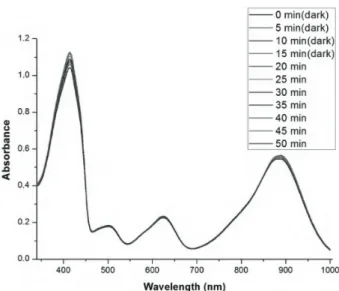
Figure 3. Absorbance decrease of 50 DPBF (50 µM) with
the increase in irradiation time in DCM in the presence of compound 4 (2 µ M). Absorbance decrease of DPBF at 410 nm. For the first 15 min the sample was kept in the dark; then it was irradiated with 890 nm light for 35 min. Absorbance was measured in 5-min intervals. The light source was an LED array with peak emission at 890 nm, fluence rate 2.5 mW/cm2.
2.3. Singlet oxygen generation experiment of compounds 4 and 5
It is noteworthy that for the first 15 min the cuvettes were kept in the dark and there was no discernible decrease in the absorbance spectrum of DPBF. The samples were then irradiated with an LED at 890 nm for 35 min (890 nm, fluence rate 2.5 mW/cm2) . Absorbance was measured in 5-min intervals and the absorption of the
trap molecule decreased. The results are given in Figures 3 and 4.
2.3.1. Singlet oxygen generation experiment of compounds 6 and 7
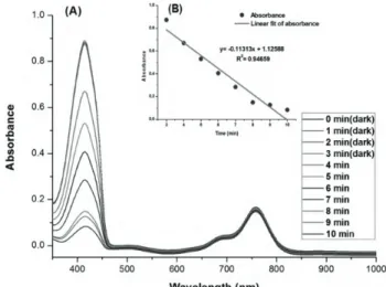
The same protocol applied to determine the singlet oxygen generation capacity of compounds 4 and 5 was carried out. For the first 3 min, the samples were kept in the dark and there was no recognizable change in the absorption of DPBF in the absence of photosensitizers. It may be clearly seen in Figure 5 for compound 7. Compound 7 was exposed to a near-IR light emitting diode (760 nm, fluence rate 2.5 mW/cm2) . The singlet
oxygen generation process of compound 7 is shown in Figure 6. Absorbance of DPBF was acquired in 1-min intervals and the absorption of the trap molecule decreased dramatically in the presence of compound 7.
2.3.2. Singlet oxygen quantum yield determination
Singlet oxygen quantum yields in DCM were calculated using a commonly used standard, methylene blue ( ϕ∆ = 0.57 in dichloromethane).36 The change in the absorption of the trap molecule at 410 nm versus irradiation
Figure 4. Absorbance decrease of 50 DPBF (50 µM) with
the increase in irradiation time in DCM in the presence of compound 5 (2 µ M). Absorbance decrease of DPBF at 410 nm. For the first 15 min the sample was kept in the dark; then it was irradiated with 890 nm light for 35 min. Absorbance was measured in 5-min intervals. The light source was an LED array with peak emission at 890 nm, fluence rate 2.5 mW/2.
Figure 5. a) Compound 7 (2 µ M) and DPBF (50 µM)
with the increase in irradiation time in DCM. b) Ab-sorbance decrease of DPBF at 410 nm. For the first 3 min the sample was kept in the dark; then it was irradiated with 760 nm light for 7 min. Absorbance was measured in 1-min intervals. The light source was an LED array with peak emission at 760 nm, fluence rate 2.5 mW/2.
Figure 6. The singlet oxygen generation process of compound 7.
time was graphically demonstrated by plotting the data for compounds 6 and 7. The results are given in Figure 7. It was noted that compounds 6 and 7 were highly effective singlet oxygen generators. As can be seen from the Table, singlet oxygen quantum yields of photosensitizers 4, 5, 6, and 7 were 0.10, 0.18, 0.52,
YEŞİLGÜL and KILIÇ/Turk J Chem
and 0.93, respectively. Compounds 6 and 7 were more effective singlet oxygen generators in comparison to the conventional singlet oxygen generator methylene blue and previously reported BODIPY-based photosensitizers.
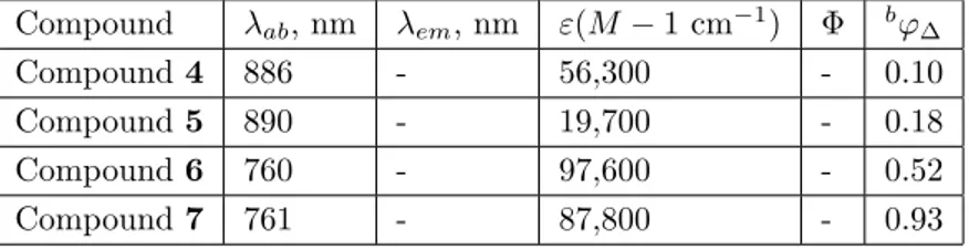
Table. Photophysical and photochemical properties of novel BODIPY derivatives.a
Compound λab, nm λem, nm ε(M − 1 cm−1) Φ bφ∆
Compound 4 886 - 56,300 - 0.10
Compound 5 890 - 19,700 - 0.18
Compound 6 760 - 97,600 - 0.52
Compound 7 761 - 87,800 - 0.93
aDichloromethane (5 × 10⁻⁶ M). bUsing methylene blue as standard.
Figure 7. Singlet oxygen mediated bleaching of the trap molecule DPBF in the presence of photosensitizers 6 and 7 (2
µM) in DCM. Absorbance at 416 nm was plotted as a function of time. The light source was an LED array with peak emission at 760 nm and a fluence rate of 2.5 mW/cm2.
2.4. Conclusion
In summary, a series of novel meso-CF3-substituted BODIPY derivatives were designed and synthesized.
Grat-ifyingly, the novel BODIPY derivatives had absorption maxima in the therapeutic window of the electro-magnetic spectrum. The introduction of electron-donating groups (4,4-dimethylaminobenzaldehyde and 4-(2 ethylhexyl)oxy benzaldehyde) at the 3- and 5-positions of the BODIPY cores and the attachment of an electron-withdrawing group to the 8-position of the BODIPY cores resulted in remarkable red shifts in the absorption and emission spectra. Compounds 6 and 7 show respectable singlet oxygen quantum yields in comparison to previously reported BODIPY-based photosensitizers.37 Further studies into the synthesis of water-soluble
meso-CF3-substituted and in vivo singlet oxygen generation experiments are ongoing at our laboratory to understand
potential PDT applications.
3. Experimental 3.1. General
1H NMR and 13C NMR spectra were recorded on a Bruker DPX-400 (operating at 400 MHz for1H NMR and
100 MHz for 13C NMR) in CDCl
at 25 °C and the coupling constants (J values) are given in Hz. Chemical shifts are given in parts per million (ppm). Absorption spectra analyses were performed using a Varian Cary-100 spectrophotometer. Fluorescence measurements were conducted on a Varian Eclipse spectrofluorometer. Mass spectra were recorded on an Agilent Technologies 6530 Accurate-Mass Q-TOF LC/MS system. The reactions were monitored by thin layer chromatography using a Merck TLC Silica gel 60 F254 set. Silica gel column chromatography was performed
with Merck Silica gel 60 (particle size: 0.040–0.063 mm, 230–400 mesh ASTM). All other reagents and solvents were purchased from Aldrich and used without further purification. Singlet oxygen quantum yield values were calculated based on the following equation:
φ∆(bod) = φ∆(ref )× (m(bod)/m(ref)) × (F (ref)/F (bod)) × (P F (ref)/P F (bod)),
where bod and ref designate the orthogonal BODIPY photosensitizer and MB, respectively. m is the slope of the difference in change in the absorbance of DPBF (414 nm) with irradiation time, F is the absorption correction factor, which is given by F = 1 – 10−OD (OD at the irradiation wavelength), and PF is the absorbed photonic flux ( µ Einstein dm−3s1) .
3.2. Synthesis of meso-CF3-substituted BODIPY (1)
Trifluoroacetic acid anhydride (5.85 mmol, 0.82 mL) was dropwise added into a 200-mL round bottom flask containing 2-methyl pyrrole (11.71 mmol, 950 mg). The reaction mixture was stirred for 20 min at room temperature. The combined organic mixture was dissolved in 200 mL of argon-degassed CH2Cl2 and was
stirred for 1 h. After that, to the reaction mixture were sequentially added 1.5 mL of Et3N and 1.5 mL of
BF3.OEt2, followed by stirring for 1 h. The resulting solution was washed three times with water and dried
over anhydrous Na2SO4. The solvent was removed in vacuo and then silica gel column chromatography was
carried out to purify the residue (as eluent CHCl3) . The orange-colored fraction was collected (495 mg, 29%). 1H NMR (400 MHz, CDCl
3) : δ H = 7.25 (s, 2H), 6.36 (s, 2H), 2.68 (s, 6H). 13C NMR (100 MHz, CDCl3) :
δ C = 161.7, 131.6, 130.3, 123.8, 121.6, 29.6, 15.4. HRMS (ESI) was calculated for C18H14BF2N3 (M-H)
287.0779, found as 287.0804 ∆ = 8.7 ppm.
3.3. Synthesis of iodinated meso-CF3−substituted BODIPY (2)
Compound 1 (100 mg, 1.47 mmol) was dissolved in 10 mL of DCM. ICl (2.21 mmol, 0.36 g) in 10 mL of DCM was added dropwise to the solution at room temperature. After stirring for 1 h, saturated Na2S2O3 solution in
water was added to the reaction mixture. The mixture was then extracted into CH2Cl2 and it was dried over
Na2SO4. The residue was purified by silica gel column chromatography (EtOAc). A brown liquid was obtained
(0.55 g, 69%). 1H NMR (400 MHz, CDCl
3, 300 K): δ H = 7.42 (s, 2H), 2.61 (s, 6H) 13C NMR (100 MHz,
CDCl3) : δ C = 162.8, 136.4, 132.2, 123.4, 120.7, 68.1, 16.0. HRMS (ESI) was calculated for C12H8BF5I2N2
(M-H) 537.8785, found as 537.8753 ∆ = 5.9 ppm.
3.4. Synthesis of brominated meso-CF3-substituted BODIPY (3)
Compound 1 (100 mg, 0.35 mmol) was dissolved in a mixture of DMF/DCM (25 mL:25 mL). Then N-bromosuccinimide (185 mg, 1.04 mmol) was dissolved in DCM (25 mL), and it was added to the reaction mixture dropwise over 15 min. The reaction mixture was stirred at room temperature for 2 h. Following
YEŞİLGÜL and KILIÇ/Turk J Chem
thin layer chromatography, the reaction was stopped, and the extraction was achieved with water and DCM. The organic phase was dried with Na2SO4 and evaporated using a rotary evaporator. The purification was
carried out with a silica gel column using a chloroform/hexane mixture (3:1 v/v). The fraction that contained brominated meso-CF3-substituted BODIPY was collected, and the solvent was then evaporated using a rotary
evaporator (120 mg, 77%). 1H NMR (400 MHz, CDCl
3) : δ H = 7.25 (s, 2H), 2.58 (s, 6H)13C NMR (100 MHz,
CDCl3) : δ C = 213.4, 160.1, 130.0, 121.0, 111.6, 29.7, 13.9. HRMS (ESI) was calculated for C12H8BBr2F5N2
(M-H) 441.9031, found as 441.8991 ∆ = 9.1 ppm.
3.5. Synthesis of 3,5-distyryl 8-CF3-substituted BODIPY (4)
Compound 3 (0.112 mmol, 50 mg) and 4,4-dimethylaminobenzaldehyde (0.336 mmol, 78.85 mg) were dissolved in benzene (20 mL). To the reaction mixture were successively added piperidine (0.5 mL) and glacial acetic acid (0.5 mL), and the mixture was refluxed for 30 min. The progress of the reaction was monitored by TLC (eluent CHCl3/hexane) (1:1). The crude product was concentrated under vacuum and purified using silica gel column
chromatography (eluent CHCl3/hexane) (1:1) (40 mg, 50.58%). 1H NMR (400 MHz, CDCl3) : δ H = 8.26 (d,
2H, J = 16.0), 7.60 (d, 4H, J = 7.52 Hz), 7.58 (d, 2H, J =16.0 Hz), 7.30 (s, 2H), 6.75 (d, 4H, J = 8.28 Hz), 3.10 (s, 12H). HRMS (ESI) was calculated for C30H26BBr2F5N4 (M-H) 705.0606, found as 705.05792 ∆ =
3.77 ppm.
3.6. Synthesis of 3,5-distyryl 8-CF3-substituted BODIPY (5)
Compound 2 (0.185 mmol, 100 mg) and 4,4-dimethylaminobenzaldehyde (0.555 mmol, 130.25 mg) were dissolved in benzene (20 mL). To the reaction mixture were successively added piperidine (0.5 mL) and glacial acetic acid (0.5 mL), and the mixture was refluxed for 30 min. The progress of the reaction was monitored by TLC (eluent CHCl3/hexane) (1:1). The crude product was concentrated under vacuum and purified using silica gel column
chromatography (eluent CHCl3/hexane) (1:1) (40 mg, 26.96%). 1H NMR (400 MHz, CDCl3) : δ H = 8.35 (d,
2H, J = 16.0), 7.65 (d, 4H, J = 8.68 Hz), 7.60 (d, 2H, J = 16.0 Hz), 7.50 (s, 2H), 6.75 (d, 4H, J = 8.68 Hz), 3.10 (s, 12H). HRMS (ESI) was calculated for C30H26BF5I2N4 (M+H) 803.0260, found as 803.0255 ∆ = 0.6
ppm.
3.7. Synthesis of 3,5-distyryl 8-CF3-substituted BODIPY (6)
Compound 3 (0.225 mmol, 100 mg) and 4-(2 ethylhexyl) oxy-benzaldehyde (0.450 mmol, 105.61 mg) were dissolved in benzene (20 mL). To the reaction mixture were successively added piperidine (0.5 mL) and glacial acetic acid (0.5 mL), and the mixture was refluxed for 10 min. The progress of the reaction was monitored by TLC (eluent CHCl3/hexane) (1:1). The crude product was concentrated under vacuum and purified using
silica gel column chromatography (eluent CHCl3/hexane) (1:1) (40 mg, 20.98%). 1H NMR (400 MHz, CDCl3) :
δ H = 8.35 (d, J = 16.4 Hz, 2H), 7.70–7.61 (m, 6H), 7.38 (s, 2H), 7.01 (d, 8.6 Hz, 4H), 3.95 (d, 5.7 Hz, 4H), 1.82–1.70 (m, 2H), 1.60–1.32 (m, 16H), 1.02–0.90 (m, 12H). 13C NMR (100 MHz, CDCl
3) : δ C = 161.6, 151.80,
141.72, 132.0, 130.6, 130.0, 129.0, 125.27, 124.15, 115.7, 115.1, 109.5, 70.7, 39.4, 30.5, 29.0, 23.8, 23.0, 14.0, 11.1. HRMS (ESI) was calculated for C42H48BBr2F5N2O2 (M+H) 877.20958, found as 877.21070 ∆ = 1.27
3.8. Synthesis of 3,5-distyryl 8-CF3-substituted BODIPY (7)
Compound 2 (0.185 mmol, 100 mg) and 4-(2-ethylhexyl) oxy-benzaldehyde (0.450 mmol, 105.61 mg) were dissolved in benzene (20 mL). To the reaction mixture were successively added piperidine (0.5 mL) and glacial acetic acid (0.5 mL), and the mixture was refluxed for 10 min. The progress of the reaction was monitored by TLC (eluent CHCl3/hexane) (1:1). The crude product was concentrated under vacuum. The progress of
the reaction was monitored by TLC (eluent CHCl3/hexane) (1:1). The crude product was concentrated under
vacuum and purified using silica gel column chromatography (eluent CHCl3/hexane) (1:1) (45 mg, 25%). 1H
NMR (400 MHz, CDCl3, 300 K): δ H = 8.36 (d, J = 16.4 Hz, 2H), 7.75–7.60 (m, 6H), 7.51 (s, 2H), 6.97 (d, 8.4
Hz, 4H), 3.97 (d, 5.7 Hz, 4H), 1.85–1.75 (m, 2H), 1.62–1.40 (m, 16H), 1.05–0.90 (m, 12H). 13C NMR (100 MHz,
CDCl3) : δ C = 161.6, 153.95, 141.77, 137.61, 136.58, 130.29, 130.01, 129.44, 128.94, 116.23, 115.14, 115.00,
70.80, 39.34, 30.51, 29.08, 23.86, 23.03, 14.06, 11.10. HRMS (ESI) was calculated for C42H48BBr2F5N2O2
(M +H) 973.18184, found as 973.18384 ∆ = 2.0 ppm.
Acknowledgments
The authors gratefully acknowledge the support from the Department of Chemistry and UNAM, Bilkent University, and the Faculty of Aviation and Aeronautical Science, Özyeğin University.
References
1. Kamat, M. A.; Hahn, N. M.; Efstathiou, J. A.; Lerner, S. P.; Malmström, P.; Choi, W.; Guo, C. C.; Lotan, Y.; Kassouf, W. Lancet 2016, 388, 2796-2810.
2. Gestaut, M. M.; Swanson, P. G. Reports of Practical Oncology and Radiotherapy 2017, 22, 77-82. 3. Taylor, C. W.; Kirby, A. M. Clin. Oncol. 2015, 27, 621-629.
4. Colagiuri, B.; Dhillon, H.; Butow, P. N.; Jansen, J.; Cox, K.; Jacquet, J. J. Pain Symptom Manag. 2013, 46, 275-281.
5. Wen, X.; Li, Y.; Hamblin, M. R. Photodiagn. Photodyn. 2017, 19, 140-152. 6. Dolmans, D. E.; Fukumura, D.; Jain, R. K. Nat. Rev. Cancer 2003, 3, 380-387.
7. Banerjee, S. M.; MacRobert, A. J.; Mosse, C. A.; Periera, B.; Bown, S. G.; Keshtgar, M. R. S. Breast 2017, 31, 105-113.
8. Richards-Kortumr, R.; Muraca, S. Annu. Rev. Phys. Chem. 1996, 47, 555-606. 9. Lu, H.; Mack, J.; Yanga, Y.; Shen, Z. Chem. Soc. Rev. 2014, 43, 4778-4823.
10. Kamkaew, A.; Lim, S. H.; Lee, H. B.; Kiew, L. V.; Chung, L. Y.; Burgess, K. Chem. Soc. Rev. 2013, 42, 77-88. 11. Loudet, A.; Burgess, K. Chem. Soc. Rev. 2007, 107, 4891-4932.
12. Ulrich, G.; Ziessel, R.; Harriman, A. Angew. Chem., Int. Ed. 2008, 47, 1184-1201.
13. Kilic, B.; Yesilgul, N.; Polat, V.; Gercek, Z.; Akkaya, E. U. Tetrahedron Lett. 2016, 57, 1317-1320.
14. Turan, I. S.; Cakmak, F. P.; Yildirim, D. C.; Cetin-Atalay, R.; Akkaya, E. U. Chem. Eur. J. 2014, 20, 16088-16092. 15. Zhang, X. F.; Yang, X. J. Phys. Chem. B 2013, 117, 5533-5539.
16. Cakmak, Y.; Kolemen, S.; Duman, S.; Dede, Y.; Dolen, Y.; Kilic, B.; Kostereli, Z.; Yildirim, L. T.; Dogan, A. L.; Guc, D. et al. Angew. Chem., Int. Ed. 2011, 50, 11937-11941.
17. Zhao, J.; Xu, K.; Yang, W.; Wang, Z.; Zhong, F. Chem. Soc. Rev. 2015, 44, 8904-8939.
YEŞİLGÜL and KILIÇ/Turk J Chem
19. Awuah, S. A.; You, Y. RSC Adv. 2012, 2, 11169-11183.
20. Zhang, J.; Jiang, C.; Longo, J. P. F.; Azevedo, R. B.; Zhang, H.; Muehlmann, L. A. Acta Pharmaceutica Sinica B
2017, 8, 137-146.
21. Li, L.; Nguyen, B.; Burgess, K. Bioorg. Med. Chem. Lett. 2008, 18, 3112-3116. 22. Awuah, S. G.; Polreis, J.; Biradar, V.; You, Y. Org. Lett. 2011, 13, 3884-3887.
23. Sobenina, L. N.; Vasiltsov, A. M.; Petrova, O. V.; Petrushenko, K. B.; Ushakov, I. A.; Clavier, G.; Meallet-Renault, R.; Mikhaleva, A. I.; Trofimov, B. A. Org. Lett. 2011, 13, 2524-2527.
24. Li, L.; Nguyen, B.; Burgess, K. Bioorg. Med. Chem. Lett. 2008, 18, 3112-3116.
25. Petrushenko, K. B.; Petrushenko, I. K.; Petrova, O. V.; Sobenina, L. N.; Trofimov, B. A. Dyes Pigments 2017,
136, 488-495.
26. Afonin, A. V.; Ushakov, I. A.; Pavlov, D. V.; Petrova, O. V.; Sobenina, L. N.; Mikhaleva, A. I.; Trofimov, B. A.
J. Fluorine Chem. 2013, 145, 51-57.
27. Yogo, T.; Urano, Y.; Ishitsuka, Y.; Maniwa, F.; Nagano, T. J. Am. Chem. Soc. 2005, 127, 12162-12163.
28. Epelde-Elezcano, N.; Martinez-Martinez, V.; Pena-Cabrera, E.; Gomez-Duran, C. F. A.; Arbeloa, I. L.; Lacombe, S. RSC Adv. 2016, 6, 41991-41998.
29. Gorbe, M.; Costero, A. M.; Sancenon, F.; Martinez-Manez, R.; Ballesteros-Cillero, R.; Ochando, L. E.; Chulvi, K.; Gotor, R.; Gil, S. Dyes Pigments 2019, 160, 198-207.
30. Davies, M. J. Biochem. Biophys. Res. Commun. 2003, 305, 761-770.
31. Xia, Q.; Boudreau, M. D.; Zhou, Y. T.; Yin, J. J.; Fu, P. P. J. Food Drug. Anal. 2011, 19, 396-402.
32. Cadet, J.; Ravanat, J. L.; Martinez, G. R.; Medeiros, M. H. G.; Mascio, P. D. Photochem. Photobiol. 2006, 82, 1219-1225.
33. Tørring, T.; Helmig, S.; Ogilby, P. R.; Gothelf, K. V. Acc. Chem. Res. 2014, 47, 1799-1806.
34. Zhang, C.; Zhao, J.; Wu, S.; Wang, Z.; Wu, W.; Ma, J.; Guo, S.; Huang, L. J. Am. Chem. Soc. 2013, 135, 10566-10578.
35. Lincoln, R.; Durantini, A. M.; Greene, L. E.; Martinez, S. R.; Knox, R.; Becerra, M. C.; Cosa, G. Photochem.
Photobiol. Sci. 2017, 16, 178-184.
36. Mirenda, M.; Strassert, C. A.; Dicelio, L. E.; Roman, E. S. ACS Appl. Mater. Inter. 2010, 2, 1556-1560. 37. Senkuytu, E.; Ecik, E. T. Spectrochim. Acta A 2017, 182, 26-31.