Full Terms & Conditions of access and use can be found at
https://www.tandfonline.com/action/journalInformation?journalCode=ibih20
Biotechnic & Histochemistry
ISSN: 1052-0295 (Print) 1473-7760 (Online) Journal homepage: https://www.tandfonline.com/loi/ibih20
Protective effect of quercetin on some
hematological parameters in rats exposed to
cadmium
H. H. Donmez, N. Donmez, I. Kısadere & I. Undag
To cite this article: H. H. Donmez, N. Donmez, I. Kısadere & I. Undag (2019) Protective effect of quercetin on some hematological parameters in rats exposed to cadmium, Biotechnic & Histochemistry, 94:5, 381-386, DOI: 10.1080/10520295.2019.1574027
To link to this article: https://doi.org/10.1080/10520295.2019.1574027
Published online: 01 Mar 2019.
Submit your article to this journal
Article views: 298
View related articles
View Crossmark data
Protective effect of quercetin on some hematological parameters in rats
exposed to cadmium
H. H. Donmez a, N. Donmez b, I. Kısadere c, and I. Undag a
aDepartment of Histology and Embryology, Faculty of Veterinary, University of Selcuk, Konya, Turkey;bDepartment of Physiology, Faculty of Veterinary, University of Selcuk, Konya, Turkey;cDepartment of Physiology, Faculty of Veterinary, University of Balıkesir, Balıkesir, Turkey
ABSTRACT
We investigated the effects of quercetin (Q) on some hematological parameters and determined the percentage of alpha-naphthyl acetate esterase (ANAE) positive lymphocytes in rats that had been exposed to cadmium (Cd). Thirty male Wistar albino rats were divided into four groups: control (C), quercetin (Q), cadmium (Cd) and Q + Cd (CdQ). Blood samples were taken to assess erythrocytes (RBC), leukocytes (WBC), hemoglobin levels (Hb), hematocrit values (Hct), platelets (PLT), alpha-naphthyl acetate esterase (ANAE) positive lymphocytes. RBC, Hb, Hct; the number of PLT significantly decreased in the Cd group. To the contrary, these parameters were increased significantly in the CdQ group compared to the Cd group. Although we found a significant increase in total WBC count and neutrophil percentage, the number of lymphocytes decreased in the Cd group compared to the other three groups. Also, the percentage of peripheral blood ANAE positive lymphocytes decreased significantly in the Cd group (p < 0.05). Q exhibits positive effects on some hematological characteristics and the percentage of ANAE positive lymphocyte in cases of acute CD toxicity.
KEYWORDS
Alpha-naphthyl acetate esterase; cadmium; hematology; quercetin; rats; toxicity
The use of toxic heavy metals, such as cadmium (Cd) in agricultural, chemical and industrial activities and smoking has increased life-threatening environmental pollution. Cd causes significant health problems in animals and humans due to consumption of contaminated food and drinking water (Schwartz and Reis 2000). Cd is an industrial and environmental pollutant that exhibits many toxic effects on living organisms (Karabulut Bulan et al. 2004). Although the liver is the principal target organ for many heavy metals, Cd accumulates also in the kidney, lung, duodenum, pancreas, testis and bone. Exposure to Cd causes loss of renal function, hepatic damage, respiratory and digestive system disorders, anemia and testicular atrophy (Schwartz and Reis2000).
Antioxidant-rich substances including lycopene, selenium, vitamin E, vitamin C, melatonin, acetylcysteine, ß-carotene are used to protect against Cd toxicity (Jahan et al. 2014). Quercetin (3,5,7,3ʹ,4ʹ-pentahydroxyflavone)
(Q) is a natural bioflavonoid polyphenolic substance that is widely present in fruits and vegetables including apples, onions, mulberries, broccoli, herbs, tea, peanuts, soybeans and red wine. In plants, Q occurs mainly in leaves and in the outer parts of the plants as aglycones and glycosides (Wach et al. 2007; Renugadevi and Prabu 2010). Q exhibits
antioxidative, anti-inflammatory, antimicrobial, antiviral, anti-ulcerogenic, antineoplastic, mutagenic, antioxidant, antihepatotoxic, antihypertensive, hypolipidemic and antiplatelet properties (Elik et al.2007; Aguirre et al.2011; Entaz et al. 2017). Q is a potent oxygen free radical scavenger and metal chelator. Morales et al. (2006) reported that Q is protective against the hemotoxic and neurotoxic effects of Cd in rats.
The hematopoietic system is sensitive to many drugs, toxins and heavy metals. Cd has been reported to disrupt hematopoietic system functions (Demir et al. 2006; El-Boshy et al. 2015). The most important effect of Cd toxicity on hematopoiesis is anemia; Cd toxicity includes microcytic anemia in rats (Waner and Gur 1993; Kocak 2004). Following intake and absorption, Cd is transported by blood, where it binds with red blood cell (RBC) membranes and plasma albumin. In the circulation, Cd stimulates the formation of metallothioneins and reactive oxygen species (ROS), which cause oxidative stress in RBC and T or B lymphocytes (Sadrzadeh et al.
1984; Kolanjiappana et al. 2002; Minetti et al. 2008). Exposure to Cd also causes temporary and rapid leukocytosis (Mackova et al. 1996). Also, acute Cd toxicity accelerates platelet aggregation, which causes
CONTACTI. Kısadere [email protected] Department of Physiology, Faculty of Veterinary, University of Balıkesir, Turkey. 2019, VOL. 94, NO. 5, 381–386
https://doi.org/10.1080/10520295.2019.1574027
pial cerebral thrombosis in mice (Fahim et al. 2012). Administration of acute high dose Cd exhibits a genotoxic effect on bone marrow, and it increases the number of polychromatic erythrocytes (Popovic-Bubujuk et al. 2013).
Alpha-naphthyl acetate esterase (ANAE) is a lysosomal enzyme that can be used to differentiate T lymphocytes, B lymphocytes and monocytes in both tissue and peripheral blood smears. ANAE is reported to be acquired during the later stages of T lymphocyte maturation in the thymus. Like other esterases, ANAE participates in the cytotoxic functions of active T lymphocytes and degradation of the phagocytic material within macrophages (Sur et al.2005).
We investigated the protective effect of Q on some hematological characteristics and ANAE positive lymphocytes that provide important information concerning the general condition of the hematopoietic system in rats that have been exposed acutely to toxic levels of Cd.
Material and methods
Animals
We used 30 healthy, 350 ± 10 g 4-month-old male Wistar albino rats obtained from Selcuk University Experimental Medicine Research and Application Center (SUEMRAC). We established four experimental groups of animals as follows: untreated control (C; n = 6), Cd (n = 8), Q (n = 8) and Cd + Q (CdQ; n = 8). Animals were housed during the study in standard plastic rat cages in the SUEMRAC. Room temperature and relative humidity were 23 ± 2 °C and 55 ± 10%, respectively, with a 12 h light:12 h dark cycle. Rats were provided access to standard feed and approximately 50 ml/day/rat fresh water ad libitum. All animal handling and procedures were approved by Experimental Medicine Research and Application Center of Selcuk University Experimental Animal Ethics Committee (2015/45).
We injected subcutaneously (s.c.) 4 mg/kg/day cadmium chloride (CdCl2) into Cd and CdQ group
animals and 50 mg/kg/day Q was injected intraperitoneally (i.p) into Q and CdQ group animals for 3 days. Cd and Q were prepared fresh before injection. At the end of treatment, blood samples were obtained by cardiac puncture under 40 mg/kg thiopental anesthesia and collected in anticoagulant tubes.
Hematology
Leukocytes (WBC), RBC, hemoglobin (Hb), hematocrit (Hct), platelets (PLT) and differential WBC counts
were assessed in blood samples using a cell counter (Mindray BC-800, Nanshan, Shenzhen, China).
Determination of the percentage of ANAE positive lymphocytes
Two blood smears were prepared from heparinized blood samples and dried at room temperature. To determine ANAE activity, smears were fixed in 50% glutaraldehyde:50% acetone, pH 4.8, at −10 °C for 3 min. The smears then were dried at room temperature and exposed to an incubation solution prepared by adding 80 ml buffered phosphate solution, pH 5.0, and 20 mg substrate (ANAE, N-8505; Sigma, Steinheim, Germany) dissolved in 0.8 ml acetone (Merck, Darmstadt, Germany) drop by drop to prevent crystallization of the substrate. Then, 4.8 ml hexazotized pararosanilin mixture, obtained by incubating 2.4 ml 4% sodium nitrite (S-3421; Merck) solution with 2.4 ml pararosanilin (P-3750; Merck) (1 g pararosanilin, 20 ml distilled water, 5 ml HCl concentrate) for 2 min, was added to the buffered phosphate solution containing α-naphthyl acetate. The solution was adjusted to pH 5.8 with 1 N NaOH and filtered. After nuclear staining with Giemsa (Donmez et al.2007), the smears were incubated in the solution for 2 h and examined by light microscopy (Leica DM 2500, Heerbrugg, Switzerland). Lymphocytes that exhibited brown cytoplasmic staining as dots were considered positive (Figure 1, arrow), while others were considered negative. Means were calculated by counting 200 lymphocytes in each smear.
Statistical analysis
Differences among the groups were evaluated by analysis of variance (ANOVA) followed by Duncan’s
Figure 1.ANAE staining in rat peripheral blood of control group. Arrow, ANAE positive lymphocyte; arrowhead, ANAE negative lymphocyte.
test using the SPSS 22.0 program (SPSS, Inc., Chicago, IL). Values for p ≤ 0.05 were considered significant.
Results
Hematology
The results of our study are presented inTables 1 and
2. We found that the number of RBCs, Hb levels, Htc values and number of PLT decreased significantly in the Cd group compared to the other three groups (p < 0.05). These reductions were ameliorated by Q in the CdQ group; the values were significantly lower than for the C and Q groups (p < 0.05). The number of WBC significantly increased in the Cd group compared to the other three groups (p < 0.05). We found a significant increase in the percentage of neutrophils in the Cd group (p < 0.05). Although the percentage of lymphocytes decreased significantly in the Cd group (p < 0.05), it was ameliorated by Q in the CdQ group compared to the Cd group (p < 0.05), shown inTable 2.
Percentage of ANAE positive lymphocytes
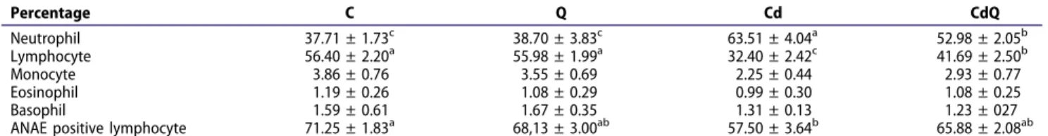
Although we found a reduction in the percentage of ANAE positive lymphocytes in the Cd group, we found no significant difference among the experimental groups, Q, CdQ and C (Table 2).
Discussion
El-Boshy et al. (2015) reported that chronic Cd toxicity caused anemia in Cd treated rats. The anemia caused by Cd is related to accumulation of toxic metals in the kidney, liver and spleen, which is related to hemolysis owing to a deformity of peripheral red blood cells
(RBC) (Kunimoto et al. 1985), iron deficiency by competition with duodenal iron absorption (Hamilton and Valberg 1974) and renal anemia caused decreased production of erythropoietin (EPO) (Horiguchi et al.
1994,2010). Ognjanović et al. (2003) also reported that the number of RBC, Htc value, Hb concentration and number of PLT were reduced significantly in rats with acute Cd toxicity. Exposure to different doses of Cd for different durations has been reported to produce deleterious effects in rats that correspond to our results (Karmakar et al. 2000; El-Demerdash et al.
2004). We found that measurements of RBC, Hb, Hct and PLT decreased significantly (Table 1) in the Cd group compared to the other three groups, C, Q and CdQ, which is consistent with earlier reports (Ognjanović et al. 2003; Kocak and Akcil 2006; Nazima et al. 2016). Exposure to Cd causes microcytic hypochromic anemia as well as changes in the membrane permeability of RBC, which leads to a decreased hematocrit. Cd also reduces the intestinal absorption of iron due to mucosal lesions, which contributes to a decreased Hct concentration (Ognjanović et al. 2003; Kocak 2004; Demir et al.
2006; El-Boshy et al.2015).
RBC, Hb, Hct, and PLT values were similar in the C and Q groups and these characteristics were increased significantly in the CdQ group compared to the Cd group (Table 1). Some antioxidants show protective activity against degradation of RBC by Cd (Nazima et al.2016). The changes that we observed can be interpreted as the protective effect of Q.
The number of WBC increased significantly in Cd group compared to the C, Q and CdQ groups. Although we found no significant change in the percentage of monocytes, eosinophils and basophils, we observed a significant increase in the percentage of Table 1.Effects of Cd and Q on some hematological characteristics.
Characteristics C Q Cd CdQ Erythrocyte (×106/mm3) 8.53 ± 0.23a 8.37 ± 0.33a 5.58 ± 0.22c 7.24 ± 0.47b Leukocyte (×103/mm3) 5.39 ± 0.38c 5.78 ± 0.37c 12.13 ± 1.59a 9.07 ± 0.53b Hematocrit (%) 44.89 ± 3.20a 42.23 ± 2.01a 22.35 ± 4.79b 27.48 ± 2.34b Hemoglobin (g/dl) 15.53 ± 0.32a 15.65 ± 0.34a 11.55 ± 1.58b 13.41 ± 1.35ab Platelet (×109/l) 627.33 ± 52.51a 646.0 ± 51.52a 259.0 ± 35.79b 414.50 ± 73.45b
Groups: C, control; Q, quercetin; Cd, cadmium; CdQ, Q + Cd.a−cMeans in the same row with different superscripts differ significantly (p < 0.05).
Table 2.Effects of Cd and Q on differential WBC and ANAE positive lymphocyte percentages.
Percentage C Q Cd CdQ Neutrophil 37.71 ± 1.73c 38.70 ± 3.83c 63.51 ± 4.04a 52.98 ± 2.05b Lymphocyte 56.40 ± 2.20a 55.98 ± 1.99a 32.40 ± 2.42c 41.69 ± 2.50b Monocyte 3.86 ± 0.76 3.55 ± 0.69 2.25 ± 0.44 2.93 ± 0.77 Eosinophil 1.19 ± 0.26 1.08 ± 0.29 0.99 ± 0.30 1.08 ± 0.25 Basophil 1.59 ± 0.61 1.67 ± 0.35 1.31 ± 0.13 1.23 ± 027 ANAE positive lymphocyte 71.25 ± 1.83a 68,13 ± 3.00ab 57.50 ± 3.64b 65.88 ± 2.08ab
neutrophils in the Cd group compared to the other groups (C, Q, Cd + Q) (p < 0. 05). To the contrary, we found a significant decrease in the percentage of lymphocytes in the Cd group compared to the other groups (p < 0. 05) (Tables 1, 2). No significant difference was found in the percentage of WBC between the C and Q groups. In addition, the number of WBC and neutrophils decreased, while the number of lymphocytes increased in the CdQ group compared to the Cd group. It has been suggested that increased number of WBC and decreased number of lymphocytes following Cd toxicity may be due to increased free radicals, decreased antioxidant activity, or suppression of nonspecific and specific immune response as well as systemic inflammation (Karuppasamy et al. 2005; Fahim et al. 2012). El-Boshy et al. (2015) reported that acute Cd administration caused neutrophilia and lymphopenia without significant changes in total WBC, monocyte, eosinophil and basophil numbers in rats; these results also confirm the neutrophilopenic and lymphopenic effects of Cd in rats. Our findings are consistent with the earlier reports. Elevation of percentage of IL-6 and TNF-α could be a consequence of the neutrophilia and lymphopenia.
El-Boshy et al. (2015) reported that interleukin-1 beta (IL-1), TNF-α, IL-6, IL-10 and number of peripheral neutrophils increased while interferon gamma and the number of lymphocytes decreased owing to Cd administration. Cd toxicity also suppresses lymphocyte proliferation and natural killer cell activity (Thomas et al.1985; Cifone et al.1990). In addition, Cd toxicity during the prenatal period causes abnormal thymocyte development by disturbing T lymphocyte production during the postnatal period (Hanson et al.2012; Holásková et al.2012).
It has been suggested that Q regulates the inflammatory responses of macrophages and T lymphocytes (Yu et al. 2008). Previous studies have shown that Q produces an anti-inflammatory response by stimulating the release of inflammatory cytokines and nitric oxide from macrophages. Q also affects T lymphocyte functions by inhibiting cell proliferation (Kumazawa et al. 2006; Yu et al. 2008). ANAE, a lymphocyte lysosomal enzyme acquired from medullary thymocytes in the thymus during the embryonic stage, has been demonstrated in mature and immunocompetent T lymphocytes activity in peripheral blood. ANAE is specific for mature T lymphocytes, but not B lymphocytes (Figure 1) (Basso et al. 1980; Donmez and Sur 2007; Donmez et al. 2007). ANAE activity also is found in monocytes and macrophages (Mueller et al.1975). We found that
the percentage of ANAE positive lymphocytes in peripheral blood decreased significantly in the Cd group compared to the C, Q, and CdQ groups (p < 0.05) (Table 2). We found that Cd decreased the percentage of peripheral blood lymphocytes significantly and administration of Q helped prevent this decrease following exposure to Cd.
We suggest that the increased percentage of peripheral blood lymphocytes might be due to the metal binding properties of Q (Blasiak 2001; Jamalan et al.2015). Simsek et al. (2009) also reported that Cd and lead reduced the percentage of peripheral blood ANAE positive lymphocytes. The use of Spirulina platensis, a blue-green alga (Oscillotoreaceae) that exhibits antioxidative and anti-inflammatory effects and binds both Cd and lead, increased the percentage of peripheral blood ANAE positive lymphocytes to near normal levels (Mao et al. 2005; Simsek et al.2009).
Q exhibited positive effects on some hematological characteristics and the percentage of ANAE positive lymphocytes following acute Cd toxicity. Q appears to exhibit protective efficacy for the hematopoietic system.
Declaration of interest
The authors declare no conflict of interest. The authors alone are responsible for the content and writing of the paper.
Disclosure statement
No potential conflict of interest was reported by the authors.
ORCID H. H. Donmez http://orcid.org/0000-0003-4664-8489 N. Donmez http://orcid.org/0000-0003-4271-598X I. Kısadere http://orcid.org/0000-0003-0732-0464 I. Undag http://orcid.org/0000-0001-8495-3930 References
Aguirre L, Arias N, Macarulla MT, Gracia A, Portillo MP. 2011. Beneficial effects of quercetin on obesity and diabetes. Open Nutraceut J. 4:189–198. doi:10.2174/ 1876396001104010189.
Basso G, Cocito MG, Semenzato G, Pezzutto A, Zanesco L. 1980. Cytochemical study of thymocytes and T lymphocytes. Br J Haematol. 44:577–582. doi:10.1111/ j.1365-2141.1980.tb08712.x.
Blasiak J. 2001. DNA-damaging effect of cadmium and protective action of quercetin. Pol J Env Stud. 10:437–442. Cifone MG, Procopio A, Napolitano T, Alesse E, Santoni G, Santoni A. 1990. Cadmium inhibits spontaneous (NK), antibody-mediated (ADCC) and IL-2-stimulated
cytotoxic functions of natural killer cells. Immun Pharmacol. 20:73–80. doi:10.1016/0162-3109(90)90009-4. Demir H, Kanter M, Coskun O, Uz YH, Koc A, Yildiz A.
2006. Effect of black cumin (Nigella sativa) on heart rate, some hematological values, and pancreatic β-cell damage in cadmium-treated rats. Biol Trace Elem Res. 110:151–162. doi:10.1385/BTER:110:2:151.
Donmez HH, Sur E. 2007. Ergin evcil ördeklerin (Anas platryhynchos) perifer kan lenfositlerinde alfa-naftil asetat esteraz ve asit fosfataz aktivitesinin belirlenmesi. Ataturk Univ Vet Bil Derg. 2:122–128.
Donmez HH, Sur E, Boydak M.2007. Determination of alpha naphthyl acetate esterase activity in peripheral blood leucocytes of Kangal fish (Garra rufa). Vet Bil Derg. 21:81–84.
El-Boshy ME, Risha EF, Abdelhamid FM, Mubarak MS, Hadda TB. 2015. Protective effects of selenium against cadmium induced hematological disturbances, immunosuppressive, oxidative stress and hepatorenal damage in rats. J Trace Elem Med. 29:104–110. doi:10.1016/j.jtemb.2014.05.009.
El-Demerdash FM, Yousef MI, Kedwany FS, Baghdadi HH. 2004. Cadmium-induced changes in lipid peroxidation, blood hematology, biochemical parameters and semen quality of male rats: protective role of vitamin E and beta-carotene. Food Chem Toxicol. 42:1563–1571. doi:10.1016/j.fct.2004.05.001.
Elik M, Serdaroglu G, Ozkan R.2007. Mirisetin ve kuersetin bileşiklerinin antioksidan etkinliklerinin DFT yöntemiyle incelenmesi. CU Fen Bil Derg. 28:53–65.
Entaz B, Geum-Hwa L, Kashi RB, Hwa-Young L, Hyun-Kyoung K, Mallikarjun H, Min-Kyung C, Sun-Young H, Han-Jung C, Hyonok Y.2017. Protective role of quercetin against manganese-induced injury in the liver, kidney, and lung; and hematological parameters in acute and subchronic rat models. Drug Des Dev Ther. 11:2605–2619. doi:10.2147/DDDT.S143875.
Fahim MA, Nemmar A, Dhanasekaran S, Singh S, Shafiullah M, Yasin J, Zia S, Hasan MY. 2012. Acute cadmium exposure causes systemic and thromboembolic events in mice. Physiol Res. 61:73–80.
Hamilton DL, Valberg LS. 1974. Relationship between cadmium and iron absorption. Am J Physiol. 227:1033–1037. doi:10.1152/ajplegacy.1974.227.5.1033. Hanson ML, Holásková I, Elliott M, Brundage KM,
Schafer R, Barnett JB. 2012. Prenatal cadmium exposure alters postnatal immune cell development and function. Toxicol Appl Pharmacol. 261:196–203. doi:10.1016/j. taap.2012.04.002.
Holásková I, Elliott M, Hanson ML, Schafer R, Barnett JB. 2012. Prenatal cadmium exposure produces persistent changes to thymus and spleen cell phenotypic repertoire as well as the acquired immune response. Toxicol Appl Pharmacol. 265:181–189. doi:10.1016/j.taap.2012.10.009. Horiguchi H, Teranishi H, Niiya K, Aoshima K, Katoh T,
Sakuragawa N, Kasuya M. 1994. Hypoproduction of erythropoietin contributes to anemia in chronic cadmium intoxication: clinical study on itai-itai disease in Japan. Arch Toxicol. 68:632–636. doi:10.1007/BF03208342. Horiguchi H, Aoshima K, Oguma E, Sasaki S, Miyamoto K,
Hosoi Y, Katoh T, Kayama F. 2010. Latest status of
cadmium accumulation and its effects on kidneys, bone, and erythropoiesis in inhabitants of the formerly cadmium-polluted Jinzu River Basin in Toyama, Japan, after restoration of rice paddies. Int Arch Occup Env Health. 83:953–970. doi:10.1007/s00420-010-0510-x. Jahan S, Zahra A, Irum U, Iftikhar N, Ullah H. 2014.
Protective effects of different antioxidants against cadmium induced oxidative damage in rat testis and prostate tissues. Syst Biol Reprod Med. 60:199–205. doi:10.3109/19396368.2014.912363.
Jamalan M, Ghaffari MA, Hoseinzadeh P, Hashemitabar M, Zeinali M. 2015. Human sperm quality and metal toxicants: protective effects of some flavonoids on male reproductive function. Int J Fertil Steril. 9:215–222. Karabulut Bulan O, Koyuturk M, Bolkent S, Yanardag R,
Oguz AT. 2004. The effects of combined therapy of vitamin C, vitamin E and selenium against cadmium-injury in thyroid gland of rats. Cerrahpasa J Med. 35:174–180.
Karmakar R, Bhattacharya R, Chatterjee M.2000. Biochemical, haematological and histopathological study in relation to time-related cadmium-induced hepatotoxicity in mice. Biometals. 13:231–239. doi:10.1023/A:1009279803842. Karuppasamy R, Subathra S, Puvaneswari S. 2005.
Haematological responses to exposure to sublethal concentration of cadmium in air breathing fish, Channa punctatus (Bloch). J Env Biol. 26:123–128.
Kocak M 2004. Kronik kadmiyum toksisitesinin hemostatik sisteme etkileri. Ankara Sağ Bil Enst. Doctoral thesis. Ankara.
Kocak M, Akcil E. 2006. The effects of chronic cadmium toxicity on the hemostatic system. Pathophysiol Haemos Thromb. 35:411–416. doi:10.1159/000102047.
Kolanjiappana K, Manoharana S, Kayalvizhi M. 2002. Measurement of erythrocyte lipids, lipid peroxidation, antioxidants and osmotic fragility in cervical cancer patients. Clin Chim Acta. 326:143–149. doi:10.1016/ S0009-8981(02)00300-5.
Kumazawa Y, Kawaguchi K, Takimoto H. 2006. Immunomodulating effects of flavonoids on acute and chronic inflammatory responses caused by tumor necrosis factor α. Curr Pharm Des. 12:4271–4279. doi:10.2174/138161206778743565.
Kunimoto M, Miura T, Kubota K. 1985. An apparent acceleration of age-related changes of rat red blood cells by cadmium. Toxicol Appl Pharmacol. 77:451–457. doi:10.1016/0041-008X(85)90185-1.
Mackova NO, Lenikova S, Fedorocko P, Brezani P, Fedorockova A. 1996. Effects of cadmium on haemopoiesis in irradiated and non-irradiated mice. 2. Relationship to the number of circulating blood cells and haemopoiesis. Physiol Res. 45:101–106.
Mao TK, Van de Water J, Gershwin ME. 2005. Effects of a spirulina-based dietary supplement on cytokine production from allergic rhinitis patients. J Med Food. 8:27–30. doi:10.1089/jmf.2005.8.27.
Minetti M, Pietraforte D, Straface E, Metere A, Matarrese P, Malorni W. 2008. Red blood cells as a model to differentiate between direct and indirect oxidation pathways of peroxynitrite. Methods Enzymol. 440:253–272. doi:10.1016/S0076-6879(07)00816-6.
Morales AI, Vicente-Sánchez C, Sandoval JM, Egido J, Mayoral P, Arévalo MA, Fernández-Tagarro M, López-Novoa JM, Pérez-Barriocanal F. 2006. Protective effect of quercetin on experimental chronic cadmium nephrotoxicity in rats is based on its antioxidant properties. Food Chem Toxicol. 44:2092–2100. doi:10.1016/j.fct.2006.07.012.
Mueller J, Brundel RG, Buerki H, Keller HU, Hess MW, Cottier H.1975. Nonspecific acid esterase activity: a criterion for differentiation of T and B lymphocytes in mouse lymph nodes. Eur J Immunol. 5:270–274. doi:10.1002/eji.1830050411. Nazima B, Manoharan V, Miltonprabu S. 2016. Oxidative stress induced by cadmium in the plasma, erythrocytes and lymphocytes of rats: attenuation by grape seed proanthocyanidins. Hum Exp Toxicol. 35:428–447. doi:10.1177/0960327115591376.
Ognjanović BI, Pavlović SZ, Maletić SD, Ikić RV, Tajn A, Radojičić RM, Saičić ZS, Petrović VM. 2003. Protective influence of vitamin E on antioxidant defense system in the blood of rats treated with cadmium. Physiol Res. 52:563–570. Popovic-Bubujuk S, Bojat N, Djelic N, Dronjak S, Kostadinovic L, Coghill-Galonja T, Andjelkovic M. 2013. The effect of different acute concentrations of cadmium chloride on the frequency of micronuclei in AO rats. Genetika. 45:727–736. doi:10.2298/GENSR1303727B. Renugadevi J, Prabu SM. 2010. Quercetin protects against
oxidative stress-related renal dysfunction by cadmium in rats. Exp Toxicol Pathol. 62:471–481. Epub 2009 Jul 16. doi:10.1016/j.etp.2009.06.006.
Sadrzadeh SM, Graf E, Panter SS, Hallaway PE, Eaton JW. 1984. Hemoglobin. A biologic fenton reagent. J Biol Chem. 259:14354–14356.
Schwartz GG, Reis IM. 2000. Is cadmium a cause of human pancreatic cancer? Cancer Epidemiol Biomark Prev. 9:139–145.
Simsek N, Karadeniz A, Kalkancı Y, Kelesci ON, Unal B. 2009. Spirulina platensis feeding inhibited the anemia-and leucopenia-induced lead anemia-and cadmium in rats. J Haz Mater. 2:1304–1309. doi:10.1016/j.jhazmat.2008.09.041. Sur E, Aydın MF, Celik I. 2005. A light microscopic
investigation on the histology and alpha-naphthyl acetate esterase (ANAE)-positive lymphocyte localization in the hemal nodes of Akkaraman sheep. Euras J Vet Sci. 21:101–108.
Thomas PT, Ratajczak HV, Aranyi C, Gibbons R, Fenters JD. 1985. Evaluation of host resistance and immune function in cadmium-exposed mice. Toxicol Appl Pharmacol. 80:446–456. doi:10.1016/0041-008X(85)90389-8.
Wach A, Pyrzyńska K, Biesaga M.2007. Quercetin content in some food and herbal samples. Food Chem. 100:699–704. doi:10.1016/j.foodchem.2005.10.028.
Waner T, Gur E. 1993. Effects of cadmium on erythrocytic parameters. Vet Clin Pathol. 22:25–27. doi: 10.1111/j.1939-165X.1993.tb00872.x.
Yu ES, Mina HJ, Ana SY, Wona HY, Hongb JH, Hwanga ES. 2008. Regulatory mechanisms of IL-2 and IFNγ suppression by quercetin in T helper cells. Biochem Pharmacol. 76:70–78. doi:10.1016/j.bcp.2008.03.020.