http://journals.tubitak.gov.tr/medical/ © TÜBİTAK
doi:10.3906/sag-1412-104
Is silicone stent insertion a clinically useful option for tracheobronchomalacia?
Levent DALAR1, Seda TURAL ÖNÜR2,*, Cengiz ÖZDEMİR2, Sinem Nedime SÖKÜCÜ2, Ahmet Levent KARASULU2, Sedat ALTIN2
1Department of Pulmonary Medicine, Faculty of Medicine, İstanbul Bilim University, İstanbul, Turkey
2Department of Chest Disease, Yedikule Chest Diseases and Thoracic Surgery Training and Research Hospital, İstanbul, Turkey
1. Introduction
Tracheobronchomalacia (TBM) occurs due to the degeneration of cartilaginous tissue and elastic fibers of the main respiratory tract. The diameters of dynamic airways decrease by 50% because of the excessive collapse of the trachea and bronchi during expiration (1,2). Dynamic airway collapse and TBM are different diseases but both may be present in the same patient group (3). Its prevalence in adults is unknown. TBM was found in 12% of patients who had bronchoscopies performed due to respiratory pathologies (4,5), in 23% of COPD patients, and in 44% of patients with chronic bronchitis (6).
Dynamic airway obstruction and air trapping may occur with TBM. Except for the asymptomatic course, TBM may also be confused with asthma or COPD; therefore, a diagnosis cannot be established for all cases (1,7). However, according to several extensive studies that were conducted on the general population, the incidence of TBM is thought to be 5%–10% (5,8).
TBM is a dynamic condition; therefore, radiography cannot be routinely used as a diagnostic method. Computerized tomography and real-time spatial-resolution three-dimensional reconstructions, especially with a multidetector scanner, are ideal methods in noninvasive scanning (9,10). Bronchoscopy is the gold standard for defining dynamic airway collapse (1,3).
Although treatment is not necessary in patients who are asymptomatic, determining the patients who are in need of treatment is difficult. There is no specific approach for TBM treatment (11,12). After treating symptoms of the underlying pathology, continuous positive airway pressure (CPAP) may be used, which functions as a pneumatic stent that prevents the airway from collapsing (13). Silicone stents may be used for stabilizing the airway as an alternative treatment method (10,11,14). Fibrous cicatrix formation and membrane stabilization can be achieved via devascularization and coagulation of tissues with yttrium aluminum garnet (YAG) laser treatment (15). For localized Background/aim: Tracheobronchomalacia (TBM) leads to the obstruction of expiratory airflow and interference with secretion
clearance. Stabilization of the airway wall using a silicon stent or laser coagulation of the posterior wall may be treatment options. This study aimed to retrospectively analyze which interventional bronchoscopic method could be used to provide airway stabilization and gain control of symptoms and for whom this method could be used.
Materials and methods: Fifteen patients who had received treatment in our interventional pulmonology unit were analyzed. We
analyzed the techniques used, stent duration, complications, and long-term treatment success retrospectively.
Results: Stents were used in 10 patients: 4 patients had silicon Y-stents and 4 patients had silicon tracheal stents. Stents were removed
due to early migration in 3 patients. In 5 of the 7 cases, the stent was removed due to frequent obstructions of the stent due to recurrent severe mucostasis. A suitable stent was not found for one patient who had an extremely enlarged trachea. Good clinical results were achieved in just two cases. The frequency of admissions to the emergency room and hospitalizations were diminished during the follow-up time in these two patients.
Conclusion: Silicon stents may be a good treatment option in selected patients with TBM and dynamic collapse. However, our patients
were high-risk; thus, the criteria for candidates for bronchoscopic treatment should be carefully defined.
Key words: Tracheobronchomalacia, interventional bronchoscopy, silicon stents, APC coagulation
Received: 25.12.2014 Accepted/Published Online: 11.05.2015 Final Version: 17.02.2016 Research Article
TBM cases, surgical techniques other than segmental tracheal resection may be performed to strengthen the cartilage wall or stabilize the posterior membrane (16,17). In TBM treatment, as an interventional bronchoscopic method, stents improve symptoms and respiratory function by restoring the diameter of the airways. In our study, we retrospectively analyzed which interventional bronchoscopic method could be used to provide airway stabilization and gain control of symptoms and for whom this method could be used.
2. Materials and methods 2.1. Patients
We analyzed the records of 944 patients who underwent rigid bronchoscopies under general anesthesia in our interventional pulmonology unit between January 2005 and January 2013. Of these 944 patients, 15 patients had TBM and underwent bronchoscopic treatment. These patients were retrospectively investigated. Thirteen of the patients with TBM had dyspnea as a cardinal symptom. The patients underwent physical examinations and radiologic and laboratory tests were undertaken. The anesthesia department was consulted for rigid bronchoscopies that were performed under general anesthesia and preoperative preparations were completed. The patients gave informed consent for the procedures.
2.2. Methods
All the patients were intubated using a rigid bronchoscope (Efer Endoscopy, La Ciotat, France) under intravenous general anesthesia using standard techniques and mechanical debridement was performed when necessary.
2.3. Laser
Diode laser or argon plasma coagulation (APC) was used during endobronchial treatment. A diode laser was used
for the endobronchial treatment at a wavelength of 980 nm with 4 to 25 W pulsed mode (Ceralas D 25; Biolitec, Germany). APC (40 W, blended mode-continuous flow) was performed using a device manufactured by ERBE Elektromedizine GBMH (Germany). Standardized protocols for appropriate power selections were used in accordance with the manufacturer’s recommendations.
2.4. Cryotherapy
Cryotherapy was performed using the ERBOKRYO system (Elektromedizine GBMH) when necessary over the granulation tissue.
We evaluated procedural techniques, stent duration, complications, and long-term treatment success. This is a retrospective data analysis. No ethical statement is needed in Turkey for studies in which there is a retrospective analysis of standard diagnostic data.
Data were collected and analyzed by descriptive and inferential statistics using MedCalc statistical software (MedCalc 12.1; MedCalc Software, Belgium).
3. Results
Ten of our 15 patients were men and the mean age was 65.07 ± 13.19 years. Nine of the patients were smokers. Thirteen patients (86.6%) had some comorbid diseases (Table 1).
Fiber-optic bronchoscopy was used once in 5 patients, twice in 3 patients, and four times in 1 patient in our patient follow-ups. The number of patients who required further rigid bronchoscopy under general anesthesia and the time (days) between procedures are shown in Table 2. The extensiveness and localization of TBM is shown in Table 3. In attempts for treatment relative to the extent and locality of TBM, Y-stents were used in 4 patients (Figure 1); silicon stents were used in the trachea of 4 patients; a silicon stent Table 1. Patient characteristics. DM: diabetes mellitus; COPD: chronic obstructive pulmonary disease; HT: hypertension, CHF:
congestive heart failure; overlap: obstructive sleep apnea syndrome + COPD.
Comorbid disease Dyspnea Smoking status
DM COPD HT CHF Malignancy Overlap + - +
-1 6 5 2 1 1 12 3 9 6
Table 2. Number of procedures performed on patients and time between procedures in days; number of patients in need of ICU and
ICU admission period in days. Number of
procedures Number ofpatients Time between procedures (days) Number of needs of ICU Number of patients ICU admissionperiod (days)
1 time 11 1 3 7.6 2 times 3 3 2 1 4 4 98 3 times 1 8 120 3 1 27
was used in the left main bronchus of 1 patient; a silicon stent was used in the right main bronchus of 1 patient; and no endobronchial treatment was used for 5 patients. Posttreatment complications and their severities are given in Table 4. Stents were removed in 2 patients during follow-ups due to early migration. Stents were removed in 5 of the 7 patients due to frequent obstructions of the stent caused by recurrent severe mucostasis (Figure 2). To provide coagulation to the tracheal posterior wall and membrane stabilization, APC was performed in 1 patient. Cryotherapy was used in 1 patient on the granulation tissue that was formed in the distal segment of the stent. A stent could not be used in 1 patient whose longitudinal
width was 45 mm because of the absence of adequately sized stents. Patients who underwent endobronchial treatment procedures were followed up in the intensive care unit (Table 2). Follow-up durations of patients whose stents were removed for various reasons are given in Table 5. Stents were not removed for the purpose of TBM treatment in 2 patients. No frequent admissions due to COPD exacerbation and respiratory distress to emergency rooms and hospitalizations were observed during the follow-up time of these patients (1141 and 849 days). One patient with a long-term stent who had no problems during follow-up died from a nonpulmonary cause. One patient is still under follow-up.
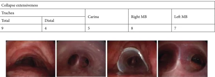
Table 3. Patient clinical characteristics. Right MB: right main bronchus. Left MB: left main bronchus.
Collapse extensiveness Trachea
Carina Right MB Left MB
Total Distal
9 4 5 8 7
Figure 1. Distal tracheal broncoscopic view of a patient before and after Y stent insertion
Table 4. Complications of patients with stent applications.
Complications Mucostasis
Migration Granulation
Mild Moderate Severe Mild Moderate Severe
1 6 5 1 2 3 0
Figure 2. The appearance of a Y-stent after cleaning the mucus plug formed in the right and left leg which was placed in the
4. Discussion
Determining the need for treatment in TBM patients and lack of a distinctive treatment approach makes treatment management more difficult. As mentioned before, previous studies have reported the results of the different treatment approaches for these patients. We evaluated 15 patients with TBM in our series in terms of treatment and reviewed the success rate of procedures with postprocedure follow-ups. After application of stents, only two patients had long-term follow-ups due to complications of the procedures, such as mucostasis, migration, and granulation. In patients who were at risk before TBM surgery, endobronchial treatment methods can be used as a tool for reducing symptoms and finding the right treatment modality. There
is a need for studies that include large numbers of patients to show a high rate of success in selected cases.
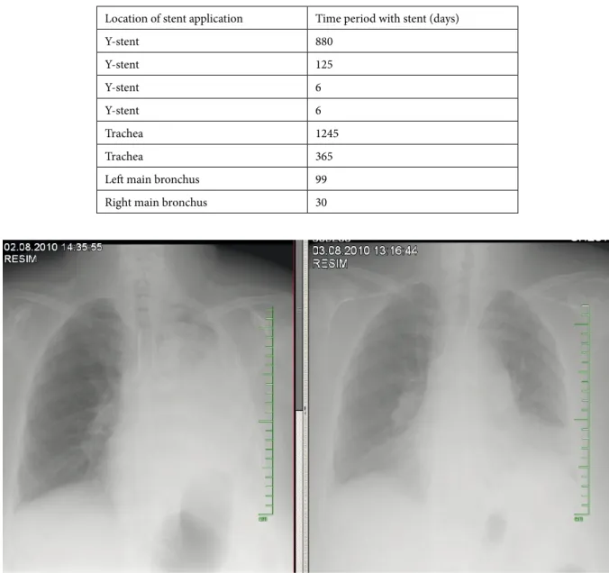
CPAP is a noninvasive method that prevents airway collapse, which may be considered after the symptoms of TBM and concomitant conditions have been managed. It reduces airway resistance and the energy spent on respiration by keeping the airways open with positive pressure. It is a method that can be both intermittently and continuously used in patients who have mild symptoms and who cannot be treated invasively. Surgical techniques other than segmental tracheal resection may be performed in patients with localized TBM to strengthen cartilage walls or stabilize the posterior membrane. Considering major and minor surgical complications, significant Table 5. Clinical follow-up data of patients with stent applications.
Location of stent application Time period with stent (days)
Y-stent 880 Y-stent 125 Y-stent 6 Y-stent 6 Trachea 1245 Trachea 365
Left main bronchus 99
Right main bronchus 30
improvements can be made with symptoms, respiratory parameters, and quality of life.
While there is no standard of care in the treatment of TBM, silicon stent application appears to be a good treatment for selected patients. Laser coagulation has also shown the same success rate. In patients who have COPD, asthma, or respiratory distress, procedures should be performed while considering high risks and bronchoscopic treatment criteria.
Ernst et al. prospectively investigated 75 patients in their study and observed that neither the respiratory function tests nor the 6-min walk test parameters showed significant improvement at the end of the first month, although there were significant improvements in terms of symptoms and quality of life (10,11). In our study, the number of presentations to emergency rooms and hospitalizations due to respiratory distress of patients who had permanent stents were significantly reduced; however, there were a limited number of patients.
YAG lasers reach into the connective tissue and submucosa and help fibrous cicatrix formation and posterior membrane stabilization. Promising results were achieved in a study conducted in Marseilles with diffuse collapse or localized collapse in posterior side (18). Prospective studies are needed to confirm the treatment efficacy of YAG laser treatment.
Stents are another method of treatment; however, there is no ideal stent available for airway stabilization in invasive bronchoscopy. There are two types of stent: metallic and silicon. Stents fix the diameters of airways. The use of silicon stents improves the clinical symptoms
of TBM (19,20). Constant change in airway diameters in TBM increases the risk of migration with silicon stents and fracture with metallic stents. With both stent types there is an increased risk of granuloma and mucus plug formation at both ends of the stents and places where there is contact with mucosa. Stents should be used in patients who have severe symptoms, whose treatment-related complications can be predicted, and who can be followed up with frequent bronchoscopies. This treatment has inadequacies, such as mucostasis formation at the distal segment of the stent and stent migration (21,22). Mucostasis, migration, and granulation tissue formation prompted stent removal in most of the patients in our study and this shows the need for large studies that include large numbers of participants.
In selected cases with TBM and dynamic airway collapse, silicon stents appear to be the ideal treatment method. Bronchoscopic procedures in selected patients should be undertaken by experienced centers because diagnosing TBM is difficult and patients who present are in respiratory distress. Stents are used as a treatment test in patients with severe symptoms who are candidates for surgery to identify the benefits of fixing the airway diameter and stabilization. Stents can be used as a long-term palliative treatment in patients who have no other treatment alternative if they provide significant symptomatic improvement, provided that stent follow-up and care are periodically undertaken.
Acknowledgments
We thank David Chapman for English language editing.
References
1. Carden KA, Boiselle PM, Waltz DA, Ernst A. Tracheomalacia and tracheobronchomalacia in children and adults: an in-depth review. Chest 2005; 127: 984–1005.
2. Murgu SD, Colt HG. Tracheobronchomalacia and excessive dynamic airway collapse. Respirology 2006; 11: 388–406. 3. Ernst A, Majid A, Feller-Kopman D, Guerrero J, Boiselle P,
Loring S, O’Donnell C, Decamp M, Herth FJ, Gangadharan S et al. Airway stabilization with silicon stents for treating adult tracheobronchomalacia: a prospective observational study. Chest 2007; 132: 609–616.
4. Feist JH, Johnson TH, Wilson RJ. Acquired tracheomalacia: etiology and differential diagnosis. Chest 1975; 68: 340–345. 5. Ikeda SH, Hanawa T, Konishi T, Adachi M, Sawai S, Chiba W,
Kosaba S, Hatakenaka R, Matsubara Y, Funatsu T. Diagnosis, incidence, clinicopathology and surgical treatment of acquired tracheobronchomalacia. Nihon Kyobu Shikkan Gakkai Zasshi 1992; 8: 1028–1035.
6. Jokinen K, Palva T, Nuutinen J. Chronic bronchitis: a bronchologic evaluation. J Otorhinolaryngol Relat Spec 1976; 38: 178–186.
7. Roberto WDN, Silvia T, Massimo G, Claudio M. Prevalence of tracheobronchomalacia and excessive dynamic airway collapse in bronchial asthma of different severity. Multidiscip Respir Med 2013; 8: 32.
8. Hasegawa I, Boiselle P, Raptopoulos V, Hatabu H. Tracheomalacia incidentally detected on CT pulmonary angiography of patients with suspected pulmonary embolism. Am J Roentgenol 2003; 8: 1505–1509.
9. Boiselle PM, Ernst A. Tracheal morphology in patients with tracheomalacia: prevalence of inspiratory lunate and expiratory ‘frown’ shapes. J Thorac Imaging 2006; 21: 190–196.
10. Lee KS, Sun MR, Ernst A, Feller-Kioman D, Majid A, Boiselle PM. Comparison of dynamic expiratory CT bronchoscopy for diagnosing airway malacia: A pilot evaluation. Chest 2007; 131: 758–764.
11. Majid A, Guerrero J, Gangadharan S, Feller-Kopman D, Boiselle P, DeCamp M, Ashiku S, Michaud G, Herth F, Ernst A. Tracheobronchoplasty for severe tracheobronchomalacia. A prospective outcome analysis. Chest 2008; 134: 801–807.
12. Majid A, Fernández L, Fernández-Bussy S, Herth F, Ernst A. Tracheobronchomalacia. Arch Bronconeumol 2010; 46: 196– 202.
13. Ferguson GT, Benoist J. Nasal continuous positive airway pressure in the treatment of tracheobronchomalacia. Am Rev Respir Dis 1993; 147: 457–461.
14. Ernst A, Odell DD, Michaud G, Majid A, Herth FFJ, Gangadharan SP. Central airway stabilization for tracheobronchomalacia improves quality of life in patients with COPD. Chest 2011; 140: 1162–1168.
15. Dutau H, Michaud G, Milhe F, Dumon JF. Laser therapy for tracheobronchomalacia. In: Ed Monduzi, editor. Proceedings of the 13th World Congress for Bronchology and 13th World Congress for Bronchoesophagology; 20–23 June 2004; Barcelona, Spain. pp. 21–26.
16. Wright CD. Tracheomalacia. Chest Surg Clin N Am 2003; 13: 349–357.
17. Gangadharan SP, Bakhos CT, Majid A, Kent MS, Michaud G, Ernst A, Ashiku SK, DeCamp MM. Technical aspects and outcomes of tracheobronchoplasty for severe tracheobronchomalacia. Ann Thorac Surg 2011; 91: 1574– 1580.
18. Dutau H, Maldonado F, Breen DP, Colchen A. Endoscopic successful management of tracheobronchomalacia with laser: apropos of a Mounier-Kuhn syndrome. Eur J Cardiothorac Surg 2011; 39: 186–188.
19. Vergnon JM, Cortes F, Bayon MC, Emonot A. Efficacy of tracheal and bronchial stent placement on respiratory functional tests. Chest 1995; 107: 741–746.
20. Zannini P, Melloni G, Chiesa G, Carretta A. Self-expanding stents in the treatment of tracheobronchial obstruction. Chest 1994; 106: 86–90.
21. Beer M, Wittenberg G, Sandstede J, Beissert M, Schmidt M, Ender J, Krahe T, Hahn D. Treatment of inoperable tracheobronchial obstructive lesions with the Palmaz stent. Cardiovasc Intervent Radiol 1999; 22: 109–113.
22. Nashef SA, Dromer C, Velly JF, Labrousse L, Couraud L. Expanding wire stents in benign tracheobronchial disease: indications and complications. Ann Thorac Surg 1992; 54: 937–940.