doi:10.3906/biy-0901-20
Siderophore, hemolytic, protease, and pyrazinamidase
activities and antibiotic resistance in motile Aeromonas isolated
from fish
Belgin ERDEM, Ergin KARİPTAŞ, Tayfun KAYA
Department of Biology, Arts and Sciences Faculty, Ahi Evran University, 40100 Kırşehir - TURKEY
Received: 29.01.2009
Abstract:A total of 120 fresh water fish samples were evaluated for the presence of Aeromonas spp. A. hydrophila, A. caviae, and A. veronii bv. sobria were isolated from the gills, intestines, livers, and skins of fish, and 78 isolated Aeromonas spp. strains were further examined for siderophore, hemolytic, protease, and pyrazinamidase activities and antibiotic resistance. Siderophore production was demonstrated in almost all of the A. hydrophila and A. caviae strains except for 2 strains of A. veronii bv. sobria. The strains identified as A. hydrophila and A. veronii bv. sobria were the stronger producers of hemolysin, whereas the A. caviae strains were nonhemolytic. It was found that 92% of A. hydrophila and 91% of A. caviae strains hydrolyzed pyrazinamide in under 48 h, whereas 5% of A. veronii bv. sobria strains hydrolyzed pyrazinamide. Protease activity was found in 100% of isolates of A. hydrophila and A. veronii bv. sobria, but in 81% of A. caviae isolates. In addition, all Aeromonas species were examined for antibiotic resistance patterns and were found to be resistant to ampicillin and tetracycline.
Key words:Aeromonas, siderophore, hemolysin, pyrazinamidase, antibiotic resistance, fish
Balıklardan izole edilen hareketli Aeromonas’larda siderofor, hemolitik, proteaz,
pirazinamidaz aktiviteleri ve antibiyotik direnç
Özet:Toplam 120 tatlı su balığı örneklerinin, solungaç, bağırsak, karaciğer ve deri numunelerinden izole edilen Aeromonas cinsine ait A. hydrophila, A. caviae ve A. veronii bv. sobria varlığı değerlendirilmiştir. İzole edilen 78 Aeromonas suşunun siderofor, hemolitik, pirazinamidaz, proteaz aktiviteleri ve antibiyotik direnç durumları incelenmiştir. Siderofor üretimi A. hydrophila, A. caviae suşlarının neredeyse tamamında görülürken A. veronii bv. sobria suşlarının yalnızca ikisinde gözlenmiştir. A. hydrophila and A. veronii bv. sobria suşları hemolizin üretirken A. caviae suşları nonhemolitiktir. A. hydrophila suşlarının % 92’si, A. caviae suşlarının % 91’i pirazinamid’i 48 saatten daha az bir zamanda hidroliz etmiş oysa A. veronii bv. sobria suşları yüzde beş oranında aktivite göstermiştir. Proteaz aktivitesi, A. hydrophila, ve A. veronii bv. sobria izolatlarının tamamında (% 100) bulunurken A. caviae izolatlarının % 81,8’de saptanmıştır. Bunlara ilaveten, tüm Aeromonas suşları ampisilin ve tetrasikline karşı dirençli bulunmuştur.
Introduction
The motile mesophilic Aeromonas species are autochthonous inhabitants of aquatic environments (1). The fact that they have been recently isolated from the acute intestinal infections identified in people has added to the importance of these microorganisms and led researchers to study this topic (2). They are increasingly being noted as significant pathogens for humans and lower vertebrates, including amphibians, reptiles, and fish (3). Some Aeromonas species are responsible for causing a variety of human infections, including septicemia, wound infections, meningitis, pneumonia, and gastroenteritis (4,5).
That motile Aeromonas are found in both chlorinated and nonchlorinated water, are transmitted from these environments to food, and can reproduce at refrigerator temperatures due to the psychrophilic characteristics of some of its types suggest that they may be pathological agents critical for human health (6-10). Several investigators reported that members of the genus Aeromonas are common contaminants of food, and they have been isolated from a wide range of food products such as fish, chicken, meat, and vegetables (2-4). Characteristically freshwater bacteria, motile types of A. hydrophila, A. caviae, and
A. veronii bv. sobria, which are dominant elements in
the natural microbial flora of the aquatic animals, can cause various diseases in freshwater fish, like carp (11,12).
Siderophores are bacterially secreted molecules with a high affinity for iron that scavenge iron from the bacterial cells’ environment for growth. Production of siderophores has been reported in different species of Aeromonas and might be related to virulence properties. However, the role of siderophores, whether enterobactins or the recently characterized and named amonobactins, in A.
hydrophila infections is not clearly determined
(13,14). Various putative virulence factors (aerolysin/hemolysin, proteases, lipases, DNases, enterotoxins, cytotoxins, and siderophores) that may play an important role in the development of diseases, either in humans or in fish, have been described in several species of the genus (15-18). Some authors reported that the production of cytotoxins and hemolysin is related to A. hydrophila and A. veronii
bv. sobria (19), and hemolytic molecules seem to be related to enterotoxigenicity (16). McMahon (20) indicated a correlation between the pathogenic potential and the hemolytic and proteolytic activity of
Aeromonas species isolated from different sources.
There are several reports dealing with pyrazinamidase activity, which has been extensively studied in
Mycobacterium spp. and is considered a differentiating feature in the classification of mycolic acid-containing actinomycetes and a virulence-associated marker in the genus Yersinia (21,22). Some isolates of Aeromonas have pyrazinamidase activity, a valuable phenotypic marker to assist in the differentiation of A. veronii bv. sobria from A.
hydrophila and A. caviae. Regarding the resistance to
antimicrobial agents, several authors have stated that
Aeromonas species are rapidly adapting to new drugs
commonly used in medicine, becoming a potential risk to public health (23,24).
In view of increasing evidence supporting the role of aeromonads in human and fish diseases, we isolated Aeromonas spp. from freshwater fish obtained from the local bazaar and characterized them with respect to their siderophore, hemolytic, protease, and pyrazinamidase activities and antibiotic resistance in in vitro conditions.
Materials and methods
Isolation and identification of motile
Aeromonas
Fish samples were randomly purchased from the local bazaar in Kırşehir, Turkey. A total of 120 samples of fish were screened for the presence of Aeromonas spp. Samples were immediately transferred to the laboratory in ice chests at 4-7 ºC. Gill, intestine, liver, and skin contents of fish samples (5 g) were aseptically swabbed using sterile cotton buds and inoculated into 45 mL of alkaline peptone water (APW, pH 8.4) containing 30 μg/mL of ampicillin (A-9393, Sigma Chemical Co., St. Louis, MO, USA) and homogenized for 2 min in sterile Stomacher bags. After 18 h of incubation at 28 ºC, 0.1 mL of the APW was streaked on the glutamate starch phenol red agar (GSP, Merck, Darmstadt, Germany). After incubation at 28 ºC for 24 h, yellow colonies surrounded by a yellow zone were picked and grown on fresh GSP agar plates for
confirmation (25). The presumptive Aeromonas colonies were identified at the genus level by testing their Gram reaction, oxidase, catalase, motility, and growth conditions in 0% and 6% NaCl, oxidation/fermentation (glucose), and resistance to vibriostatic agent O/129 (2,4-diamino-6,7-diisopropylpteridine) (150 μg/mL) (1,26). All
Aeromonas spp. were reidentified biochemically at the
species level by using tests chosen from those described by Joseph and Carnahan (26), that is, esculin hydrolysis, production of H2S from cysteine, production of gas from glucose, growth in KCN broth, salicin and arabinose fermentation, Voges-Proskauer reaction, and lysine decarboxylase. As reference strains, A. hydrophila (ATCC 7966), A. veronii bv.
sobria (ATCC 43979), and A. caviae (ATCC 15468)
were used. The strains were stored in trypticase soy broth (TSB) with 15% (v/v) sterile glycerol at -70 ºC until assayed.
Determination of siderophore production The chrome azurol siderophore detection agar (CAS agar) (27), prepared as described by Barghouthi et al. (14), was used to detect siderophore production. Single colonies of each isolate were transferred to this medium, and the plates were incubated at 28 °C for 48 h. The presence of an orange halo around the colonies was recorded as positive for siderophore production.
Determination of hemolytic activity
The strains were tested for hemolytic activity by streaking them onto trypticase soy agar (TSA) plates containing 7% sheep blood for 48 h at 37 °C. Beta hemolytic zones of 2 mm or more around the colonies were regarded as the sign of positive hemolytic activity (28). Hemolytic activity for the Aeromonas spp. strains was categorized as alpha, beta, or gamma (29).
Determination of proteolytic activity
Protease activity was determined on the surface of skim milk agar, in which skim milk was added just before pouring the medium into the petri plates. The plates were incubated at 28 °C for 4 days. After the incubation period, the clear zones of hydrolysis were measured and recorded. The presence of a transparent zone around the colonies indicated protease activity (30).
Pyrazinamidase activity assays
The following tests were additionally performed on all Aeromonas strains. Pyrazinamidase activity was assayed by using a supplied medium that contained trypticase soy agar (15 g; Difco), yeast extract (1.5 g; Sigma Chemical Co.), and Tris-maleate buffer (0.2 M, pH 6, 500 mL) and pyrazinamide (0.5 g; Sigma), and was dispensed in 5 mL aliquots into screw-cap tubes (16 × 150 mm), autoclaved at 121 °C for 15 min. These media were inoculated with 24-48 h of bacterial growth taken from tryptic soy agar (Difco) and incubated for 48 h at 37 °C. One milliliter of 1% (wt/vol) freshly prepared ferrous ammonium sulfate aqueous solution was flooded over each slant, and a positive (pinkish rust color) or negative (colorless) reaction was recorded after 15 min. Positive pyrazinamidase activity indicated the presence of pyrazinoic acid resulting from the action of the enzyme pyrazinamidase (21).
Antimicrobial resistance
The resistance of all strains to different antimicrobial agents was determined by the disk diffusion method (31). Bacteria were cultured in TSB and spread onto the surface of Mueller-Hinton agar. The isolates were individually tested against 9 antimicrobial agents. The antibiotics and concentration ranges tested were as follows: ampicillin, 10 μg; amoxicillin, 10 μg; streptomycin, 10 μg; ceftazidime, 30 μg; ciprofloxacin, 5 μg; kanamycin, 30 μg; tetracycline, 30 μg; gentamicin, 10 μg; and trimethoprim, 5 μg.
All antibiotics were obtained from Oxoid Limited (Hampshire, England). Plates with bacteria inoculum were incubated at 28 °C for 4 days. Inhibition zones that formed in the culture media at the end of this period were evaluated in millimeters. The resistance breakpoints were those defined by the National Committee for Clinical Laboratory Standards (NCCLS, 2004) for gram-negative bacteria. Escherichia
coli (ATCC 25922), Pseudomonas aeruginosa (ATCC
27853), A. hydrophila (ATCC 7966), and A. caviae (ATCC 15468) were used as controls.
Results and discussion
Aeromonas spp. were isolated from 78 (65%) of the
120 samples tested. As seen in Table 1, presumptive
Aeromonas spp. were identified and confirmed as A. hydrophila, A. veronii bv. sobria, and A. caviae
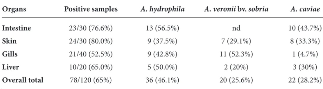
morphologically, physiologically, and biochemically. In general, 65% (78/120) of the fish samples examined were positive for Aeromonas spp.: 46.1% (36/78) were positive for A. hydrophila, the predominant species; 28.2% (22/78) for A. caviae; and 25.6% (20/78) for A. veronii bv. sobria (Table 2). The incidence of Aeromonas spp. varied depending on the samples (gills, intestines, skins, or livers) examined. The results indicate that the highest Aeromonas incidence is observed on the skin (80%) of fish samples.
As shown in Table 2, A. hydrophila was the dominant species (46.1%), followed by A. caviae (28.2%) and A. veronii bv. sobria (25.6%). A.
hydrophila and A. veronii bv. sobria, which have been
linked with human infections, were isolated from fish examined in this study. More recent investigations on the prevalence of Aeromonas species in sources such as environmental, clinical, food, and veterinary origins have focused on 3 mesophilic species, namely
A. hydrophila, A. caviae, and A. veronii bv. sobria,
respectively. Several studies showed pronounced variations in their occurrence (2,7,8,25,32-37).
When the isolates were grown on a CAS agar, on which siderophore production can be detected, all isolates produced a clear zone (18). In our study, siderophore production was found in 54 (69.2%) of
Table 1. Biochemical properties of isolates.
Phenotypic expression (%) Biochemical testa
A. hydrophila A. veronii bv. sobria A. caviae
Esculin hydrolysis 100 0 100
Growth in KCN broth 100 0 100
Gas from glucose 100 100 0
H2S from cysteine 100 100 0
Fermentation of salicin 100 0 100
Fermentation of arabinose 100 0 100
Voges-Proskauer 98 100 0
Lysine decarboxylase 100 100 0
a: Joseph and Carnahan (1994)
Table 2. The distributions of Aeromonas species in different body parts of fish samples examined.
Organs Positive samples A. hydrophila A. veronii bv. sobria A. caviae
Intestine 23/30 (76.6%) 13 (56.5%) nd 10 (43.7%) Skin 24/30 (80.0%) 9 (37.5%) 7 (29.1%) 8 (33.3%) Gills 21/40 (52.5%) 9 (42.8%) 11 (52.3%) 1 (4.7%) Liver 10/20 (65.0%) 5 (50.0%) 2 (20%) 3 (30%) Overall total 78/120 (65%) 36 (46.1%) 20 (25.6%) 22 (28.2%) nd: none detected
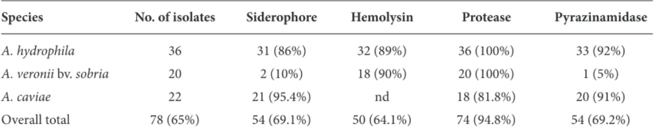
78 isolates. As shown in Table 3, production of siderophores was detected in all aeromonad species. The highest producer of siderophores was A. caviae, at a rate of 95.4% (21/22), followed by 86% of the A.
hydrophila (31/36) and 10% of the A. veronii bv. sobria
(2/20).
Another virulence factor is the ability to scavenge required nutrients such as iron. Many pathogenic bacteria utilize iron uptake pathways, such as the production of siderophores, to access iron for growth. These uptake pathways are considered to be one of the virulence factors, since they facilitate the growth of pathogenic bacteria and the subsequent production of other virulence factors (18). Earlier studies have reported that the ability to produce siderophores correlated with higher virulence in Aeromonas species (38).
This study of siderophore synthesis by 78 isolates classified as phenospecies A. hydrophila, A. caviae, and A. veronii bv. sobria showed that more than 86% of the A. hydrophila isolates synthesized siderophores, while about 21% of A. caviae and 10% of A. veronii bv.
sobria may have produced siderophores. This
indicates that A. hydrophila and A. caviae were higher producers of siderophores than A. veronii bv. sobria, a rare siderophore producer. Some investigators (13,16) indicated that A. hydrophila, A. caviae, and A. veronii bv. sobria isolated from fish produced siderophores. Gierer et al. (39) showed that a total of 355 strains of fish-pathogenic bacteria, including A. hydrophila, A.
caviae, Pseudomonas fluorescens, and P. putida
isolates, were determined as siderophore producers. However, a previous investigation mentioned above indicated that the pathogenicity of A. hydrophila was not always accompanied by the presence of
siderophores. Whether or not siderophore production is an important virulence factor is not clear (13). As defined by Alavandi and Ananthan (40), there was no significant difference between the clinical and environmental aeromonads with respect to their enterotoxigenicity, cytotoxicity, and ability to produce siderophores. Although it is known that bacteria can produce some siderophore compounds simply for survival, the cross-feeding assays demonstrated that at least aerobactin and enterobactin biosynthesis are not essential virulence determinants in motile
Aeromonas strains (12). As can be seen in Table 3, our
results show no significant relation between siderophore and hemolytic activities. We could propose a minor relation between the production of siderophores and hemolysin. Furthermore, it was shown that Aeromonas strains play an important role in iron acquisition. Molecular studies are needed to actually reveal the correlation between these 2 factors.
The hemolysin production by Aeromonas strains is shown in Table 3. Among the Aeromonas isolates, 90% of A. veronii bv. sobria, followed by 89% of A.
hydrophila, from fish samples were beta-hemolytic on
sheep blood agar plates, while none of the A. caviae strains were hemolytic. This indicates that A.
hydrophila and A. veronii bv. sobria were higher
producers of hemolysin than A. caviae. No type of hemolysis, except for beta-hemolysis, was found in the isolates.
The production of hemolytic toxins has been regarded as strong evidence of pathogenic potential in aeromonads (16,19). Beta hemolysin has been reported as a virulence factor in motile aeromonads (41). In this study, A. hydrophila and A. veronii bv.
sobria strains exhibited beta-hemolytic activity to
Table 3. Incidence of some virulences in motile Aeromonas spp. isolated from fish samples.
Species No. of isolates Siderophore Hemolysin Protease Pyrazinamidase
A. hydrophila 36 31 (86%) 32 (89%) 36 (100%) 33 (92%)
A. veronii bv. sobria 20 2 (10%) 18 (90%) 20 (100%) 1 (5%)
A. caviae 22 21 (95.4%) nd 18 (81.8%) 20 (91%)
Overall total 78 (65%) 54 (69.1%) 50 (64.1%) 74 (94.8%) 54 (69.2%)
different extents, but not A. caviae strains. Our findings agree with the results recorded by Janda et al. (42), who showed similar percentages among 121
Aeromonas strains: 93% of A. veronii bv. sobria and
95% of A. hydrophila were hemolytic. These results are substantiated by the findings of Farag (43) and Yucel and Çitak (44), who showed a significant difference between the hemolysin production of A.
hydrophila and A. veronii bv. sobria as compared with
that of A. caviae. This represents that hemolysin production may be related to A. veronii bv. sobria and
A. hydrophila, but not A. caviae. Therefore, hemolysin
can be one of several factors determining pathogenicity, but are not required for virulence in all
Aeromonas species.
Table 3 presents protease activity, which was found in 74 (94.8%) of 78 Aeromonas isolates from fish. Specifically, 100% of A. hydrophila and A. veronii bv.
sobria were producers of protease, followed by 81.8%
of A. caviae. The majority of the isolates obtained in the present study produced protease.
Proteases are important factors in the spoilage of foods, and the presence of proteases and hemolysins are used as an indicator of potential pathogenicity (20). Both the quantitative and qualitative production of protease is important in establishing the virulence of a particular strain (45). The proteolytic activity of
A. hydrophila has been correlated with its ability to
induce pathology in fish (46). Our findings revealed that proteases, more than hemolysin, may be important virulence factors in Aeromonas infections. Proteases are thought to contribute to the virulence of aeromonads for fish and other hosts; however, their contribution to human pathogenicity still needs to be determined.
The summarized results in Table 3 show 78 well-characterized Aeromonas strains representing 3 species from fish for pyrazinamidase activity. This activity was detected in 92% of A. hydrophila, 91% of
A. caviae, and 5% of A. veronii bv. sobria in under 48
h (Table 3).
The presence of pyrazinamidase in A. hydrophila and A. caviae, but its absence in representatives of other species of Aeromonas (A. veronii bv. sobria, A.
schubertii, and A. veronii), was described 19 years ago
(47). Because the taxonomy of aeromonads has been
in a state of constant flux, we considered it imperative to start with strains that were well characterized at the species level. The present study has demonstrated that all (36 of 36) strains of A. hydrophila, 21 of 22 A.
caviae, and 1 of 20 A. veronii bv. sobria hydrolyzed
pyrazinamide. These findings are consistent with the observations by Carnahan et al. (47) and Wakabongo et al. (48). The given results in Table 3 show that pyrazinamidase activity could be a valuable phenotypic marker to aid in the identification of A.
veronii bv. sobria from A. hydrophila and A. caviae.
Several virulence factors have been stated as indicators of pathogenicity in Aeromonas spp. However, pyrazinamidase activity by Aeromonas is not clearly determined. It has been reported that negative pyrazinamidase might also be a new virulence-associated marker for A. veronii bv. sobria,
A. schubertii, and A. veronii (47). Further studies are
necessary on the determination of the nature of this association.
The antibiotic resistance patterns of the
Aeromonas species isolated from the fish samples are
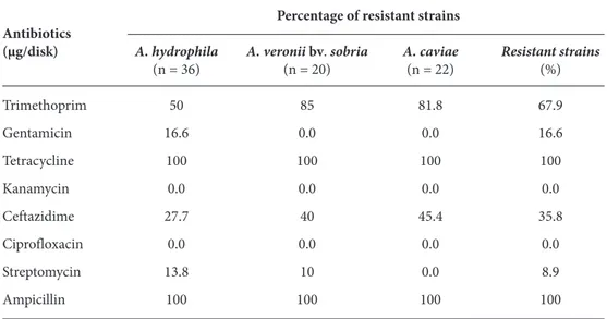
shown in Table 4. All strains of A. hydrophila, A.
veronii bv. sobria, and A. caviae were resistant to
ampicillin and tetracycline. Furthermore, the highest resistances encountered were 67.9% to trimethoprim, 44.8% to amoxicillin, and 35.8% to ceftazidime. The lowest resistance was detected for streptomycin (8.9%). None of the Aeromonas strains encountered in the present study were resistant to ciprofloxacin or kanamycin.
The frequent occurrence of multiple antimicrobial resistances among Aeromonas spp. isolates that are both fish pathogens and emerging opportunistic human pathogens has been monitored over the past decades (25,37,44,45,49-57). In this study, all strains were 100% resistant to ampicillin/beta-lactam and tetracycline. Aeromonas spp. have been reported to be intrinsically resistant to ampicillin (49). As defined in our experiment, 44.8% of the strains were resistant to amoxicillin/beta-lactam, which is similar to the findings of Hatta et al. (50), who reported that 33.3%-52.7% of the Aeromonas strains from freshwater fish were resistant to amoxicillin. Obtained results indicate that beta-lactam agents should be avoided in the treatment of Aeromonas spp. infections. As mentioned, 100% of the Aeromonas strains exhibited
resistance against tetracycline in our study. In a previous paper, Ko et al. (51) found as many as 49% tetracycline-resistant Aeromonas spp., compared with 14% in the data of Goni-Urizza et al. (52). Additionally, the resistance to tetracycline has been reported to be acquired and encoded by plasmids or transposons (53,54). The resistance level to trimethoprim was comparable to the findings of Yucel and Çitak (44), who reported 63%-100% of trimethoprim resistant strains, and these results are supported by Vivekanandhan et al. (55) and Ansary et al. (56). However, resistance of Aeromonas spp. to ceftazidime was 35.8% in this study, compared to the findings Radu et al. (25) and Ceylan et al. (57), who observed no resistance at all (0%) in retail fish and livestock, respectively. We found that around 8.9% of the strains were resistant to streptomycin, but none of the A. caviae demonstrated resistance to streptomycin. This result is in contrast to the findings of Hatha et al. (50), who reported that none of the
Aeromonas strains from different freshwater fish were
resistant to this antibiotic. Strains of Aeromonas isolated from frozen fish (36) showed 58.4% resistance against gentamicin, while we encountered 16.6% resistance to gentamicin. However, Yucel et al. (37) reported that 10%-54% of the Aeromonas strains from retail fish were resistant to this antibiotic. These results are in contrast to the findings of Goni-Urizza
et al. (52), who reported that 1% of Aeromonas strains from rivers were resistant to gentamicin.
Results obtained in this study indicate that multiple resistance, particularly to ampicillin and tetracycline, is often seen in Aeromonas spp. isolated from fish, since geographic locations and local selective pressure influence the antibiotic resistance levels.
There has been no study about the presence of motile Aeromonas types in carp sold in public outdoor bazaars of Kırşehir, Turkey. To our knowledge, this is the first report on the existence of putative virulence factors and antimicrobial resistance of foodborne
Aeromonas. The present study isolated motile Aeromonas spp., which have been recently identified
as an important food pathogen, from carp supplied for public consumption in Kırşehir. In order to determine whether they posed a risk to public health, they were tested for putative virulence properties like siderophore, hemolysin, protease, and pyrazinamidase activity, and their antibiotic resistance was examined.
In conclusion, the present work has clearly emphasized an important incidence of Aeromonas spp. with virulence and antibiotic resistance in fish intended for human consumption. Consuming raw or undercooked fish increases the risk of developing
Table 4. Prevalence of antibiotic resistance among motile aeromonads isolated from fish.
Percentage of resistant strains Antibiotics
(μg/disk) A. hydrophila A. veronii bv. sobria A. caviae Resistant strains
(n = 36) (n = 20) (n = 22) (%) Trimethoprim 50 85 81.8 67.9 Gentamicin 16.6 0.0 0.0 16.6 Tetracycline 100 100 100 100 Kanamycin 0.0 0.0 0.0 0.0 Ceftazidime 27.7 40 45.4 35.8 Ciprofloxacin 0.0 0.0 0.0 0.0 Streptomycin 13.8 10 0.0 8.9 Ampicillin 100 100 100 100 n: number of isolates
human infections because of the high occurrence of pathogenic Aeromonas strains. Pyrazinamidase activity, which is more commonly considered a phenotypic marker in the differentiation of some types of Mycobacterium, was used in the characterization of Aeromonas. This activity has almost never been employed in such studies in Turkey. An increasing presence of multiple drug-resistance among Aeromonas species may become a potential human health hazard. Therefore, the results of the present investigation indicate that additional studies in this field are warranted to elucidate the public health significance of aeromonads in the aquatic environment.
Acknowledgment
This work at AEU was supported by grants from the Gazi University Scientific Research Projects Department.
Corresponding author:
Belgin ERDEM
Department of Biology, Arts and Sciences Faculty, Ahi Evran University, 40100 Kırşehir - TURKEY E-mail: [email protected]
1. Popoff M. Genus III. Aeromonas. In: Krieg NR. ed. Bergey’s Manual of Systematic Bacteriology, Volume 1. Williams & Wilkins. Baltimore; 1994: pp. 545-548.
2. Altwegg M, Steigerwalt AG, Altwegg-Bissing R et al. Biochemical identification of Aeromonas genospecies isolated from humans. J Clin Microbiol 28: 258-265, 1990.
3. Davies A, Capell C, Jehanno D et al. Incidence of foodborne pathogens on European fish. Food Control 12: 67-71, 2001. 4. Galindo CL, Sha J, Fadl AA et al. Host immune responses to
Aeromonas virulence factors. Current Immun Rev 2: 13-26,
2006.
5. Galindo CL, Chopra AK. Aeromonas and Plesiomonas species. In: Doyle MP, Beuchat LR. eds. Food Microbiology: Fundamentals and Frontiers. ASM Press. Washington, D.C.; 2007: pp. 381-400.
6. Havelaar AH, During M, Versteeg JFM. Ampicillin-dextrin agar medium for the enumeration of Aeromonas species in water by membrane filtration. J Appl Bacteriol 62: 279-287, 1987. 7. Massa S, Altieri D, D’Angela A. The occurrence of Aeromonas
spp. in natural mineral water and well water. Int J Food Microbiol 63: 169-173, 2001.
8. Martins LM, Marquez RF, Yano T. Incidence of toxic Aeromonas isolated from food and human infection. FEMS Immun Med Mic 32: 237-242, 2002.
9. Falcao JP, Dias AMG, Correa EF et al. Microbiological quality of ice used to refrigerate foods. Food Microbiol 19: 269-276, 2002.
10. Papageorgiou DK, Melas DS, Abrahim A et al. Growth and survival of Aeromonas hydrophila in rice pudding (milk rice) during its storage at 4 °C and 12 °C. Food Microbiol 20: 385-390, 2003.
11. Aoki T. Motile Aeromononads (Aeromonas hydrophila). In: Woo PTK, Bruno DW. eds. Fish Diseases and Disorders, Volume 3. Viral, Bacterial and Fungal Infections Cab International; 1999: pp. 427-453.
12. Santos Y, Toranzo AE, Barja JC et al. Virulence properties and enterotoxin production of Aeromonas strain isolated from fish. Infect Immun 56: 3285-3293, 1998.
13. Zywno SR, Arceneaux JEL, Altwegg M et al. Siderophore production and DNA hybridization groups of Aeromonas spp. J Clin Microbiol 30: 619-622, 1992.
14. Barghouthi S, Young R, Olson MOJ et al. Amonabactin, a novel tryptophan- or phenylalanine containing phenolate siderophore in Aeromonas hydrophila. J Bacteriol 171: 1811-1816, 1989. 15. Pemberton JM, Kidd SP, Schmidt R. Secreted enzymes of
Aeromonas. FEMS Microbiol Lett 152: 1-10, 1997.
16. Santos JA, Gonzalez CJ, Otero A et al. Hemolytic activity and siderophore production in different Aeromonas species isolated from fish. Appl Environ Microb 65: 5612-5614, 1999. 17. Soler L, Figueras MJ, Chacon MR et al. Potential virulence and
antimicrobial susceptibility of Aeromonas popoffii recovered from freshwater and seawater. FEMS Immunol Med Mic 32: 243-247, 2002.
18. Lee S, Kim S, Oh Y et al. Characterization of Aeromonas
hydrophila isolated from rainbow trouts in Korea. J Microbiol 1:
1-7, 2000.
19. Turnbull PCB, Lee JV, Miliotis MD et al. Enterotoxin production in relation to taxonomic grouping and source of isolation of Aeromonas species. J Clin Microbiol 19: 175-180, 1984.
20. McMahon MAS. The expression of proteinases and haemolysins by Aeromonas hydrophila under modified atmospheres. J Appl Microbiol 89: 415-422, 2000.
21. Kandolo K, Wauters G. Pyrazinamidase activity in Yersinia
enterocolitica and related organisms. J Clin Microbiol 21:
980-982, 1985.
22. Carnahan A, Hammontree L, Bourgeois L et al. Pyrazinamidase activity as a phenotypic marker for several Aeromonas spp. isolated from clinical specimens. J Clin Microbiol 28: 391-392, 1990.
23. Burgos A, Quindos G, Martinez R et al. In vitro susceptibility of
Aeromonas caviae, A. hydrophila and A. sobria to fifteen
antibacterial agents. Eur J Clin Microbiol Infect Dis 9: 413-417, 1990.
24. Overman TL, Janda JM. Antimicrobial susceptibility patterns of Aeromonas jandaei, A. veronii biotype veronii, A. trota and A.
shubertii. J Clin Microbiol 37: 706-708, 1999.
25. Radu S, Ahmad N, Ling FH et al. Prevalence and resistance to antibiotics from Aeromonas species from retail fish in Malaysia. Int J Food Microbiol 81: 261-266, 2003.
26. Joseph SW, Carnahan A. The isolation, identification and systematics of the motile Aeromonas species. Ann Rev Fish Dis 4: 315-343, 1994.
27. Schwyn B, Neilands JB. Universal chemical assay for the detection and determination of siderophores. Anal Biochem 160: 47-56, 1987.
28. Güven K, Mutlu MB. Properties of Bacillus cereus collected from different food sources. Turk J Biol 33: 101-108, 2009. 29. Brender R, Janda JM. Detection, quantitation and stability of
the beta haemolysin of Aeromonas spp. J Med Microbiol 24: 247-251, 1987.
30. Gudmundsdottir BK. Comparison of extracellular proteases produced by Aeromonas salmonicida strains, isolated from various fish species. J Appl Bacteriol 80: 105-113, 1996. 31. Bauer AW, Kirby WMM, Sherris JC et al. Antibiotic
susceptibility testing by a standardized single disk method. Am J Clin Pathol 45: 493-496, 1966.
32. Gray SJ, Stickler DJ, Bryant TN. The incidence of virulence factors in mesophilic Aeromonas species isolated from farm animals and their environment. Epidemiol Infect 105: 277-294, 1990.
33. Carnahan AM, Joseph SW. Systematic assessment of geographically and clinically diverse aeromonads. Syst Appl Microbiol 16: 72-84, 1993.
34. Sugita H, Nakamura T, Tanaka K et al. Identification of
Aeromonas species isolated from freshwater fish with the
microplate hybridization method. Appl Environ Microbiol 60: 3036-3038, 1994.
35. Huys G, Kersters I, Vancanneyt M et al. Diversity of Aeromonas spp. in Flemish drinking water production plants as determined by gas-liquid chromatographic analysis of cellular fatty acid methyl esters (FAMEs). J Appl Bacteriol 78: 445-455, 1995.
36. Castro-Escarpullia G, Figueras MJ, Aguilera-Arreola G et al. Characterisation of Aeromonas spp. isolated from frozen fish intended for human consumption in Mexico. Int J Food Microbiol 84: 41-49, 2003.
37. Yucel N, Aslim B, Beyatli Y. Prevalence and resistance to antibiotics for Aeromonas species isolated from retail fish in Turkey. J Food Quality 28: 313-324, 2005.
38. Janda JM, Guthertz LS, Kokka RP et al. Aeromonas species in septicemia: laboratory characteristics and clinical observations. Clin Infect Dis 19: 77-83, 1994.
39. Gierer W, Rabsch W, Reissbrodt R. Siderophore pattern of fish-pathogenic Vibrio anguiliarum, Aeromonas spp. and
Pseudomonas spp. from the German Baltic coast. J Fish Dis 15:
417-423, 2006.
40. Alavandi SV, Ananthan S. Biochemical characteristics, serogroups, and virulence factors of Aeromonas species isolated from cases of diarrhoea and domestic water samples in Chennai. Indian J Med Microbiol 21: 233-238, 2003.
41. Majeed KN, MacRae IC. Effect of pH on the growth and exotoxin production by Aeromonas at refrigeration temperature. Microbios 73: 281-288, 1993.
42. Janda JM, Reitano M, Bottone EJ. Biotyping of Aeromonas isolates as a correlate to delineating a species-associated disease spectrum. J Clin Microbiol 19: 44-47, 1984.
43. Farag HESM. Incidence of hemolysin producing motile
Aeromonas in some shellfish and their public health significance
in Port-Said City. J Appl Sci Res 2: 972-979, 2006.
44. Yucel N, Çitak S. The occurrence, hemolytic activity and antibiotic susceptibility of motile Aeromonas spp. isolated from meat and milk samples in Turkey. J Food Sci 23: 189-200, 2003. 45. Scoaris Dde O, Colacite J, Nakamura CV et al. Virulence and antibiotic susceptibility of Aeromonas spp. isolated from drinking water. Anton Leeuw Int J G 93: 111-122, 2008. 46. Austin B, Adams C. Fish pathogens. In: Austin B, Altwegg M,
Gosling PJ, Joseph S. eds. The Genus Aeromonas. Wiley. Chichester; 1996: pp. 198-243.
47. Carnahan A, Hammontree L, Bourgeois L et al. Pyrazinamidase activity as a phenotypic marker for several Aeromonas spp. isolated from clinical specimens. J Clin Microbiol 28: 391-392, 1990.
48. Wakabongo M, Bortey E, Meier FA et al. Rapid identification of motile Aeromonas. Diagn Micr Infec Dis 15: 511-515, 1992. 49. Aziz KMS, Rahim Z, Faruque ASG et al. Aeromonas hydrophila:
its isolation from acute diarrhoeal illness in rural Bangladesh. Bangladesh Med Res Counc Bull 12: 49-58, 1986.
50. Hatha M, Vivekanandhan AA, Joice GJ et al. Antibiotic resistance pattern of motile Aeromonads from farm raised fresh water fish. Int J Food Microbiol 98: 131-134, 2005.
51. Ko WC, Lee HC, Chuang YC et al. Clinical features and therapeutic implications of 104 episodes of monomicrobial
52. Goni-Urizza M, Pineau L, Capdepuy M et al. Antimicrobial resistance of mesophilic Aeromonas spp. isolated from two European rivers. J Antimicrob Chem 46: 297-301, 2000. 53. Rhodes G, Huys G, Swings J et al. Distribution of
oxytetracycline resistance plasmids between aeromonads in hospital and aquaculture environments: implication of Tn1721 in dissemination of the tetracycline resistance determinant. Appl Environ Microbiol 66: 3883-3890, 2000.
54. Casas C, Anderson EC, Ojo KK et al. Characterization of pRAS1-like plasmids from atypical North American psychrophilic Aeromonas salmonicida. FEMS Microbiol Lett 242: 59-63, 2005.
55. Vivekanandhan G, Savithamani K, Hatha AAM et al. Antibiotic resistance of Aeromonas hydrophila isolated from fish and prawns. Int J Food Microbiol 76: 165-68, 2002.
56. Ansary A, Haneef RM, Torres JL et al. Plasmids and antibiotic resistance of Aeromonas hydrophila. J Fish Biol 15: 191-196, 1992.
57. Ceylan E, Berktas E, Ağaoglu Z. The occurrence and antibiotic resistance of motile Aeromonas in livestock. Trop Anim Health Pro 9175-9179, 2008.