Review
Photosensitization and controlled photosensitization with BODIPY dyes
Abdurrahman Turksoy
a, Deniz Yildiz
a, Engin U. Akkaya
a,b,⇑a
Department of Chemistry, Bilkent University, 06800 Ankara, Turkey
bUNAM-National Nanotechnology Research Center, Bilkent University, 06800 Ankara, Turkey
a r t i c l e i n f o
Article history:Received 18 July 2017
Received in revised form 27 September 2017
Accepted 29 September 2017 Available online 26 October 2017 Keywords:
Photosensitization BODIPY
Photodynamic therapy Reactive oxygen species Selective photosensitizers
a b s t r a c t
Highly versatile BODIPY dyes proved themselves to be very useful as photosensitizers. These dyes can be derivatized to absorb essentially anywhere in the visible the near IR region of the spectrum. As a result of their diverse reactivity, singlet oxygen generation efficiency can be modulated very precisely, leading to a number of selective photosensitizers for photodynamic therapy. Among the biologically relevant modu-lators, glutathione concentration and pH received particular attention. In this review, we highlight mod-ulatable BODIPY-based photodynamic photosensitizers, and various synthetically useful chemical reactions triggered by singlet oxygen and other reactive oxygen species generated by BODIPY-based photosensitizers.
Ó 2017 Elsevier B.V. All rights reserved.
Contents
1. Introduction . . . 48
2. Photodynamic sensitization. . . 48
2.1. Efficient BODIPY derivatives for PDT . . . 48
2.2. Dual effective designs . . . 50
2.3. PDT-imaging duality . . . 52
2.4. Antimicrobial PDT . . . 52
3. Modulation of photosensitization activity . . . 54
3.1. Activation through pH responsive designs . . . 54
3.2. Activation by biological thiols . . . 55
3.3. Activation by enzymatic degradation. . . 56
3.4. Activation through ROS responsive design . . . 56
3.5. Logic gates . . . 56
3.6. Control on PDT through PS delivery. . . 57
3.7. Control on PDT through subcellular localization. . . 59
4. Photocatalysis by BODIPY dyes . . . 60
4.1. Photooxidation of sulfides . . . 60
4.2. Photooxidation of dihydroxynaphthalenes . . . 60
4.3. Photocatalytic synthesis of 2-aryl benzothiazole . . . 61
4.4. BODIPY–fullerene dyads in photooxidation . . . 62
5. Conclusion . . . 62
Acknowledgments . . . 63
Appendix A. Supplementary data . . . 63
References . . . 63
https://doi.org/10.1016/j.ccr.2017.09.029 0010-8545/Ó 2017 Elsevier B.V. All rights reserved.
⇑ Corresponding author at: Department of Chemistry, Bilkent University, 06800 Ankara, Turkey. E-mail address:[email protected](E.U. Akkaya).
Contents lists available atScienceDirect
Coordination Chemistry Reviews
1. Introduction
Although structurally related to porphyrins, the parent BODIPY compound is not particularly efficient as a photosensitizer. Typi-cally these dyes are known to be highly fluorescent, and the inter-system crossing to the triplet manifold is ineffective. However, intersystem crossing rate can be enhanced by heavy atom substitu-tion, or by the special arrangement of the BODIPY chromophores. Bromine or iodine substitution at the BODIPY core is very straight-forward to enhance singlet oxygen generation. The interplay of various excited state processes such as fluorescence, intersystem crossing, through-bond and through space energy transfers, pho-toinduced electron transfer and non-radiative relaxations make modulatable photosensitization possible. Reversible or irreversible changes in the structures of photosensitizers as a result of interac-tion with chemical and/or biological modulators open a path for such control in activity. Thus, a spatial and temporal control of photosensitizer action becomes possible, which is an important issue in the photodynamic therapy of cancers.
Reactive oxygen species (ROS), and specifically singlet oxygen is also important in synthetic transformations. BODIPY based photo-sensitizers proved useful in this way as well, because of the fact that the excitation wavelength and other properties can be adjusted in a very straightforward manner.
2. Photodynamic sensitization
Starting from the beginning of 20th century photodynamic ther-apy (PDT) has been recognized as an alternative treatment modality for various types of cancer[1–4]. Even though the first experiments to cure cancer date back to 1903, modern treatment of cancer cells with photodynamic therapy started in mid-20th century with the synthesis and application of compound called ‘haematoporphyrin’ known as HPD[3]. In 1975, Thomas Dougherty et al. reported the elimination of mammary tumor growth with HPD and red light in mice [5]. Following this initial demonstration of PDT in mice, patients of bladder cancer became first subjects of human trials in 1976[6]. Afterwards, lung tumors[7], esophageal cancer[8]and gastric carcinoma[9]patients were treated with PDT and promising responses were obtained from early-stage patients[3]. In subse-quent studies PDT was utilized as a treatment method for following tumors and cancer types: brain tumors[10–12], intraocular cancer
[13–15], breast cancer[16–18], head and neck tumors[19,20], pan-creatic cancer[21], gynecological tumors[22,23].
Photodynamic therapy requires basically three elements: pho-tosensitizer, light and oxygen. Effective treatment requires the combination of each element at the cellular level. The prerequisite of PDT imposed several properties on photosensitizers, evolving into the concept of ideal photosensitizer characterized by follow-ings: low dark toxicity, red or Near-IR absorption, high absorption coefficient (>20,000 M 1
cm 1), ease of synthesis, known composi-tion, long shelf-life, easy elimination from patient, and high singlet oxygen quantum yield. Therefore, photodynamic therapy modality relies heavily on the properties of administered photosensitizer. The first approved photosensitizer in clinical use was PhotofrinÒ which has several disadvantages ranging from shorter wavelength absorption compared to optical window to prolonged skin sensitiv-ity[24]. This has motivated researchers to develop advanced pho-tosensitizers having superior photophysical properties. BODIPY dyes stand out in means of preferable photophysical properties, yet without imperative functionalization BODIPY dyes are not appropriate for PDT application due to their highly fluorescent nat-ure. It was envisioned that analysis of BODIPY derivatives with dis-tinct functionalization methods is imperative, yet discussion is constrained to only phototherapeutic cases.
2.1. Efficient BODIPY derivatives for PDT
The highly fluorescent BODIPY (4,4-difluoro-4-borata-3a-azo nia-4a-aza-s-indacene) unit which has similar structure with por-phyrin was designed and synthesized by Treibs and Kreuzer in 1968[25]. The parent BODIPY unit, having a major absorption peak corresponding to S0–S1transition near 500 nm, can be
functional-ized with heavy halogen atoms such as bromine and iodine so as to enhance intersystem crossing efficiency, increasing singlet oxy-gen quantum yield, through increase in spin–orbit coupling as reported by O’Shea in aza-BODIPY and Nagano in BODIPY deriva-tives[26,27]. However, incorporation of halogen and other heavy atoms to enhance intersystem crossing efficiency to produce sin-glet oxygen leads to increase in dark toxicity. That is, photothera-peutic agent induces damage to normal cells as well. Akkaya and co-workers reported orthogonal BODIPY structure 1 which gener-ates singlet oxygen in the absence of heavy atoms due to peculiar nature of first excited state with tetraradicalic character as dis-cussed in detailed manner in follow-up theoretical studies
[28,29]. Further cell culture experiments of micelle-embedded dyes with K562 human erythroleukemia validated likely usage in phototherapeutic applications with EC50(median effective
concen-tration) value of 50 nM after PDT treatment without observable dark toxicity. Zhang and Yang reported the dimer, which is no longer necessarily being orthogonal, produced singlet oxygen only in nonpolar solvents due to fast internal charge transfer which ren-ders intersystem crossing noncompetitive, indicating possible
con-trol mechanism on phototherapeutic treatment modality [30].
However, the major drawback of orthogonal BODIPYs in PDT application is being incompetent singlet oxygen generator under longer wavelength of light exposure. Moreover, extension of
p
-conjugation on one of the BODIPY dyes was found to disrupt symmetry, leading formation of non-degenerate HOMOs and LUMOs which eventually gives rise to poor ISC[31]. Akkaya and co-workers also reported merging orthogonal BODIPYs with upconverting nanoparticles (UCNPs) enabled generation of singlet oxygen at comparably longer wavelength of light; 980 nm[32]. Working principle relied on the emission from UCNPs at shorter wavelengths upon exposure to NIR light through multitude of metastable states with relatively long lifetimes. Covalent function-alization of UCNPs yielding energy transfer from UCNPs to orthog-onal BODIPYs resulted in the excitation of BODIPY dyes with NIR light source, producing singlet oxygen with biocompatible BODIPY functionalized nanoparticles. Another approach toward formation of singlet oxygen without utilizing heavy atoms is dimericstruc-ture of BODIPY dyes reported by Krüger and co-workers [33].
Dimeric structure 2 was reported to have comparable singlet oxy-gen quantum yields in toluene and dichloromethane; however, negligible singlet oxygen quantum yield was observed in acetoni-trile, exhibiting solvent dependence of photophysical properties of 2. Besides, orthogonal BODIPY trimer 3 was shown to generate singlet oxygen as well with working principle similar to orthogonal BODIPY dimers[34](Fig. 1).
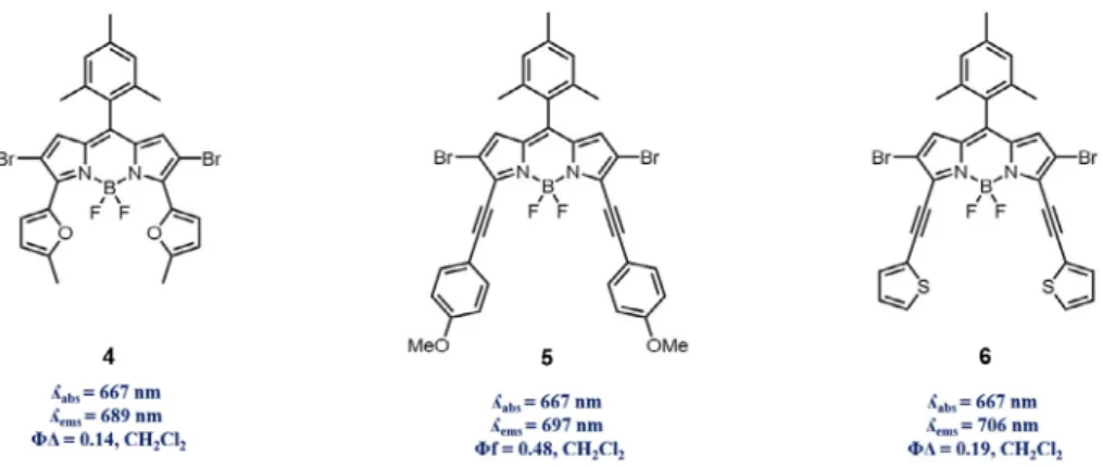
Photosensitizers need to be absorbing in therapeutic window for clinical application purposes. There are several methods to modify BODIPY dyes, one of which is fusion of aromatic groups into the structure through several mechanisms. Recently Hao and co-workers reported facile, region-selective and stepwise synthe-sis of several BODIPY derivatives through C–H arylation with Suzuki, Stille, Heck and Sonogashira coupling reactions[35]. All
a
-arylated structures showed red-shifted absorption/emission, but 2-methylfuran 4, 4-ethynylanisole 5 and 2-ethynylthiophene 6 were reported have absorption maxima around 670 nm due to their electron rich and higherp
-conjugation capabilities compared to thiophene and phenyl derivatives placed at 3 and 5 positions of the dye. Singlet oxygen quantum yield experiments with1,3-diphenylisobenzofuran (DPBF) indicated comparable singlet oxygen generation ability of these dyes (Fig. 2).
Also, Jiao and Hao reported pyrrole fused at 3 and 5 positions of BODIPY dyes with several modifications in terms of types of pyr-role and substituted groups on BODIPY[36]. Addition of each pyr-role unit shifted absorption/emission maxima towards NIR region such that structure 7 has absorption maxima at 745 nm. It was also reported that addition of bromine atoms into the structure not only pronounces heavy atom effect but also shifts absorption max-ima towards NIR region as can be realized in the comparison of 7 and 8. Moreover, alkyl substitutions on pyrrole moieties were reported to decrease singlet oxygen generation efficiency and fluo-rescence. However, singlet oxygen generation efficiency of the dyes 9 and 10 were found to be fairly comparable with methylene blue along with good fluorescence quantum yields, indicating their potential usage in PDT and imaging purposes (Fig. 3).
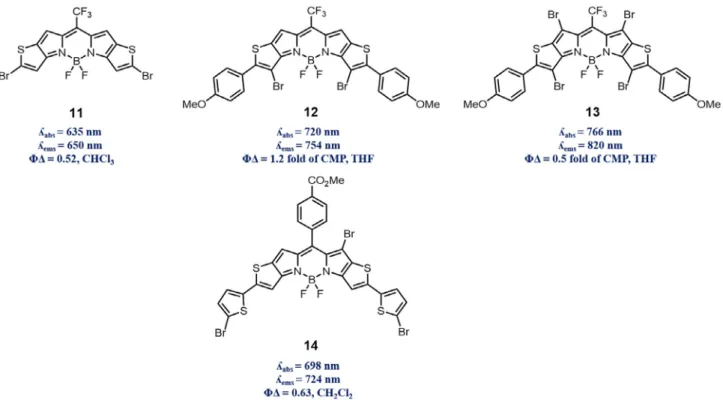
Another versatile functionalization of BODIPY derivatives was reported by You and co-workers [37]. In this particular study thieno-pyrrole-fused BODIPY 11 was utilized as a platform to obtain NIR region absorbing/emitting dyes. Participation of 11 in Suzuki, Stille and Heck coupling reaction enabled wide range of functionalization desired for NIR dyes. Functional group tolerating nature of Suzuki coupling reactions offered preparation of highly conjugated systems in wide scope of chemical scaffolds. Besides, Heck coupling enabled to reach extended
p
-conjugation for further red-shift. Resultant structures from such chemistry displayed absorption maxima ranging from 650 nm to 850 nm which is rare case in BODIPY dye derivatives. Along with such spectral shift, derivatives of thieno-pyrrole-fused BODIPY 11, exhibiting compa-rable singlet oxygen generation and fluorescence emission that can also be utilized in imaging purposes, provide promising future. You and co-workers also reported that similar BODIPY derivativesFig. 1. Heavy atom free singlet oxygen producing BODIPY derivatives 1–3 (orthogonal BODIPY derivatives).
Fig. 2. Longer wavelength absorbing/emitting, aromatic group fused (through various coupling methods) BODIPY derivatives 4–6.
12 and 13 produced singlet oxygen upon exposure to light above 700 nm along with being relatively high resistant to photo-bleaching, demonstrating desirable properties for use in PDT
[38]. Moreover, Shen and co-workers reported unsymmetrical
thieno-pyrrole-fused BODIPY derivative 14, absorbing above 700 nm, as a powerful example of PDT agent supported with HeLa cell culture experiments, resulting in IC50(half maximal inhibitory
concentration) value of 7.12mM without observable dark toxicity
[39](Fig. 4).
Direct functionalization of BODIPY dyes, obtained from 2,4-dimethyl pyrrole, with styryl moieties due to acidic nature of hydrogen atoms on 3 and 5 positions was achieved via Knoeve-nagel condensation [40,41]. Further efforts enabled to obtain tetra-styryl BODIPY derivatives[42]. Addition of each styryl unit shifted S0–S1absorption band of BODIPY toward NIR region which
is imperative for PDT applications. To illustrate possible usage of such derivatives in vitro Akkaya and co-workers reported water soluble di-styryl BODIPY 15 decreased viability of K562 human erythroleukemia cells with EC50value of less than 200 nM without
observable dark toxicity [43]. Moreover, in order to obtain NIR absorption/emission nitrogen atom can be incorporated on meso position of BODIPY dyes, forming so called aza-BODIPY dye. In fact, changing meso atom from carbon to nitrogen tends to narrow the HOMO–LUMO band gap, enabling longer wavelength absorption. O’Shea and co-workers reported in vitro photo-therapeutic effects of aza-BODIPY 16 by exhibiting cell viability decrease of HeLa cer-vical carcinoma and MRC5-SV40 transformed fibroblast cancer cell lines with EC50values of 41 ± 3 nM and 14 ± 1 nM at higher light
dosages, respectively[26]. Additionally, it has been reported that incorporation of aryl substituents on aza-BODIPY dyes shifted absorption maxima above 700 nm for practical applications in PDT[44](Fig. 5).
2.2. Dual effective designs
Photodynamic therapy is a well-established treatment modality depending on formation of reactive oxygen species through
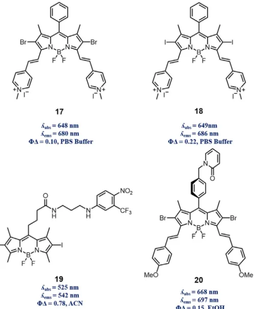
competitive Type I and II mechanisms. It has been reported that vast majority of photosensitizers follow Type II mechanism, form-ing sform-inglet oxygen as main cytotoxic agent[45]. BODIPY type dyes have been considered to perform with same mechanism until the Wang and co-workers reported first observation of DNA cleavage through formation of reactive oxygen species other than singlet oxygen with BODIPY type dyes[46]. Prepared styryl and di-halogenated BODIPY dyes 17 and 18, proven to bind CT DNA with absorption titration, circular dichroism and viscosity experiments,
successfully cleaved CT DNA. In contrast to 18, NaN3 and KI
restricted photo-activity of 17, indicating exertion of photoactivity through both Type I and Type II mechanisms. Even though these mechanisms are competitive in nature, Wang et al. pointed that BODIPY dyes can perform as both types. Such duality stems from intrinsic property of photosensitizers; however, rational designs can enhance PDT with release of other reactive species in a syner-getic manner. Sortino and co-workers designed BODIPY derivative
Fig. 4. Longer wavelength absorbing/emitting, thieno-pyrrole fused BODIPY derivatives 11–14.
Fig. 5. BODIPY 15 functionalized with Knoevenagel condensation. Longer wave-length absorbing/emitting, heavy atom incorporated aza-BODIPY 16.
19 with NO photodonor (NOPD); trifluoromethyl substituted
nitroaniline [47]. Mechanism of NOPD relies on
photo-rearrangement prior to NO release with resultant formation of phe-nol derivatives. Excitation of structure with blue and green light sources produced NO and singlet oxygen, respectively. Cell viabil-ity experiments conducted with melanoma carcinoma cells proved the dual nature of this design such that depending on irradiation wavelength formed reactive species decreased cell viability greatly. On the other hand, in order to increase the efficiency of PDT instead of using combinatory effects of reactive species, intrin-sic limitations of PDT can be overcome. It has been reported that PDT diminishes oxygen level inside the cell by simply consuming dissolved oxygen to generate singlet oxygen, and also destroying vasculature of tumor tissue feeding cells, making cell environment even more hypoxic[48–50]. Such induction of more hypoxic envi-ronment renders PDT self-limiting. Fractional photodynamic ther-apy, which delivers light fractionally to allow cells replenish oxygen levels during dark cycle of treatment, is promising so as to eliminate such intrinsic limitation. Akkaya and co-workers reported that singlet oxygen storage unit (2-pyridone) containing, longer wavelength absorbing/emitting BODIPY dye derivative 20 produces singlet oxygen under light irradiation (light cycle of FPDT) and stores some of it in the storage unit[51]. During dark
cycle of FPDT, which is not utilized for treatment purposes, with thermal cycloreversion formed endoperoxide structure released singlet oxygen while cells replenish their oxygen level to keep treatment ongoing. Such dual effective nature of 20, being vali-dated with HeLa cancer cell culture experiments indicating CC50
(50% cytotoxic concentration) value of 8.6 nM, represents promis-ing future as s FPDT drug (Fig. 6).
Photodynamic therapy requires penetration of light through turbid environment in tumor tissue. Several reports have agreed upon best penetration of light through tissue is observed wave-lengths between approximately 700–1000 nm; therapeutic win-dow [52,53]. Considering light penetration issue and intrinsic hypoxia-induction by PDT as mentioned before, Chen and co-workers reported dual active agent that can go through PDT along with PTT[54]. That is, BODIPY embedded into polymeric vesicles can absorb light at/under 660 nm. Even though, such design can produce singlet oxygen efficiently upon exposure to light at 660 nm, vesicle structure can absorb light at 785 nm which is much more penetrable through tissue and produces lower dosages of PDT along with hypothermia through non-radiative relaxation of BODIPY dyes from first excited singlet state to ground state. PDT-synergized PTT treatment induced total tumor ablation along with several characteristic features to address some forenamed issues;
preferable cellular uptake, enhance tumor accumulation, economy in usage of cellular oxygen and deeper light penetration. Another synergetic demonstration of PDT-PTT dual treatment method based on aza-BODIPY reported by Dong and co-workers[55]. Pho-totherapeutic performance of self-assembled cubic nanoparticles
from IABDP with DSPE-mPEG2000were exploited by using Xenon
lamp (indicating no specific light source is required such as laser) as the light source in vivo and in vitro studies, leading to the conclu-sion of this dual therapeutic treatment modality being remarkably phototoxic such that tumor growth potently can be suppressed without any apparent side effect. In addition, photodynamic ther-apy can be employed with other treatment modalities like chemotherapy to synergistically destroy tumor tissue. Hu and Wang reported very recent demonstration of such duality. Vesicles formed from BODIPY dye and pillar[5]arene with host–guest chemistry can be efficaciously loaded with chemotherapy drug DOX[56]. The cytotoxicity analysis of vesicles with A549 lung ade-nocarcinoma cell line pointed out that, due to intrinsic low-pH nat-ure, cancer cell leads release of DOX with destruction of vesicle along with phototherapeutic effect of photosensitizer, causing reduction in cell viability with IC50values of 1.40mM under light
irradiation and 5.50mM under dark. Another PDT/chemotherapy
treatment duality was reported by Yan and co-workers depending on release of DOX and phototherapeutic agent in cancer cells due to acidic nature of microenvironment of tumor tissue and cancer cells. DOX polymeric micelles entrapped BODIPY dyes were pre-pared and evaluated with HepG2 cell lines[57]. Cell culture
exper-iments validated treatment duality based on release of
chemotherapy drug and PDT drug inside cancer cells by indicating immense difference in cell viability under light and dark conditions.
2.3. PDT-imaging duality
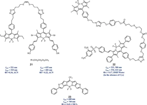
In previous section, synergetic treatment designs were dis-cussed for enhancement of PDT, yet it is of utmost importance to be able to measure the effect of phototherapeutic modalities directly rather than measuring biological effect on a cellular level which is the cumulative result; cell death. Therefore, structural construction can be accomplished by combining photosensitizer and imaging units since triplet state accumulation required for PDT will eventually quench fluorescence of chromophore, reducing its imaging competence. Erbas-Cakmak and Akkaya reported proof of principle PDT/imaging design 21 consisting of a photosensitizer and the fluorophore[58]. Covalently combined BODIPY units per-formed electronic energy transfer from fluorophore (donor) to pho-tosensitizer (acceptor) such that upon excitation of fluorophore at shorter wavelength of light yielded characteristic fluorescence emission of photosensitizer. However, exposure to longer wave-length of light, that can excite photosensitizer and generate singlet oxygen, resulted recovery of fluorescence of the donor unit, indi-cating the working principle. That is, upon fracture of singlet oxy-gen labile linker enhancement of fluorescence of donor indicated formation of main cytotoxic agent in a self-reporting manner.
Employment of biological thiols (GSH or Cys) in activation of several designs can also be exploited in PDT-imaging modality. Wu and co-workers reported the BODIPY dyad 22 was designed to contain photosensitizer and fluorophore as being donor and acceptor, respectively [59]. Therefore, singlet oxygen generation capability of photosensitizer was quenched since accumulation on triplet state is not allowed through FRET mechanism. On the other hand, fluorescence of acceptor unit was caged with DNBS (2,4-dinitrobenzenesulfonyl), leading that PDT and imaging capa-bility are not pronounced; however, dyad contains disulfide linker which can be cleaved by biologicals thiols. Wu et al. reported that in HeLa cells existent biological thiol derivatives uncaged PDT and
imaging units of 22 through cleavage of di-sulfide bond (inacti-vates FRET) and removal of DNBS (restores fluorescence) such that singlet oxygen and fluorescence quantum yield increased from 16.7% and 1.3% to 71.5% and 47.6%, respectively. Based on these reports, it is worth mentioning that combination of a reporter and a photosensitizer much more auspicious method, yet usage of single chromophore cannot be underestimated. However, for PDT/imaging duality, it is compulsory to balance the capability of singlet oxygen generation and fluorescence of photosensitizers along with high extinction coefficients at relatively long wave-length compared to optical window. One of recent examples of sin-gle chromophore containing PDT/imaging duality was reported by You and co-workers. Halogen atom free BODIPY dye 23 having sin-glet oxygen quantum yield of 0.42 and moderate fluorescence quantum yield 0.22 was proven to be phototoxic in a cell culture experiments with colon-26 cells (IC50value of 3.0mM after
irradi-ation without any dark toxicity up to 5.0mM) and an antitumor agent in demonstration with Balb/c tumor-bearing mice along with live mice optical imaging, indicating achievement of PDT/imaging duality with single chromophore[60](Fig. 7).
2.4. Antimicrobial PDT
Novel treatment strategies are highly demanding for the infec-tious diseases despite the focused continuous research on antibi-otics and vaccines, such as modifying the structures of traditional antibiotics, because of the fact that the prokaryotic organisms can easily develop resistance against the new agents. Antimicro-bial–PDT, which is also called as PACT (photodynamic antimicro-bial chemotherapy) is a promising alternative to traditional chemotherapies. Since photodynamic action is based on oxidative damaging in bacterial membrane resulting the cytotoxic effects, it can be used for all types of microorganisms regardless of resis-tance obstacle. There are several examples of APDT with chlorine6
or porphyrin derivatives [61–63]; however, BODIPY dyes are
indeed promising due to low dark-toxicity and ease of functional-ization. Some cationic BODIPY derivatives are developed for that purpose as shown inFig. 8.
The idea behind the cationic structure is that positive charge on the photosensitizer can promote stronger electrostatic interaction with negatively charged bacterial cell wall. To ensure selectivity towards pathogens, human MDA-MB-231 cell lines are also incu-bated with compound 24, by O’Shea and co-workers and results showed only minimal binding to human cell lines compared to Gram-positive Staphylococcus aureus, Gram-negative Escherichia coli and Candida albicans yeast cells[64]. Additionally, compound 24 showed no dark toxicity in the absence of light with 1mg/mL dosage; however, with introduction of light, reduction in the viabil-ity was observed (2, 4, 8, 16 J/cm2irradiation causes log
10reduction
of 0.1, 0.3, 1.4, 3.0, respectively). Among others, compound 25 showed the lowest photodynamic minimal bactericidal concentra-tion value, 1.66mM, for the E. coli. (whereas intrinsic toxicity was observed up to 40mM.) Banfi and his co-workers compared the effi-ciency of compound 25 with compound 26, only methyl pyridinium cationic moiety is changed with benzyl pyridinium, concluding that masking the positive charge by benzylic unit prevents the photo-sensitizer from coordinating tightly on the bacterial cell wall result-ing around 4-fold less efficiency[65]. Due to the excellent APDT efficiency of compound 25 on E. coli and Staphylococcus xylosus, Ghiladi and his co-workers conducted the experiments on eight bacteria strains including Gram-positive, Gram-negative and drug-resistant bacteria, three yeast species and three viruses with promising results so that compound 25 is able to mediate the photodynamic inactivation at nanomolar concentrations with short light dosages, meaning it is more efficient compared to methylene blue and TMPyP[66]. Barbieri and Orlandi tested the photodynamic
activity of compound 25 on Pseudomonas aeruginosa which is a widespread Gram-negative planktonic microorganism that has the ability to colonize and form biofilms[67]. Those biofilms can locate in many body parts such as lungs and diabetic wounds as well as in household and medical related surfaces. P. aeruginosa within the lower layers of biofilms exhibit slower metabolic activity and high resistance to antibiotics. Since APDT is a viable method where antibiotic treatment is inefficient, experiments with com-pound 25 showed decrement in the cell viability at even 2.5mM and prevention of regrowth due to photoinactivation. Control experiment resulted that the photosensitizer has dark toxicity in
the higher concentrations (80mM); however, additional growth
for 24 h fully overcome the intrinsic toxicity. Cationic photosensi-tizer structures can be adapted to potential medical applications like self-sterilizing surfaces and other equipment which need to have anti-infective feature.
Li and co-workers designed a polymer based macromolecular photosensitizer with antibacterial activity toward both Gram-negative and positive bacterial strains [68]. Introduction of the
polymeric conjugation to photosensitizer overcomes the problems related to aggregation of BODIPY’s hydrophobic structure in aque-ous based biological media, enhancing the cytocompatibility. For that purpose, iodinated BODIPY derivative was copolymerized with (dimethylamino)ethyl methacrylate and galactose. 2-(Dimethylamino)ethyl methacrylate has cationic character in aqueous media so that it increases the binding efficiency to bacte-rial cell walls, on the other hand galactose unit decreases the cyto-toxic and pro-inflammatory effects of positively charged polymer, increasing the macromolecules biocompatibility. The results indi-cated that the macromolecule disturbs the cell membrane of the S. aureus and P. aeruginosa bacteria leading the apoptotic cell death
as shown in the SEM images in Fig. 9. Dark experiments have
shown that the antibacterial activity of the macromolecules was poor compared with under light irradiation experiments.
Similar approach was demonstrated with the conjugation of porphyrin and BODIPY based photosensitizers to the cellulose fibers in order to form self-sterilizing, antibacterial paper sheets by Ghiladi and his co-workers[69]. An alternative targeting unit,
Fig. 7. BODIPY designs 21–23 for PDT/imaging modality.
zinc(II)–dipicolylamine (ZnDPA), was utilized by Smith and co-workers in the place of cationic module for the targeted imaging and photodynamic action for eradicating pathogens as seen in
Fig. 10. ZnDPA can coordinate with anionic groups like phosphate containing units on the bacterial cell membrane preferentially, whereas remain nearly inert to the neutral surface of healthy
mammalian cells [70] (Fig. 11).mDestroy exhibited 99–99.99%
inactivation of four bacterial stains with selectivity in the presence of mammalian cells, besides mSeek, non-halogenated derivative of mDestroy, used for fluorescence imaging and detection with a CCD camera. As proposed in the aforementioned examples, photosensi-tization with BODIPY dye is an effective and selective options for the inactivation of pathogens on non-in vivo surface sterilization due to applicability in wide range of bacterial strains, which is independent of drug resistance.
3. Modulation of photosensitization activity
As described in previous sections, photodynamic therapy has highly desirable features in various applications in medicinal areas, including tumor diagnostics and treatments, or antibacterial/ antiviral activities. In clinical research, PDT has the ease of the localization of irradiation on desired area, thus it has great poten-tial in cancer treatment techniques due to its non-invasive nature.
Still, some normal tissue and organ damaging causing edema around the treated tissues are reported[71–73]. Therefore besides the spacial selectivity, tumor-associated treatment strategies are required to decrease the side-effects. In the past decade, ‘‘turn off–on” photosensitizers which remain inert in bloodstream and normal tissues, but activated only when cancerous tissue or cells are encountered were reported[74–76]. Specific targeting to the cancerous tissues can be performed in many ways, because cancer-ous tissues have different chemical environments due to changes in metabolic/catabolic activities and over expression of some bio-logical parameters and proteins. Novel designs regarding to the cancer selective PDT has been reported with BODIPY dyes via uti-lization of cancer specific moieties.
3.1. Activation through pH responsive designs
In order to especially target the tumor in the tissues or organs, one can take the advantage of characteristics of the microenviron-ment of tumor tissue. As studied on many cancerous cells, there is a difference in the concentrations of some ions or molecules between normal and cancer cells. As a consequence, those param-eters are promising ways to access for the activation of therapeutic actions, including PDT. One of the parameters is the acidity of the media: the extracellular pH of the cancer cells is around 6.0,
Fig. 9. (Left) Structure of p(GEMA–co-DMAEMA–co-BODIPYMA)-2I (Right) SEM images of (A and B) S. aureus and (C and D) P. aeruginosa before (A and C) and after (B and D) illumination. Adapted with permission from Ref.[68]. CopyrightÓ 2015, Royal Society of Chemistry.
Fig. 10. (Left) Bacteria cell surface targeting by a ZnDPA–BODIPY fluorophore (mSeek) or photosensitizer (mDestroy). The prefix ‘‘m” designates ‘‘microbial”. (Right) Fractions of killed bacterial cells treating with increasing concentrations of mDestroy and one hour of green light irradiation (50 mW cm 2
). Adapted with permission from Ref.[70]. CopyrightÓ 2015, Royal Society of Chemistry.
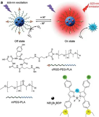
whereas normal cells have slightly alkaline pH around at 7.4[77]. Thus, many pH-activable systems including polymeric materials, nanocarriers besides photosensitizers are being developed. Ju and co-workers reported an aza-BODIPY derivative which is encapsu-lated in a cRDG-functionalized micelles which can be selectively taken up by
a
vb3overexpressed tumor cells into lysosomes[78].The usage of the polymer based agents for the encapsulation of photosensitizer are far more convenient than inorganic and metal based nanostructures due to biocompatibility and non-toxic degra-dation products in the body. In addition, the substituted pH-responsive diethylaminophenyl group shows excellent switch-on selectivity in tumor imaging and photodynamic action in both in situ and real time experiments. The fluorescence and singlet oxygen quantum yield enhanced in the acidic media because of
the fact that PET from the aniline group to BODIPY is no longer available. The system has also the advantage of absorbing in NIR region (kabs = 822 nm) which is essential for the penetration into the biological tissues.
Yan and co-workers, proposed similar design for pH activation of photodynamic therapy designing a polypeptide micelle which entrapped the photosensitizer, BODIPY-Br2, and has
diisopropy-lamine group on the hydrophobic end regulating the on–off activa-tion mechanism[79]. The core–shell structure of the nano micelles would degrade under acidic conditions (pH 5.5) due to the proto-nation of the DIPEA groups conjugated to polypeptide side chains as shown in theFig. 12, releasing the encapsulated BODIPY-Br2into
the desired area.
MTT assay with HepG2 (liver hepatocellular carcinoma) cells verified the selective photodynamic action in cancerous cells suc-cessfully under low energy density (12 J/cm2) and low concentra-tions (5.4mM) with 45% higher mortality compared to the cells incubated under dark.
3.2. Activation by biological thiols
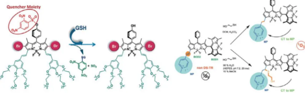
The intracellular glutathione concentration is another success-ful approach to the modulation of activatable PDT since glu-tathione (GSH) concentration in cancerous cells was reported up to 1000-fold higher compared to normal cells[80–82]. Considering this tremendous difference in concentration of GSH between nor-mal and cancer cells, a water-soluble BODIPY derivative was devel-oped by Akkaya and co-workers which contains GSH-cleavable quencher moiety, so that ‘‘caged photosensitizer” can only gener-ate cytotoxic singlet oxygen in GSH over-expressed medium[83]. While singlet oxygen generation capability is restored, fluores-cence is also reinstated by the removal of the electron-sink quencher in 30 min with 80% yield, so that the progress of the cleavage reaction can be monitored. The behavior of the design was also validated by the cell culture experiments with human epithelial cancer cells (Huh7, MCF7, HCT116). It was proven that the photosensitizer is nontoxic and not capable of generation of singlet oxygen at its caged form in human fetal lung fibroblast cells (MRC-5) where GSH level is low, however with the uncaging in high GSH levels of cancer cells, photosensitization is active and causes the apoptosis resulting the death. For HCT116 cell lines, IC50value under irradiation is 20.0 nM, on the other hand under
dark, IC50value is much higher (4.38mM), showing that the
photo-sensitizer has comparably less dark toxicity. Another perspective to the GSH activation for PDT is to alter the properties of energy states of the photosensitizer to turn on–off the singlet oxygen
Fig. 11. Structures and pH-activatable generation of fluorescence and1
O2by cRGD–
NEt2Br2BDP NP. Reprinted from Ref.[78]. Copyright Ó 2015, Royal Society of
Chemistry.
Fig. 12. Structure of polypeptides loading BODIPY–Br2, micellization, and pH triggered Drug release followed by NIR PDT. Reprinted with permission from Ref.[79]. Copyright
production by reacting with GSH [31]. Proposed dissymmetric dimer of BODIPY dyes shown inFig. 13, has nearly no photosensi-tization of molecular oxygen into singlet oxygen as a consequence of not having a degenerate set of HOMO and LUMO resulting lower intersystem crossing. The BODIPY dimer remains in its passive state until introduction into high-GSH level. GSH cuts the exten-sion of conjugation and isolates the methylpyridinium group by either reduction or conjugate addition to the styryl double bond resulting the formation of the similarity and orthogonality which increases the ISC efficiency because dimeric BODIPY has a special electronic arrangement that similar subunits possesses a degener-ate pair of HOMO and LUMO. Cell culture with HeLa cells studies also showed revealing results: photodynamic action of 28 causes the apoptotic death and HeLa cells have 38% survival rate upon the treatment of 28 (IC50: 280 ng/mL for 28, 32.6 ng/mL for 28r),
however normal NIH 3T3 cells have 80% survival rate under same conditions. Also, no significant dark toxicity was observed without irradiation. All the finding showed that the design can stay passive even it absorbs the light, and regains photosensitization ability when cancerous tissue is encountered which has high concentra-tions of glutathione.
3.3. Activation by enzymatic degradation
Another promising attempts toward specific targeting is the uti-lization of enzyme-activated treatment of the cancerous tissues. Besides targeting the tumors, the on-site activated photosensitiza-tion was developed by Dong and his co-workers recently[84]. A diiodostyryl BODIPY substituted hyaluronic acid nanoparticles
(DBHA-NPs, Fig. 14a) were introduced, which shows enhanced
hydrophilicity and biocompatibility due to the presence of hya-luronic acid. The nanoparticles are composed of self-assembled aggregation of DBHA, which can disaggregate inside the lysosomes (hyaluronidase cause the enzymatic degradation of the HA poly-mer) and the singlet oxygen generation ability, under irradiation at 588 nm, is restored as a consequence of disassembly. Besides its photodynamic efficiency, it has ability to regain fluorescence after disaggregation which makes it beneficial for diagnostic pur-poses. In vivo and in vitro experiment demonstrated that the design has excellent targeting ability to CD44 receptors which are overex-pressed on the surface of HCT-116 cells without dark toxicity (Fig. 14b and c), resulting reduction of side effects in photodynam-ically activatable cancer therapy.
3.4. Activation through ROS responsive design
Utilization of activation with reactive oxygen species is an alter-native way of targeting since cancer cells are generally under oxidative stress due to the increased metabolic activity resulting ROS generation. A two segment photosensitizer-trap which can be activated in an increased ROS was shown by Cosa recently
[85]. A naturally occurring antioxidant, chromanol ring of
a
-tocopherol, which is an efficient scavenger of ROS, was modulated on brominated BODIPY core as photosensitizer. The trap moiety is both an efficient PET donor which quenches the singlet oxygen generation capability of BODIPY unit and a good physical 1O2
quencher itself. Besides, when it reacts with ROS, PET is deacti-vated, and photosensitizer regains the singlet oxygen generation ability by around 40-fold (Fig. 15). Since singlet oxygen is a ROS indeed, the activation of1O
2generation is an autocatalytic process,
enhancing cytotoxic singlet oxygen generation by itself. The use-fulness of this approach was demonstrated on Gram negative E. coli strain. One part of the cell lines were stressed with hydrogen peroxide which stimulates ROS production in Gram-negative cells and those cells showed an excessive drop in colony forming units compared to non-stressed healthy cells in the presence of com-pound and light irradiation. The PD-MBC (Photodynamic Minimum Bactericidal Concentration) value for the pre-activated compound
by ROS was determined as 10mM, and PD-MIC (Photodynamic
Minimum Inhibitory Concentration) was reported as 10mM under
light irradiation and 40mM in dark conditions for the same com-pound. This study is a successful example for ROS mediated activa-tion of photodynamic therapy which can also amplify cytotoxic singlet oxygen generation by itselfFig. 16.
3.5. Logic gates
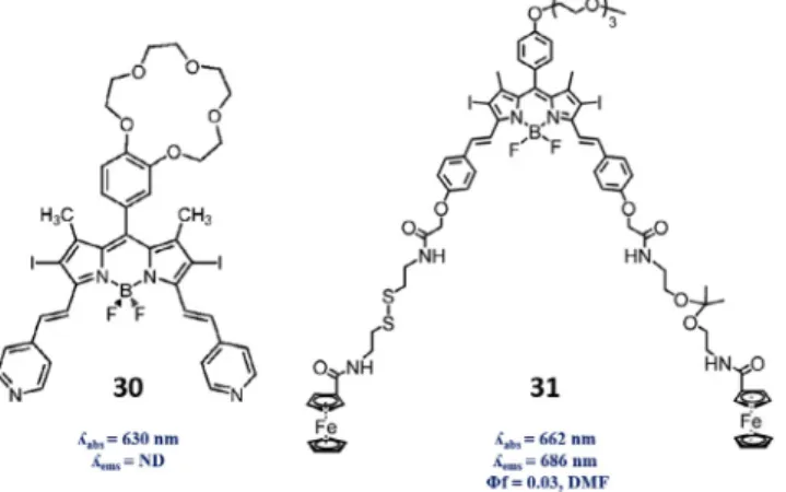
To enhance the tumor selectivity, one can take advantage of more than one characteristics of intercellular and intracellular chemical environment of cancerous tissues. For that purpose, Boo-lean logic, which is a key facet of computer programming, can also have significant potential in molecular systems which was intro-duced by de Silva in 1993[86]. Molecular logic gates have been adapted to several examples including photodynamic therapy so that the photosensitizer would generate singlet oxygen as a response to two characteristic parameters together in the chemical environment. This system was first demonstrated with a BODIPY dye 30 in Akkaya group with a crown-ether-based PET modulator for Na+and pyridylethenyl module for H+input because both Na+
and H+concentrations are found significantly higher in cancer
tis-sues than normal tistis-sues[87]. When Na+concentration is high, PET
is quenched and singlet oxygen generation is enhanced, on the other hand, in acidic medium, a red shift from 630 nm to 660 nm in absorption spectra is observed. Under irradiation at 660 nm, the dye produces singlet oxygen most efficiently – about 6-fold-only when pyridine groups are protonated in acidic media and PET is quenched in the presence of Na+ ions, thus an AND logic gate is proposed where singlet oxygen is the output. This design was improved in Akkaya group later, integrating GSH and
acidic response to form another AND logic gate [88]. Another
aspect of thiol and pH activable photosensitization was demon-strated by Ng and Lo[89]. A halogenated BODIPY 31 derivative
Fig. 13. (Left) Mode of operation for the GSH-mediated activation of caged photosensitizer 27. (Right) Conversion of compound 28 into 28r or 28m on reaction with mercaptoethanol in DCM and in aqueous buffer solutions, respectively. Adapted with permission from Refs.[31,83]. CopyrightÓ 2014 & 2015, Wiley-VCH Verlag GmbH & Co. KGaA, Weinheim.
was functionalized with two ferrocenyl groups which are employed as quencher through PET mechanism. Ferrocenyl moi-eties are linked through a pH-cleavable ketal and a thiol-labile disulfide linkers so that the fluorescence and singlet oxygen gener-ation efficiency will be enhanced by the coexistence of two-tumor associated parameters. In vitro results with MDF-7 breast cancer cells which are pretreated with dithiothreitol (DTT) were in agree-ment with the dual activation mechanism for the both fluorescence imaging and photodynamic activity of the design (IC50 value of
140 nM in 2mM DTT). Moreover, nude mice having a HT29 human
colorectal carcinoma was treated with this compound and the
restoration of fluorescence was observed inside the tumor within 9 h, concluding that the cleavage of the two linkers by cutting the photoinduced electron transfer from ferrocenyl group to the dye was demonstrated which has great potential for theranostic and imaging of cancerous tissues.
3.6. Control on PDT through PS delivery
Besides the spacial selectivity of photosensitization, tumor-associated treatment can also be improved by introducing drug delivery systems for the theranostics of cancer. The incorporation
Fig. 14. (a) Diiodostyryl BODIPY conjugated hyaluronic acid nanoparticles (DBHA-NPs). (b) Comparison of cell viabilities by MTT assay (l >600 nm). (c) The relationship between tumor volume and treatment time for the DBHA-NPs in the HCT-116 mouse model tumor cells via tail vein injection. Twenty-four mice were randomly assigned into four groups (6 mice per group), including saline with light, HA with light, DBHA–NPs without light and DBHA-NPs with light. (d) Typical photographs of tumor-bearing mice treated at different times. Adapted with permission from Ref.[84]. CopyrightÓ 2016, Royal Society of Chemistry.
Fig. 15. Proposed Reaction of 29 with1O
of the photosensitizer into nanomaterials enhances the permeabil-ity and retention effect along with biocompatibilpermeabil-ity thus
therapeu-tic accuracy and efficiency increase [90,91]. Those carrier
nanomaterials can be both organic based, such as polymers, lipo-somes etc. or inorganic structures like metallic composites [92– 94]. BODIPY derivatives substituted with n-decyloxyphenyl and pentadecyl groups which can embed themselves into micellar form
and BODIPY chemisorbed on NaYF4:Er3+, Yb3+ up-conversion
nanoparticles absorbing NIR region are both water soluble designs having desirable properties convenient for the biological media
[95,96]. The enhanced permeability and retention (EPR) effect can be facilitated by incorporation of active targeting ligands onto the surface of nanostructures so that specific receptors on the cancerous cell membrane can be targeted selectively. Conventional targeting groups are based on galactose, folic acid, oligopeptides, and antibody derivatives[97,98]. For instance, HepG2 (liver hepa-tocellular carcinoma) cells overexpress ASGP-R galactose receptors so that it can be targeted by galactose functionalized amphiphilic copolymer micelles (PMAGP–POEGMA–PLys) as nanocarriers for
BODIPY, reported by Yan and co-workers[99]. POEGMA is the
hydrophilic part of the polymer whereas PLys forms the hydropho-bic core where hydrophohydropho-bic BODIPY-PS is located (Fig. 17). PMAGP is the galactose containing targeting unit for the hepatoma carci-noma cells.
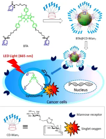
These nanostructures formed spherical micellar structure in aqueous media with 173 nm average size with the PS loading con-tent about 2.5%. Cell viability experiments were carried out for testing the dark toxicity and targeting ability of nanostructures. The nanostructures didn’t exhibit dark toxicity in the absence of light irradiation and when they were exposed to light, cell viability decreased up to 50% for HepG2 cell lines while HeLa cells only decreased by 28% because endocytosis of the BODIPY containing micelles into the cells are more likely compared to HeLa cells which has lower ASGP-R galactose receptors. Similarly, Yin and co-workers designed mannose functionalized BODIPY nanoparti-cles for the selective targeting of lectin-type mannose-receptor
overexpressing MDA-MB-231 breast cancer lines [100]. Long
wavelength absorbing BODIPY derivative having three adamantane
units assembled to a heptamannosylatedb-cyclodextrin through
supramolecular host–guest interactions forming spherical
nanoparticles with 117 nm average size as given inFig. 18. MDA-MB-231 breast cancer lines were incubated with those nanoparticles, resulting photodynamic cellular damage and apop-totic cell death. On the other hand, the nanoparticles without man-nose groups on theb-cyclodextrin didn’t exhibit cellular damage under irradiation in same conditions. In vivo experiments also ver-ified the selective targeted PDT for breast cancer cell lines without damaging normal cells and exhibiting very low dark toxicity. Usage of folate ligands for targeting purposes is an alternative convenient method for some cancer cell lines such as 4T1 breast cancer cells. The NIR absorbing dicarbazole functionalized BODIPY derivative was encapsulated by PLA–PEG–FA polymer to form a water soluble
micellar structure [101]. TEM measurements indicated that
the size of the dye loaded nanomicelles are around 14 nm which are quite smaller compared to related nanostructures. The tumor targeting ability were studied with HeLa cells in comparison with
Fig. 16. Designs of the double-stimulus-responsive photosensitizers 30 and 31.
Fig. 17. Galactose-targeting and light-confined NIR imaging and photodynamic therapy on hepatoma cells with 32. Reprinted with permission from Ref.[99]. CopyrightÓ 2016, Royal Society of Chemistry.
healthy human skin epidermal Greengo cells. MTT assay and in vivo demonstration results verified the folate ligand improve the selectivity in cancer cells and no obvious organ dysfunction, dark toxicity, inflammation lesion abnormal results in serum anal-ysis was observed. Those novel designs with the employment of nanoscale structures provide promising results for the platform for selective tumor photodynamic action for clinical applications. 3.7. Control on PDT through subcellular localization
Photodynamic therapy has been shown to operate in various mechanisms to destroy cancer cells, containing direct cell death among destruction of tumor vasculature and triggering immune response[45,102–105]. Even though each mechanism cooperates, direct cell death of tumor is beneficiary in terms of control on
pho-todynamic effect. That is to say, cellular uptake can be utilized to effectively direct treatment on oxidative damage sensitive subcel-lular parts; organelles. For instance, it has been reported that nucleus is far more vulnerable to oxidative damage compared to plasma membrane[106,107]. Therefore, rational designs targeting oxidative damage sensitive subcellular organelles are far more likely to outperform.
Very first example of application of BODIPY dye 15 as a photo-sensitizer on K562 human erythroleukemia cells reported by Akkaya et al. revealed water soluble 15 decreases cell viability even under low fluence rate irradiation[43]. Following such photother-apeutic demonstration, unsymmetrical di-styryl BODIPY dyes 33–37 were analyzed in terms of photo-toxicity and subcellular localization [108]. Cellular uptake and photo-toxicity analysis of 33–37 on HT29 human colorectal carcinoma cells by intracellular fluorescence indicated greater uptake of 36 and 37, leading enhanced cytotoxicity with IC50values around 15–17 nM. Detailed
subcellular localization analysis showed localization of 37 on plasma membrane attributed to positively charged nature of the drug. On the other hand, 36 was found to localize on lysosome which isn’t considered to be high priority target since other orga-nelles localized photosensitizers outperform due to aggregation of dyes in lysosome (Fig. 19).
Endoplasmic reticulum has been reported to lead apoptotic cell death through caspase cascade upon being exposed to PDT. Com-pound 38, found to specifically localize on ER and lesser extent to mitochondria, outperformed in photo-cytotoxicity compared to its counterpart 39, which exhibited no subcellular localization, due to oxidative sensitivity of endoplasmic reticulum (EC50values
for HeLa, A549 and MRC-5 of 4.7, 6.8, and 18.5mM without observ-able dark toxicity, respectively)[109]. In addition, Borggraeve and co-workers reported that BODIPY dye 40 and 41 co-localized in lysosome and endoplasmic reticulum of with T24 human urinary bladder carcinoma cell line; however, staining imaged of nuclear sequence localization peptide embedded 40, even though showed absence of 40 in nucleus, showed enhanced localization on ER
compared to 41, explaining 2.6 times lower IC50 value of 40
compared to control structure 41 [110]. Aforementioned studies conducted with BODIPY dyes lead to similar results in terms of higher oxidative sensitivity of endoplasmic reticulum.
Instead of ER or nucleus, mitochondria regulating oxidative stress, metabolic energy and apoptotic machinery serves as a bet-ter target since mitochondrial integrity is essential for viability of cells. Compound 42 was prepared by Chakravarty and co-workers for a combinatory effect of PDT and chemotherapy such that mitochondria localized complex was expected to operate in such a way that release Pt from the complex is capable of inhibiting replication of mt-DNA (mitochondrial DNA) and BODIPY moiety embedded into the complex produces singlet oxygen for oxidative damage[111]. Further analysis on HaCaT human skin keratinocyte
Fig. 18. Schematic illustration for the preparation of BTA@CD–Man7nanoparticles
and their application for targeted PDT. Reprinted with permission from Ref.[100]. CopyrightÓ 2016, American Chemical Society.
cells indicated localization of BODIPY in mitochondria via confocal imaging as rationally designed. In another study, ternary copper(II) complex embedded with curcumin and BODIPY dye with similar working principle was shown to be preferentially localized in mito-chondria of HeLa cancer cells, leading decrease in viability of can-cer cells (IC50 values of 3.8 ± 0.2mM after PDT treatment and
around 30mM under dark conditions) being comparable to first
approved cancer drug PhotofrinÒ [112]. That is to say, rational design of PDT agents with particular subcellular localization may enhance the control on phototherapeutic effect (Fig. 20).
4. Photocatalysis by BODIPY dyes
In the steadily developing research on photooxidation reactions, many catalyst species are reported in literature such as rubidium
and iridium based photocatalysts [113–118]. Considering
drawbacks of organometallic catalysts such as requirement of UV-light, high toxicity and cost, scientists tend to find new organic catalysts for oxidation reactions. 9,10-dicyanoanthracene, tri-phenylpyrylium tetrafluoroborate, n-methylquinolinium tetrafluo-roborate as metal-free photocatalysts working in visible range gave promising results for that purpose[119–121]. Organic dyes, like BODIPY dyes, also play a significant role acting as photocatalysts
instead of metal containing catalysts ascribed to having advanta-geous properties such as low toxicity, ease of functionalization and low cost. Photocatalytic efficiency of organic dyes are exam-ined by Zeitler for photoredox transformations with various sim-ple, inexpensive organic dyes such as rhodamine B, alizarin red S, perylene and xanthene derivatives[122]. With the great degree of functionalization feasibility, excellent thermal and photochemi-cal stability, high singlet oxygen quantum yield and good solubil-ity, BODIPY dyes are also possible candidates for photooxidation reactions, yet in this context only direct singlet oxygen involved catalytic systems are discussed.
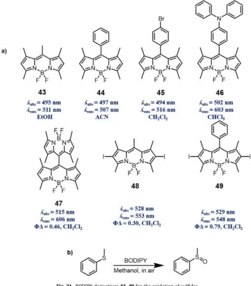
4.1. Photooxidation of sulfides
Preparation of organic sulfoxides from sulfides are essential for organic synthesis not only for rudimentary research but also for multifarious applications, especially for biological and synthetic considerations[123–126]. Due to its significance, oxidation of sul-fides has been drawing attention for many decades. Mild and high yielding conditions, which prevent side reactions including sulfone production have been desired and investigated in the synthetic and manufacturing manner. Herein, photocatalytic organic reactions introduce environmentally friendly, mild reactions with relatively high yields and lower production of by-products. Singlet oxygen forms the persulfoxide intermediate to form sulfoxide[127,128]. Xie first reported the photooxidation of sulfides, here thioanisole, with four BODIPY derivatives which are highly effective photocat-alysts with 89–99% conversion in methanol under visible light irra-diation[129](Fig. 21).
Then the results were improved by optimizing the reaction con-ditions and introducing four new BODIPY derivatives, concluding that the catalytic activity of BODIPY is highly dependent on singlet oxygen quantum yield and the protic solvent (methanol/ethanol) is beneficial to this photooxidation reaction[130]. Cui and He exam-ined this catalytic reaction with ten various BODIPY derivatives using six thioanisole substrates[131]. Given BODIPY dyes in pho-tocatalytic oxidation of sulfides, the ones which have higher singlet oxygen quantum yields: orthogonal dimeric BODIPY and halo-genated derivatives, demonstrated higher oxidation yields, sup-porting the findings by Jing. Indeed, this work proved that BODIPY dyes are successful alternatives for known photosensitiz-ers such as Nile Red and Ru-based organometallic catalysis. 4.2. Photooxidation of dihydroxynaphthalenes
Singlet oxygen can also form endoperoxide intermediate with hydroxynaphthalene substrates yielding dihydroxynaphthalene (DHN) derivatives. One studied example is juglone which is a chemical used as herbicides or coloring agent in cosmetics, food industry and cloth (Fig. 22).
BODIPY derivatives conjugated with 2-(2-hydroxyphenyl)benzo thiazole and benzoxazole subunits for excited state intramolecular proton transfer (ESIPT) were prepared by Zhao and co-workers and used as1O
2photosensitizer for the photocatalytic oxidation of
1,4-dihydroxynaphtalene [132]. These photosensitizers exhibited
more efficiency compared to the traditional transition metal com-plexes, like ([Ir(ppy)2(phen)][PF6]). Zhao and co-workers also
pre-pared iodo-BODIPY derivatives with strong absorption of visible light and efficient long-lived triplet excited state for1O
2mediated
dihydroxynaphthalenes photooxidation to produce specific naph-thalenedione derivatives which are further used for production of some anticancer and antibiotic reagents[133]. The yields were relatively lower (73–87%), however the reactions took place in just 30 min which is considerably fast compared to other photoox-idation reactions. Mechanism of those reactions rely on oxidiza-tion of naphthol by singlet oxygen (1O
2) which is produced by
photosensitizing of the triplet oxygen (3O
2) by the
photosensitiz-ers, BODIPY. Since, the aza-BODIPY derivatives showed high singlet oxygen generation capability, Ramaiah and co-workers synthe-sized halogenated aza-BODIPY dyes and explored their use as
photooxygenation catalysts for 1-naphthol oxidation into juglone
[134]. The highest yield was achieved under focused sunlight to yield 100% conversion in 30 min. For comparison, the traditional photosensitizers, tetraphenylporphyrin, rose bengal and methy-lene blue, were used. Core and peripheral halogenated aza-BODIPY derivative showed higher conversion efficiency than the known photosensitizers, despite having comparable singlet-oxygen efficiencies, because aza-BODIPY has better photostability under the irradiation conditions (more than 8 h).
4.3. Photocatalytic synthesis of 2-aryl benzothiazole
Singlet oxygen generation capacity of BODIPY type dyes makes them highly desirable also for a photocatalyzed oxidative conden-sation reaction to produce 2-aryl benzothiazoles, which play crucial roles in various applications, containing biologically active products, drugs, fibers, plastics, liquid crystals, off-color materials along with several others. Zhou and Yang has reported that BODIPY 50 can enable the condensation reaction between amines and aminothiophenol via production of singlet oxygen to produce
2-aryl benzothiazole [135]. Reaction pursues oxidation of
amine with singlet oxygen prior to condensation of intermediate with 2-aminothiophenol. In the follow up study, Yang et al. reported that BODIPY polymer, prepared from BODIPY 51 and 1–4-phenylenebisboronic acid through Suzuki coupling, loaded on silica powder showed enhanced photocatalytic activity and thermal
Fig. 21. BODIPY derivatives 43–49 for the oxidation of sulfides.
Fig. 22. Photooxidation of DHN with a triplet photosensitizer; BDP = BODIPY compounds.
and photo stability along with improved dispersibility in the con-densation reaction yielding 2-aryl benzothiazoles[136](Fig. 23). 4.4. BODIPY–fullerene dyads in photooxidation
BODIPY dyes have been manipulated in several ways for photo-catalytic applications, one of which is formation of dyads with full-erene. In this type of light harvesting designs, BODIPY derivatives are utilized as light harvesting antenna, and fullerene structure is designed as spin converter. This relies on the fact that fullerene structure has low-lying singlet excited state, enabling the energy transfer from chromophore to fullerene. Intramolecular energy transfer eventually populates S1 state of C60, resulting population of T1 state of C60 due to intrinsic ISC property of fullerene unit; however, this doesn’t necessarily mean that triplet state localizes on fullerene unit. In contrast, due low lying triplet excited state of BODIPY unit compared to fullerene lead triplet energy transfer toward BODIPY unit. Zhao and co-workers reported BODIPY–fuller-ene dyads 52, 53 and 54 based on ‘‘ping–pong” BODIPY–fuller-energy transfer
[137](Fig. 24).
Application of these dyads on oxidation of 1,5-dihydroxy naph-thalene to form juglone demonstrated their enhanced singlet oxy-gen production capability. Zhao and co-workers also reported
BODIPY–fullerene triads and tetrads which contain two and three antennas with varying absorption wavelengths, indicating broad-band absorption nature of such designs[138]. Based on similar energy transfer processes, triads and tetrads were found to be even more effective in singlet oxygen production as observed in photo-catalytic oxidation of DHN compared to TPP and MB. Therefore, without using heavy atoms, rationally designed energy harvesting systems can produce singlet oxygen for photocatalytic purposes. 5. Conclusion
BODIPY dyes are highly versatile, and a bewildering number of applications with BODIPY dyes playing essential roles were reported. As presented in this review, BODIPY dyes are very amen-able to structural modifications making them viamen-able alternatives for porphyrins and phthalocyanines as photosensitizer. It is also noteworthy that in recent years, most creative works on photosen-sitizers were based BODIPY dyes, most likely because of their syn-thetic and photochemical versatility. It is possible to precisely fine-tune the photosensitization characteristics of BODIPY derivatives. We are confident that future advances in photodynamic therapy, and photosensitizer based chemistry in general, will benefit tremendously for this upsurge of interest in BODIPY chemistry.
Fig. 23. General synthesis reaction of 2-aryl benzothiazole derivatives catalyzed by BODIPY 50.
Acknowledgments
The authors acknowledge support from Bilkent University. A.T. is also grateful for a scholarship from TUBITAK (B_IDEB-2210 E). Appendix A. Supplementary data
Supplementary data associated with this article can be found, in the online version, athttps://doi.org/10.1016/j.ccr.2017.09.029. References
[1]N.C. Zeitouni, A.R. Oseroff, S. Shieh, Mol. Immunol. 39 (2003) 1133–1136. [2]Z. Huang, Technol. Cancer Res. Treat. 4 (2005) 283–293.
[3]D.E. Dolmans, D. Fukumura, R.K. Jain, Nat. Rev. Cancer 3 (2003) 380–387. [4]S.B. Brown, E.A. Brown, I. Walker, Lancet Oncol. 5 (2004) 497–508. [5]T.J. Dougherty, G.B. Grindey, R. Fiel, K.R. Weishaupt, D.G. Boyle, J. Natl Cancer
Inst. 55 (1975) 115–121.
[6]J.F. Kelly, M.E. Snell, J. Urol. 115 (1976) 150–151.
[7]Y. Hayata, H. Kato, C. Konaka, J. Ono, N. Takizawa, Chest 81 (1982) 269–277. [8]J.S. McCaughan Jr., W. Hicks, L. Laufman, E. May, R. Roach, Cancer 54 (1984)
2905–2910.
[9]Y. Hayata, H. Kato, H. Okitsu, M. Kawaguchi, C. Konaka, Semin. Surg. Oncol. 1 (1985) 1–11.
[10]J.S. Hill, A.H. Kaye, W.H. Sawyer, G. Morstyn, P.D. Megison, S.S. Stylli, Neurosurgery 26 (1990) 248–254.
[11]E.A. Popovic, A.H. Kaye, J.S. Hill, J. Clin. Laser Med. Surg. 14 (1996) 251–261. [12]M.A. Rosenthal, B. Kavar, J.S. Hill, D.J. Morgan, R.L. Nation, S.S. Stylli, R.L. Basser, S. Uren, H. Geldard, M.D. Green, S.B. Kahl, A.H. Kaye, J. Clin. Oncol. 19 (2001) 519–524.
[13]C.J. Gomer, D.R. Doiron, J.V. Jester, B.C. Szirth, A.L. Murphree, Cancer Res. 43 (1983) 721–727.
[14]I. Favilla, M.L. Favilla, A.D. Gosbell, W.R. Barry, P. Ellims, J.S. Hill, J.R. Byrne, Melanoma Res. 5 (1995) 355–364.
[15]I.M. Landau, B. Steen, S. Seregard, Acta Ophthalmol. Scand. 80 (2002) 531– 536.
[16]T.J. Dougherty, G. Lawrence, J.H. Kaufman, D. Boyle, K.R. Weishaupt, A. Goldfarb, J. Natl Cancer Inst. 62 (1979) 231–237.
[17]T.S. Mang, R. Allison, G. Hewson, W. Snider, R. Moskowitz, Cancer J, Sci. Am. 4 (1998) 378–384.
[18]A. Dimofte, T.C. Zhu, S.M. Hahn, R.A. Lustig, Lasers Surg. Med. 31 (2002) 305– 312.
[19]V.G. Schweitzer, Otolaryngol. Head Neck Surg. 102 (1990) 225–232. [20]M.A. Biel, Laryngoscope 108 (1998) 1259–1268.
[21]S.G. Bown, A.Z. Rogowska, D.E. Whitelaw, W.R. Lees, L.B. Lovat, P. Ripley, L. Jones, P. Wyld, A. Gillams, A.W. Hatfield, Gut 50 (2002) 549–557. [22]R. Hornung, Curr. Drug Targets Immune Endocr. Metabol. Disord. 1 (2001)
165–177.
[23]M.K. Fehr, R. Hornung, V.A. Schwarz, R. Simeon, U. Haller, P. Wyss, Gynecol. Oncol. 80 (2001) 62–66.
[24]A. Kamkaew, S.H. Lim, H.B. Lee, L.V. Kiew, L.Y. Chung, K. Burgess, Chem. Soc. Rev. 42 (2013) 77–88.
[25]A. Treibs, F.-H. Kreuzer, Liebigs Ann. Chem. 718 (1968) 208–223.
[26]A. Gorman, J. Killoran, C. O’Shea, T. Kenna, W.M. Gallagher, D.F. O’Shea, J. Am. Chem. Soc. 126 (2004) 10619–10631.
[27]T. Yogo, Y. Urano, Y. Ishitsuka, F. Maniwa, T. Nagano, J. Am. Chem. Soc. 127 (2005) 12162–12163.
[28]Y. Cakmak, S. Kolemen, S. Duman, Y. Dede, Y. Dolen, B. Kilic, Z. Kostereli, L.T. Yildirim, A.L. Dogan, D. Guc, E.U. Akkaya, Angew. Chem. Int. Ed. Engl. 50 (2011) (1941) 11937–11941.
[29]S. Duman, Y. Cakmak, S. Kolemen, E.U. Akkaya, Y. Dede, J. Org. Chem. 77 (2012) 4516–4527.
[30]X.F. Zhang, X. Yang, J. Phys. Chem. B 117 (2013) 9050–9055.
[31]S. Kolemen, M. Isik, G.M. Kim, D. Kim, H. Geng, M. Buyuktemiz, T. Karatas, X.F. Zhang, Y. Dede, J. Yoon, E.U. Akkaya, Angew. Chem. Int. Ed. Engl. 54 (2015) 5340–5344.
[32]S.D. Topel, G.T. Cin, E.U. Akkaya, Chem. Commun. (Camb.) 50 (2014) 8896– 8899.
[33]B. Ventura, G. Marconi, M. Bröring, R. Krüger, L. Flamigni, New J. Chem. 33 (2009) 428–438.
[34]T. Ozdemir, J.L. Bila, F. Sozmen, L.T. Yildirim, E.U. Akkaya, Org. Lett. 18 (2016) 4821–4823.
[35]Z. Feng, L. Jiao, Y. Feng, C. Yu, N. Chen, Y. Wei, X. Mu, E. Hao, J. Org. Chem. 81 (2016) 6281–6291.
[36]E. Dai, W. Pang, X.F. Zhang, X. Yang, T. Jiang, P. Zhang, C. Yu, E. Hao, Y. Wei, X. Mu, L. Jiao, Chem. Asian J. 10 (2015) 1327–1334.
[37]S.G. Awuah, S.K. Das, F. D’Souza, Y. You, Chem. Asian J. 8 (2013) 3123–3132. [38]S.G. Awuah, J. Polreis, V. Biradar, Y. You, Org. Lett. 13 (2011) 3884–3887. [39]Y. Yang, Q. Guo, H. Chen, Z. Zhou, Z. Guo, Z. Shen, Chem. Commun. (Camb.) 49
(2013) 3940–3942.
[40]K. Rurack, M. Kollmannsberger, J. Daub, New J. Chem. 25 (2001) 289–292. [41]Z. Dost, S. Atilgan, E.U. Akkaya, Tetrahedron 62 (2006) 8484–8488.
[42]O. Buyukcakir, O.A. Bozdemir, S. Kolemen, S. Erbas, E.U. Akkaya, Org. Lett. 11 (2009) 4644–4647.
[43]S. Atilgan, Z. Ekmekci, A.L. Dogan, D. Guc, E.U. Akkaya, Chem. Commun. (Camb.) (2006) 4398–4400.
[44]X.-D. Jiang, D. Xi, B. Le Guennic, J. Guan, D. Jacquemin, J. Guan, L.-J. Xiao, Tetrahedron 71 (2015) 7676–7680.
[45]P. Agostinis, K. Berg, K.A. Cengel, T.H. Foster, A.W. Girotti, S.O. Gollnick, S.M. Hahn, M.R. Hamblin, A. Juzeniene, D. Kessel, M. Korbelik, J. Moan, P. Mroz, D. Nowis, J. Piette, B.C. Wilson, J. Golab, CA Cancer J. Clin. 61 (2011) 250–281. [46]J. Wang, Y. Hou, W. Lei, Q. Zhou, C. Li, B. Zhang, X. Wang, ChemPhysChem 13
(2012) 2739–2747.
[47]A. Fraix, M. Blangetti, S. Guglielmo, L. Lazzarato, N. Marino, V. Cardile, A.C. Graziano, I. Manet, R. Fruttero, A. Gasco, S. Sortino, ChemMedChem 11 (2016) 1371–1379.
[48]Z. Xiao, S. Halls, D. Dickey, J. Tulip, R.B. Moore, Clin. Cancer Res. 13 (2007) 7496–7505.
[49]L. Yang, Y. Wei, D. Xing, Q. Chen, Lasers Surg. Med. 42 (2010) 671–679. [50]S.L. Gibson, K.R. VanDerMeid, R.S. Murant, R.F. Raubertas, R. Hilf, Cancer Res.
50 (1990) 7236–7241.
[51]I.S. Turan, D. Yildiz, A. Turksoy, G. Gunaydin, E.U. Akkaya, Angew. Chem. Int. Ed. Engl. 55 (2016) 2875–2878.
[52]B.C. Wilson, W.P. Jeeves, D.M. Lowe, Photochem. Photobiol. 42 (1985) 153– 162.
[53]A.P. Castano, T.N. Demidova, M.R. Hamblin, Photodiagnosis Photodyn. Ther. 1 (2004) 279–293.
[54]H. He, S. Ji, Y. He, A. Zhu, Y. Zou, Y. Deng, H. Ke, H. Yang, Y. Zhao, Z. Guo, H. Chen, Adv. Mater. 29 (2017).
[55]Q. Tang, W. Si, C. Huang, K. Ding, W. Huang, P. Chen, Q. Zhang, X. Dong, J. Mater. Chem. B 5 (2017) 1566–1573.
[56]L.B. Meng, W. Zhang, D. Li, Y. Li, X.Y. Hu, L. Wang, G. Li, Chem. Commun. (Camb.) 51 (2015) 14381–14384.
[57]Z. Ruan, L. Liu, W. Jiang, S. Li, Y. Wang, L. Yan, Biomater. Sci. 5 (2017) 313–321. [58]S. Erbas-Cakmak, E.U. Akkaya, Org. Lett. 16 (2014) 2946–2949.
[59]J. Zhao, L. Huang, X. Cui, S. Li, H. Wu, J. Mater. Chem. B 3 (2015) 9194–9211. [60]R.L. Watley, S.G. Awuah, M. Bio, R. Cantu, H.B. Gobeze, V.N. Nesterov, S.K. Das,
F. D’Souza, Y. You, Chem. Asian J. 10 (2015) 1335–1343.
[61]G.P. Tegos, M. Anbe, C. Yang, T.N. Demidova, M. Satti, P. Mroz, S. Janjua, F. Gad, M.R. Hamblin, Antimicrob. Agents Chemother. 50 (2006) 1402–1410. [62]Y.Y. Huang, T. Balasubramanian, E. Yang, D. Luo, J.R. Diers, D.F. Bocian, J.S.
Lindsey, D. Holten, M.R. Hamblin, ChemMedChem 7 (2012) 2155–2167. [63]T. Dai, Y.Y. Huang, M.R. Hamblin, Photodiagnosis Photodyn. Ther. 6 (2009)
170–188.
[64]D.O. Frimannsson, M. Grossi, J. Murtagh, F. Paradisi, D.F. O’Shea, J. Med. Chem. 53 (2010) 7337–7343.
[65]E. Caruso, S. Banfi, P. Barbieri, B. Leva, V.T. Orlandi, J. Photochem. Photobiol., B 114 (2012) 44–51.
[66]B.L. Carpenter, X. Situ, F. Scholle, J. Bartelmess, W.W. Weare, R.A. Ghiladi, Molecules 20 (2015) 10604–10621.
[67]V.T. Orlandi, M. Rybtke, E. Caruso, S. Banfi, T. Tolker-Nielsen, P. Barbieri, Biofouling 30 (2014) 883–891.
[68]Z. Lu, X. Zhang, Y. Zhao, Y. Xue, T. Zhai, Z. Wu, C. Li, Polym. Chem. 6 (2015) 302–310.
[69]B.L. Carpenter, F. Scholle, H. Sadeghifar, A.J. Francis, J. Boltersdorf, W.W. Weare, D.S. Argyropoulos, P.A. Maggard, R.A. Ghiladi, Biomacromolecules 16 (2015) 2482–2492.
[70]D.R. Rice, H. Gan, B.D. Smith, Photochem. Photobiol. Sci. 14 (2015) 1271– 1281.
[71]J. Golab, D. Olszewska, P. Mroz, K. Kozar, R. Kaminski, A. Jalili, M. Jakobisiak, Clin. Cancer Res. 8 (2002) 1265–1270.
[72]M.S. Mathews, D. Chighvinadze, H.M. Gach, F.A. Uzal, S.J. Madsen, H. Hirschberg, Lasers Surg. Med. 43 (2011) 892–900.
[73]H. Hirschberg, F.A. Uzal, D. Chighvinadze, M.J. Zhang, Q. Peng, S.J. Madsen, Lasers Surg. Med. 40 (2008) 535–542.
[74]S.O. McDonnell, M.J. Hall, L.T. Allen, A. Byrne, W.M. Gallagher, D.F. O’Shea, J. Am. Chem. Soc. 127 (2005) 16360–16361.
[75]T. Yogo, Y. Urano, A. Mizushima, H. Sunahara, T. Inoue, K. Hirose, M. Iino, K. Kikuchi, T. Nagano, Proc. Natl. Acad. Sci. U.S.A. 105 (2008) 28–32. [76]J. Chen, K. Stefflova, M.J. Niedre, B.C. Wilson, B. Chance, J.D. Glickson, G.
Zheng, J. Am. Chem. Soc. 126 (2004) 11450–11451. [77]R.A. Gatenby, R.J. Gillies, Nat. Rev. Cancer 4 (2004) 891–899.
[78]J. Tian, J. Zhou, Z. Shen, L. Ding, J.-S. Yu, H. Ju, Chem. Sci. 6 (2015) 5969–5977. [79]L. Liu, L. Fu, T. Jing, Z. Ruan, L. Yan, A.C.S. Appl, Mater. Interfaces 8 (2016)
8980–8990.
[80]Z.B. Zheng, G. Zhu, H. Tak, E. Joseph, J.L. Eiseman, D.J. Creighton, Bioconjug. Chem. 16 (2005) 598–607.
[81]R.A. Cairns, I.S. Harris, T.W. Mak, Nat. Rev. Cancer 11 (2011) 85–95. [82]C.C. Yeh, M.F. Hou, S.H. Wu, S.M. Tsai, S.K. Lin, L.A. Hou, H. Ma, L.Y. Tsai, Cell
Biochem. Funct. 24 (2006) 555–559.
[83]I.S. Turan, F.P. Cakmak, D.C. Yildirim, R. Cetin-Atalay, E.U. Akkaya, Chemistry 20 (2014) 16088–16092.
[84]H. Shi, W. Sun, C. Liu, G. Gu, B. Ma, W. Si, N. Fu, Q. Zhang, W. Huang, X. Dong, J. Mater. Chem. B 4 (2016) 113–120.
[85]A.M. Durantini, L.E. Greene, R. Lincoln, S.R. Martinez, G. Cosa, J. Am. Chem. Soc. 138 (2016) 1215–1225.
[86]P.A. de Silva, N.H. Gunaratne, C.P. McCoy, Nature 364 (1993) 42–44. [87]S. Ozlem, E.U. Akkaya, J. Am. Chem. Soc. 131 (2009) 48–49.
![Fig. 15. Proposed Reaction of 29 with 1 O 2 . Reprinted with permission from Ref. [85]](https://thumb-eu.123doks.com/thumbv2/9libnet/5948355.124016/11.892.97.801.105.623/fig-proposed-reaction-o-reprinted-permission-ref.webp)