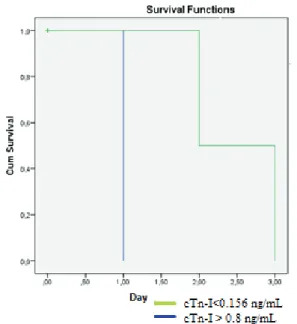
Serum Cardiac Troponin-I in dogs with CPV-2 infection
Idil BASTAN1, Arif KURTDEDE1, Tevhide SEL2 , Doğukan ÖZEN3, Nihat YUMUŞAK4,
M. Özkan TIMURKAN5, Ahmet BAYDIN1
1 Department of Internal Medicine, Faculty of Veterinary Medicine, Ankara University, Ankara, Turkey; 2Department of
Biochemistry, Faculty of Veterinary Medicine, Ankara University, Ankara, Turkey; 3Department of Biostatistics, Faculty of
Veterinary Medicine, Ankara University, Ankara, Turkey; 4Department of Pathology, Faculty of Veterinary Medicine, Harran
University, Şanlıurfa, Turkey; 5Department of Virology, Faculty of Veterinary Medicine, Atatürk University, Erzurum, Turkey.
Summary: The objective of study was to determine the clinical significance of serum cTn-I level in dogs infected with Canine parvovirus-2 (CPV-2). The study material consisted of 4- to 10-week-old puppies of either sexes and 8 different breeds of twenty five dogs with CPV-2. Control group consisted of 4- to 10-week-old puppies of either sexes and 7 different breeds of twenty five dogs. Although treatment, 12 of the dogs with serum cTn-I level > 0.8 ng/mL died within 24 hours of admission. Remaining 13 dogs, had the serum cTn-I level <0.156 ng/mL (cut-off). Of 13 dogs, 4 died within 24-72 hours of admission and 9 were recovered. In terms of cTn-I concentration, whether below 0.156 ng/mL or above 0.8 ng/mL there was a statistically significant difference between survivor and non-survivor animals (p<0.001). Construction of Kaplan-Meier, survival curves revealed dogs with a cTn-I concentration > 0.8 ng/mL had survival time of 1 day, whereas dogs with a cTn-I concentration <0.156 ng/mL had a median survival time of 2.5 days (p<0,001). No statistically significant difference was determined between the mean ages of two groups (dogs with cTn-I concentration <0.156 and > 0.8 ng/mL) (p>0.05). At necropsy, the histopathological examination of the heart revealed the presence of pathological myocarditis findings in 12 of the non-survivor dogs (cTn-I > 0.8 ng/mL), whilst in the other non-survivor 4 dogs (cTn-I<0.156), no pathological alteration was observed in the myocardium. The present study demonstrated that serum cTn-I concentrations > 0.8 ng/mL were indicators of poor prognosis and increased serum cTn-I concentrations were consistent with short survival times in dogs with CPV-2.
Key words: Canine parvovirus, cardiac troponin I, myocarditis.
CPV-2 infeksiyonlu köpeklerde Serum Kardiyak Troponin-I
Özet: Bu çalışmada, CPV-2’li köpeklerde cTn-I konsantrasyonunun klinik öneminin değerlendirilmesi amaçlandı. Çalışmanın materyalini değişik ırk ve her iki cinsiyetten 4-10 haftalık yaşta 25’i hasta ve 25’i sağlıklı olmak üzere 50 yavru köpek oluşturdu. Sağaltım uygulamasının başlatılmasına rağmen serum cTn-I konsantrasyonu > 0.8 ng/mL olan hasta köpeklerin 12’si kliniğe getirildikten sonraki 24 saat içinde öldü. Kliniğe getirildikten sonra 24-72. saatlerde ölen 4 köpek ile sağaltım sonucu iyileşen 9 köpekte cTn-I konsantrasyonları <0.156 ng/mL (cut-off değeri) bulundu. Sağlıklı köpeklerde serum cTn-I konsantrasyonu 0,156 ng/ml değerinin (cut-off) altında saptandı. CTn-I konsantrasyonu karşılaştırıldığında ölen ve iyileşen köpekler arasında önemli farklılık saptandı (p<0.001). Serum cTn-I konsanrasyonu> 0.8 ng/mL olan köpeklerde yaşam zamanı 1 gün iken cTn-I konsantrasyonı <0.156 ng/mL olan köpeklerde ortalama yaşam süresi 2.5 gün (p<0,001) olarak hesaplandı.Serum cTn-I konsantrasyonu açısından değerlendirildiğinde yaş ortalamaları arasında istatiksel olarak önemli bir farklılık bulunmadı (p>0.05). Ölen 12 köpeğin (cTn-I > 0.8 ng/mL) nekropsisinde kalbin histopatolojik incelemesinde myokarditise bağlı patolojik değişiklikler belirlenirken ölen diğer 4 köpekte (cTn-I<0.156) kalp kasında herhangi bir patolojik değişikliğe rastlanmadı. Sonuç olarak bu çalışma serum I konsantrasyonunun 0.8 ng/mL’ nin üstünde olması kötü prognozun belirleyicisi olduğunu ve yüksek serum cTn-I konsantrasyonu ile kısa yaşam süresi arasında ilişki olduğunu gösterdi.
Anahtar sözcükler: Kanin parvovirus, kardiyak troponin- I, miyokarditis
Introduction
In dogs, parvoviral (CPV-2) infection is a fatal disease that is generally associated with acute, fibrinous and haemorrhagic enteritis, and occasionally with non-suppurative myocarditis (6). The disease is observed in 3 forms: 1) Leukopenia, vomiting and diarrhoea in dogs almost 8 weeks of age 2) Mortality in 3- to 8-week-old dogs due to non-suppurative myocarditis and heart
failure 3) Systemic infection characterized by focal necrosis in multiple tissues in puppies younger than 2 weeks of age (9). In dogs, myocarditis caused by parvoviral infection is characterized by peracute heart failure and is observed mostly in puppies aged 3-8 weeks. Puppies that develop myocarditis, display sudden lethargy and die within a very short time period. Those that survive, present with symptoms arising from heart
failure, including tachycardia, weakening of the pulse, cyanosis and dyspnoea. Some of the dogs that develop myocarditis may survive and lead a relatively normal life. However, several months after recovery, some of these animals may present with symptoms related to heart failure (11).
Cardiac specific troponin-I (cTn-I) is a sensitive and specific biomarker that can be used to determine myocardial injury (1, 4, 16). This protein is released from the myocardium as a result of myocardial injury (1,22). Upon cardiac injury, troponins are released from damaged myocytes into the blood circulation and reach a measurable level within 4 hours and a peak level within 12-14 hours. Subsequently, according to the severity of initial cardiac damage, their blood level decreases progressively to an undetectable level within 5-20 days (23). In veterinary medicine cTn-I has been proposed as an indicator of myocardial damage in dogs with a variety of diseases including gastric dilatation volvulus (20), babesiosis (10), erhlichiosis (4), precapillary and postcapillary pulmonary hypertension (7), blunt trauma (21), infaction (18), congestive heart failure (3), dilated cardiomyopathy (DCM) (15) and boxers with arhythmogenic right ventricular cardiomyopathy (2). Some studies demonstrated that there were correlation between high cTn-I level and poor prognosis in dogs with cardiomyocyte injury (5,15,26).
The primary objective of this study was to determine whether CPV-2 infection correlated with myocardial damage assessed by serum cTn-I. Secondary objective was to evaluate whether the survival time of dogs with CPV-2 infection is associated with serum cTn-I levels.
Materials and Methods
Animals and clinical diagnosis: This study was conducted in a total of 50 puppies. Twenty-five were 4- to 10-week-old young animals of either sexes and 8 different breeds, which were referred to the clinics of the Department for Internal Medicine of Ankara University Faculty of Veterinary Medicine with any of the complaints of lethargy, appetence, vomiting or diarrhoea, and were diagnosed with CPV-2 infection based on the examination of blood and rectal smears by conventional PCR between April 2009 and December 2009. The other 25 animals were also 4- to 10-week-old CPV-2 negative puppies of both sexes and 7 different breeds, but did not present with any disease symptom and were confirmed to be healthy upon clinical examination and haematological parameters. These animals had been referred to the clinics for routine vaccination (control group). While the diseased animals were physically examined on a daily basis, in the healthy animals physical examination was performed only once.
Concurrent Babesia spp., Hepatozoon spp and Ehrlichia spp. were excluded by blood smear evaluation.
Dogs were tested for giardia and coccidial oocyst and confirmed to be negative before study. Dogs were excluded if they presented clinical sings of canine distemper (conjunctival or nasal discharge or neurological sings).
The Research and Ethics Committees of the University of Ankara approved the study, and written consent was obtained from all dogs' owners.
Laboratory diagnosis: The diagnosis of CPV was performed with a PCR method described by Ozkul et al. (14). The resulting DNA amplicons were separated on 1.0% agarose gels containing 25 µg ethidium bromide after electrophoresis at 80 V for 30 min. The DNA bands were observed under UV light and records were made and 2245 bp bands as positive.
Laboratory Analyses: Blood samples were collected on the day of admission (0) and on days 1, 2, 3, 5, 7 and 10 in the diseased group and only once in the control group.
The serum samples that had been stored at -70° C until analysis were thawed at room temperature. Measurements were performed using canine cardiac troponin-I ELISA kit (Life Diagnostic, Inc, PA, USA; cat. no. 2010-3-HS) and an ELISA Reader (TECAN) according to manufactures instructions, with values less than 0.156 ng/ml were considered to be cTn-I negative.
Standart treatment: After admission, all dogs were rehydrated over 6 hours with Lactated Ringer’s solution (Eczacıbası-Baxter Ltd) with added dextrose 5% (Eczacıbası-Baxter Ltd), potassium chloride 20 mEq/L (Potassium chloride, %7.5, 10 ml, Drogsan) and Duphalate (Phizer). Volumes of fluids administered intravenous (IV) for estimated dehydration deficit. After rehydration, fluid requirements were maintained based on the clinical assessments.
The antibiotic therapy consisted of IV 22 mg/kg ampicillin (Duocid, 250 mg/Flk, Pfizer) every eight hours. As an antiemetic, 0,5 mg/kg metoclopramide (Metpamid 10mg/ 2ml, Yeni) given as IV bolus or constant rate infusion every 8 hours until vomiting had ceased. Pyrantel pamoate 10 mg/kg, PO (Kontil 250/5 ml, Husnu Arslan Ilaçları AS.) was used to eradicate intestinal parasites.
The day of discharge was assigned as the day when hemogram of dog was normal and the animal was consistently eating and drinking sufficiently to maintain hydration.
The necropsies of the animals that died were performed at the Pathology Department of Ankara University, Faculty of Veterinary Medicine. At necropsy, the heart tissue was harvested and placed into 10% buffered formaldehyde. After being trimmed, the tissues were subjected to routine tissue processing and embedded in paraffin. Five-micron-thick sections were
cut from the blocks, passed through alcohol and xylol series, and stained with haematoxylin-eosin.
Statistical Analysis: The Mann-Whitney U test was used to compare the differences for age between cTn-I concentration >0.8 ng/mL and cTn-I concentration <0.156 ng/mL patients among the non-survivors after checking the data for normality with the Shapiro-Wilk test and the homogeneity of variances with Levene’s test as parametric test assumptions. Survival analysis was performed with Kaplan-Meier method. For all comparisons, p values <0,05 were considered as significant. All statistical analysis were performed by using SPPS®14.1 for Windows.
Results
Anamnesis revealed that, of the 25 dogs that were referred to the clinics of the Department for Internal Medicine of Ankara University, Faculty of Veterinary Medicine and diagnosed with CPV-2 infection, 2 were 4-week-old, 9 were 6-4-week-old, 11 were 8-4-week-old, and 3 were 10-week-old. Two of the dogs were Huskies, 6 were Anatolian Shepherd Dogs, 5 were German Shepherd Dogs, 2 were Terriers, 2 were Pointers, 1 was a Rottweiler, 1 was a Cocker, and 6 were mongrels. Seventeen of the animals were male and 8 were female.
The first examination showed that all of the animals suffered from vomiting, and that the patients were depressed and lethargic with a dull and matted hair coat. It was observed that 15 of the dogs presented with watery faeces (blood was also observed in the faeces of 11) and dehydration. In 3 of the remaining 10 dogs, 24 hours after the first examination, blood was observed in the faeces.
After treatment, 12 of the dogs with cTn-I concentration > 0.8 ng/mL died within 24 hours of admission. In these animals, the mean serum cTn-I level was determined as 42.35±37.2 (ng/mL) (min: 0.8; max: 451.5; median: 1.52). Reaming 13 dogs, had the serum cTn-I level <0.156 ng/mL (cut-off value) 13 dogs, 4 does within 24-72 hours admission, 9 were recovered.
Construction of Kaplan-Meier, survival curves revealed dogs with a cTn-I concentration > 0.8 ng/mL had survival time of 1 day, whereas dogs with a cTn-I concentration <0.156 ng/mL had a median survival time of 2.5 days (p<0,001).
In the healthy dogs, the cTn-I level was determined to be below the cut-off value (0.156 ng/mL) and these animals were also considered to be negative for cTn-I.
The percentages of dogs with a cTn-I concentration >0.8 ng/mL and dogs with a cTn-I concentration <0.156 ng/mL were statistically different between the survivor and non-survivor groups (p<0.001) (Table 1).
Table 1. Kaplan-Meier Survival Curves. Table 1. Kaplan-Meier Yaşam Eğrisi.
In the non-survivor group, the mean age of dogs with cTn-I > 0.8 ng/mL was 7.83±0.39 weeks and the mean age of dogs with cTn-I <0.156 ng/mL was 6.50±0.96 weeks. No statistically significant difference existed for mean age between two groups (p>0.05) (Table 2).
At the heart necropsy of the 12 non-survivor dogs with cTn-I > 0.8 ng/mL, it was observed that, in the cardiac tissue, cells forming muscle bundles had lost their striated appearance in certain foci. In two of the animals, the normal appearance had disappeared completely due to coagulation necrosis (Figure 3). In one of these two animals, calcium deposits were observed in some regions (Figure 1). In 8 of the animals, the nucleus of muscle cells had a granular appearance and lacked chromatin. In some cases, in addition to the findings described above, small haemorrhages and few inflammatory cell infiltrations composed of neutrophil leukocytes, lymphocytes and macrophages were observed, in the cardiac tissue (Figure 2). In three of the 4 non-survivor dogs with cTn-I <0.156 ng/mL, no pathological finding was observed. One of the animals was not able to be necropsied.
Table 2. Descriptive statistics for age of serum cTn-I concentration <0.156 ng/mL and > 0.8 ng/mL patients among the non-survivors.
Tablo 2. Serum cTn-I konsantrasyonu <0.156 ng/mL and > 0.8 ng/mL görünümlü ölen hastalarda yaşa bağlı tanımlayıcı istatistikler N Mean Standard Error Standard Deviation Median Minimum Maximum p
cTn-I > 0.8 ng/mL 12 7,83 0,39 1,34 8,00 6,00 10,00
>0.05
Figure 1. Accumulation of calcium due to necrosis in heart muscle cells. 20, HXE
Şekil 1. Kalp kasında nekroza bağlı kalsiyum çöküntüleri. 20, HXE
Figure 2. Mononuclear cell infiltration between perivascular spaces and heart muscle cell. 20, HXE.
Şekil 2. Kalp kası hücreleri arasında ve perivasculer alanlarda mononüklear hücre infiltrasyonu. 20, HXE.
Figure 3. Degeneration and necrosis in heart muscle cells. 20, HXE.
Şekil 3. Kalp kas hücrelerinde dejenerasyon ve nekroz. 20, HXE. Discussion and Conclusion
Enteritis and myocarditis are two symptoms associated with CPV-2 infection. In recent years, myocarditis has been observed only rarely in CPV-2 infections (8). Myocarditis occurs neonates which are
affected during the period of rapid myocardial cell proliferation (17). Furthermore myocarditis may develop in puppies younger than 8 weeks of age, which are born to non-vaccinated bitches (8). The disease is progressive, and affected animals may be found dead or may die within the first 24 hours after the onset of clinical symptoms (8,19). Mucosal intestinal injures result in haemorrhagic enteritis and Gram-negative bacteraemia. Gram-negative bacteria into peripheral circulation lead to endotoxemia, systemic inflammatory response syndrome (SIRS) and sepsis (24, 13) Mortality was higher in dogs which met the criteria of SIRS on admission (24,25). Myocardial depressant factor has described in SIRS-induced myocarditis in mice and dogs (4). In the present study, all of dogs with serum cTn-I > 0.8 ng/mL which had myocardial damage died within the first 24 hours after admission. Anamnesis revealed that these animals were born to bitches that had not been vaccinated against CPV-2. Viral myocarditis, SIRS or combination can result in myocarditis.
In compliance with previous reports indicating canine CPV-2 infection to be associated with non-suppurative interstitial myocarditis, as well as with the presence of diffuse necrosis and mineralisation in areas of granulomatous myocarditis, and also multifocal fibrosis, and inflammatory cell infiltrations composed of lymphocytes and macrophages (9) in the present study, similar findings were detected in the myocardium of dogs with cTn-I level > 0.8 ng/mL.
Serum cTn-I is an independent factor having an impact on the survival of dogs with DCM and the 0,1 ng/ml increase in cTn-I elevates the risk of death by as much as 8,5 times (15). Low serum cTn-I concentrations are associated with long survival times of dogs, indicating absence of significant active cardiomyocyte death (5). Furthermore, a correlation between increased cTn-I concentrations and shorter survival times has been detected in dogs with acquired cardiac diseases (26). Twelve of the 16 non-survivor dogs having been determined to be serum cTn-I level > 0.8 ng/mL and to display signs of myocarditis at necropsy suggested that damage of the heart muscle played a major role in mortality. This result is in support of the major causes of mortality in dogs infected with CPV-2 is myocarditis. Furthermore, the present study demonstrated that, the prognosis of the disease was poor in CPV-2-infected dogs, which were assessed as serum cTn-I level > 0.8 ng/mL.
In conclusion, serum cTn-l level uses in the confirmation of myocarditis. Measurement of cTn-I have the potential to provide valuable poor prognostic information in dogs with CPV-2. Furthermore, increased serum cTn-I concentrations were consistent with short survival times.
References
1. Babuin L, Jaffe AS (2005): Troponin: the biomarker of
choice for the detection of cardiac injury. Canadian
Medical Association , 173: 1191–1202.
2. Baumwart RD, Orvalho J, Meurs KM (2007).
Evaluation of serum cardiac troponin I concentration in
Boxers with arrhythmogenic right ventricular
cardiomyopathy. Am J Vet Res, 68: 524-8.
3. DeFrancesco TC, Atkins CE, Keene BW, Coats JR, Hauck ML (2002). Prospective clinical evaluation of
serum cardiac troponin T in dogs admitted to a veterinary teaching hospital. J Vet Intern Med, 1: 553–557.
4. Diniz PPVP, Morais HSA, Breitschwerdt EB, Schwartz (2008): Serun Cardiac Troponin I Concentration in Dogs
with Erlichiosis. J Vet Intern Med, 22: 1136-1143.
5. Fonfara S, Loureiro J, Swift S, James R, Cripps P, Dukes-McEwan J (2010): Cardiac troponin I as a marker
for severity and prognosis of cardiac disease in dogs. Vet
J, 184: 334-9.
6. Goddard A, Leisewitz MM (2010). Canine Parvovirus Vet Clin North Am Small Anim Pract. 4: 1041-53. 7. Guglielmini C, Civitella C, Diana A, Di Tommaso M,
Cipone M, Luciani A (2010): Serum Cardiac troponin I
concentration in dogs with precapillary and postcapillary pulmonary hypertension. J Vet Intern Med, 24: 145-52.
8. Hoskins JD (1997): Update on canine parvoviral enteritis. Vet Med, 92, 694–709.
9. Lenghaus C, Studdert MJ (1982): Acute and chronic
viral myocarditis. Acute diffuse nonsuppurative myocarditis and residual myocardial scarring following infection with canine parvovirus. Am J Pathol, 115: 316–319.
10. Lobetti R, Dvir E, Pearson J (2002). Cardiac troponins
in canine babesiosis. J Vet Intern Med, 16:63–68.
11. McCandlish IAP (1999): Specific Infactions of the dog. 921-958. In: Dunn J Textbook Small Animal Medicine. 12. Mellor PJ, Richard JM, Elizabeth AB, Elizabeth JV,
Joy A, Michael E H (2006). High serum troponin I
concentration as a marker of severe myocardial damage in a case of suspected exertional heatstroke in a dog. Journal
or Veterinary Cardiology, 8: 55-62.
13. Nappert G, Duphy E, Ruben D, Mann FA (2002).
Determination of serum organic acids in puppies with naturally acquired parvoviral enteritis. The Canadian
Journal of Veterinary Research, 66: 15-18.
14. Nelson RW (2009) :Small Animal Internal Medicine, fourt edition, Elsevier, China.
15. Noszczyk-Nowak A (2011): NT-pro-BNP and troponin I
as predictors of mortality in dogs with heart failure. Pol J
Vet Sci, 14: 551-6.
16. Porciello F, Rishniw WM, Herndon W, Birettoni F, Antognooni MT, Simpson KW (2008): Cardiac troponin
I is elevated in dogs and cats with azotemia renal failure and in dogs with non-cardiac systemic disease. Australian
Veterinary Journal, 86, 10: 390-394.
17. Prittie J (2004): Canine parvoviral enteritis: a review of
diagnosis, management, and prevention. J Vet Emerg Crit
Care, 14: 167-176
18. Ricchiuti V, Sharkey SW, Murakami MM, Voss EM, Apple FS (1998). Cardiac troponin I and T alterations in
dog hearts with myocardial infarction: correlation with infarct size. Am J Clin Pathol, 110: 241–247.
19. Robinson WF, Huxtable CR, Pass DA, Howell JM (1979). Clinical and electrocardiographic findings in
suspected viral myocarditis of pups. Aust Vet J, 55: 3.
20. Schober KE, Cornand C, Kirbach B, Aupperle H, Oechtering G (2002): Serum cardiac tro-ponin I and
cardiac troponin T concentrations in dogs with gastric dilatation-volvulus. J Am Vet Med Assoc, 221: 381–388.
21. Schober KE, Kirbach B, Oechtering G (1999).
Noninvasive assessment of myocardial cell injury in dogs with suspected cardiac contusion. J Vet Cardiol,1: 17–25.
22. Shih AC, Maisenbacher HW, Barreirinha A, Adin DB, Schmidt MK, Prosek R, Estrada AH (2009): Effect of
routine cardiovascular catheterization in dogs. Journal of
Veterinary Cardiology, 11: 87-92.
23. Spratt DP, Mellanby RJ, Drury N, Archer J (2005):
Cardiac troponin I: evaluation of a biomarker for the diagnosis of heart disease in the dog. J Small Anim Pract,
46: 139-145.
24. Turk J, Fales W, Miller M, Pace L, Fischer J, Johnson G, Kreeger J, Turnquist S, Pittman L, Rottinghaus A (1992). Enteric Clostridium perfringens infection
associated with parvoviral enteritis in dogs: 74 cases (1987-1990). J Am Vet Med Assoc, 200: 991-4.
25. Turk J, Miller M, Brown T, Fales W, Fischer J, Gosser H, Nelson S, Shaw D, Solorzano R (1990). Coliform
septicemia and pulmonary disease associated with canine parvoviral enteritis: 88 cases (1987-1988). J Am Vet Med
Assoc, 196: 771-3.
26. Oyama MA, Sisson DD (2004). Cardiac troponin-I
concentration in dogs with cardiac disease. J Vet Intern
Med, 18: 831-9.
27. Ozkul A, Keles I, Karaoglu T, Çabalar M, Burgu I (2002): Detection and rflp analysis of canine parvovirus
(cpv) dna by polymerase chain reaction (pcr) in a dog.
Turk J Vet Anim Sci, 6: 1201–1203.
Geliş tarihi: 28.01.2013 / Kabul tarihi: 22.04.2013
Address for correspondence:
İdil Baştan, DVM, Ph.D.
Ankara University, Faculty of Veterinary Medicine, Department of Internal Medicine, 06110,
Diskapi, Ankara, Turkey e-mail: idilbastan@yahoo.com