221
http://journals.tubitak.gov.tr/medical/
Turkish Journal of Medical Sciences Turk J Med Sci
(2015) 45: 221-224 © TÜBİTAK
doi:10.3906/sag-1312-38
The effect of extracorporeal shock wave lithotripsy on distribution of interstitial
cells of Cajal in rabbit renal pelvis and proximal ureter
Özlem BOYBEYİ1,*, Mine FEDAKAR ŞENYÜCEL1, Ebru Şebnem AYVA2, Tutku SOYER1, Mustafa Kemal ASLAN1, Mehmet Murad BAŞAR3, Ahmet Murat ÇAKMAK4
1Department of Pediatric Surgery, Faculty of Medicine, Kırıkkale University, Kırıkkale, Turkey 2Department of Pathology, Faculty of Medicine, Başkent University, Ankara, Turkey 3Department of Urology, Faculty of Medicine, Kırıkkale University, Kırıkkale, Turkey 4Department of Pediatric Surgery, Faculty of Medicine, Ankara University, Ankara, Turkey
* Correspondence: [email protected]
1. Introduction
Urolithiasis in the pediatric population is increasing in frequency (1,2). Management in children is very similar to that in adults in recent years. Among several management options, extracorporeal shock wave lithotripsy (ESWL) has become established for the management of stones located in the calyces or the renal pelvis of up to 2 cm in diameter. Although the efficacy of ESWL in children is higher than in adults due to easy passage of stone fragments through shorter and more elastic ureters, it still has some adverse effects (3). Besides renal and extrarenal tissue damages, incomplete fragmentation, residual stone fragments, and obstruction of urinary flow may also be seen after ESWL treatment (4,5). It is reasonable to assume that clearance of stone fragments after ESWL depends on contractility of the ureter, but to the best of our knowledge, the effect of ESWL on the contractility of the ureter has not been demonstrated before.
The control of peristalsis in the ureter is still unclear, but recent studies have demonstrated that there are autonomous pacemaker activities in the upper urinary tract (UUT) (6). After physiological studies demonstrating the pacemaker cells called interstitial cells of Cajal (ICC) in the gut, several studies were performed to investigate the pacemaker cell distribution in the UUT (6). The pacemaker cells in the UUT have been shown to be positively stained with c-kit (CD117) and negatively stained with mast cell tryptase, the features of Cajal-like cells (6). The relation between ESWL and ICC distribution in the UUT has not been studied before, so an experimental study was performed to evaluate the effect of ESWL on the distribution of ICCs in rabbit renal pelvis and proximal ureter.
2. Materials and methods
Six New Zealand rabbits weighing 2500–3000 g were included in the study. After being fasted overnight, Background/aim: An experimental study was performed to evaluate the effect of extracorporeal shock wave lithotripsy (ESWL) on the
distribution of interstitial cells of Cajal (ICC) in rabbit renal pelvis and proximal ureter.
Materials and methods: Six New Zealand rabbits were included. Right kidneys were exposed to a total of 3000 shock waves (14 kV)
by using an electrohydraulic-type ESWL device. Right sides were allocated as the ESWL group (EG, n = 6) and left sides as the control group (CG, n = 6). Tissues were harvested on day 7. Tissues were examined histopathologically for the presence of edema, inflammation, congestion, hemorrhage, fibrosis, and vascularization. Mast cell tryptase and CD117 (c-kit) staining was performed for ICC distribution.
Results: Although increased tissue edema in renal pelvises and increased inflammation in ureters were observed in EG, no statistical
difference was detected between groups (P > 0.05). In CG, positive CD117 staining was detected in 2 renal pelvises and ureters. None of the EG samples showed CD117 staining and no statistical difference was detected between groups (P > 0.05).
Conclusion: Rabbit does not appear to be a good model for investigating ICCs. ESWL may cause histopathological alterations in the
renal pelvis and ureter. Since it has not been statistically proven, reduced contractility of the ureter after ESWL may not be attributed to altered distribution of ICCs in the renal pelvis and ureter.
Key words: Extracorporeal shock wave lithotripsy, proximal ureter, renal pelvis, interstitial cells of Cajal
Received: 06.12.2013 Accepted: 20.02.2014 Published Online: 12.01.2015 Printed: 09.02.2015 Research Article
222
BOYBEYİ et al. / Turk J Med Sci rabbits were anesthetized with intramuscular ketamine
hydrochloride (50 mg/kg Ketalar, Eczacıbaşı, Turkey). Right kidneys were exposed to 3 sessions of ESWL with 1000 shock waves at 14 kV (total of 3000 shock waves) by using an electrohydraulic-type third-generation Stonelith V5 ESWL device (PCK, Turkey). Each session of ESWL was performed for 20 min (rate: 50 shock waves/min) and 48 h after the previous treatment. The ESWL was performed by the same investigator each time. Ultrasonographic probing was performed to localize the right kidney. Since the focus distance of the ESWL device is 130 mm, an elevating shelter was put under the subjects. During the treatment period the rabbits were kept warm and in the same conditions with standard feeding.
Right side ureters and renal pelvises were allocated as the ESWL group (EG, n = 6), whereas left sides were allocated as the control group (CG, n = 6). Both pelvises and proximal ureters were harvested on the seventh day after ESWL. Tissues were examined histopathologically for presence of edema, inflammation, congestion, hemorrhage, fibrosis, and vascularization. Mast cell tryptase and CD117 (c-kit) staining was performed for evaluation of ICC distribution in samples.
The data obtained from the experiments were analyzed with the Mann–Whitney U test and median test since the variables were nonparametric (not normally distributed) as confirmed by ANOVA test (SPSS 15.0). P-values of lower than 0.05 were considered significant. The experiments were performed in adherence to the Declaration of Helsinki and by approval of the Local Ethics Committee of Kırıkkale University (2011/11). This study was supported by the Kırıkkale University Scientific Research Council (2011/59).
2.1. Histopathological evaluations
The preserved tissues were fixed with 10% formalin. All segments were embedded in paraffin blocks after tissue processing. Tissues were sectioned in pieces of 4–5 µm and stained with routine hematoxylin and eosin staining. The specimens were examined under a light microscope (Olympus CX31; Mason Technology, Ireland) by the same pathologist blinded to the study.
For both the EG and the CG, presences of edema, inflammation, congestion, hemorrhage, fibrosis, and vascularization were evaluated. Histopathologic findings were graded semiquantitatively for each parameter separately as follows: grade 0, normal; grade 1; mild, grade 2; moderate, and grade 3; severe.
2.2. Immunohistochemistry
Sections of 5 µm were prepared from each paraffin block. Each section was deparaffinized in xylene and rehydrated in graded serial ethanol baths, and was then placed in 3% H2O2 for 5 min to block endogenous peroxidase activity. Antigen retrieval was performed using a citrate buffer
solution (pH 6) for CD117 and mast cell tryptase antibodies. After 3 rinses of 5 min each in phosphate-buffered saline, sections were incubated for 1 h with primary antibodies in an automatized immunohistochemical staining machine (Leica Bond Max, Leica Biosystems, Newcastle Ltd., UK). Anti-CD117 (CD117-R-7-CE, Novocastra, Leica Biosystems) and antimast cell tryptase (PA0019, Novocastra, Leica Biosystems) were used as primary antibodies. A Novocastra Bond Polymer Refine Detection system including peroxide block, postprimary, polymer reagent, DAB chromogen, and hematoxylin counterstain was used to detect positive staining. Phosphate-buffered saline was used as the negative control.
2.3. Determination of ICC distribution
Cytoplasmic immunostaining for CD117 and mast cell tryptase were considered as positive immunoreactivity. Since double-staining could not be performed, immunohistochemically stained sections were evaluated separately. First, positive staining of mast cells with mast cell tryptase was evaluated. Cells with polygonal-shaped cytoplasm having rough granules that were stained positive with CD117 at the same location were considered as mast cells in the immunohistochemical examination. The other cells that were negative for mast cell tryptase, positive for CD117, and had spindle-shaped cytoplasm were estimated as ICCs. The ICCs were counted manually in renal pelvis and proximal ureter sections under light microscope.
3. Results
Histopathologically, tissue edema was increased in renal pelvises and inflammation was increased in ureters in the EG compared to the CG. However, no statistical difference was detected between exposed and unexposed samples (P > 0.05) (Table). There was no statistical difference between groups regarding other parameters including congestion, hemorrhage, fibrosis, and vascularization (P > 0.05).
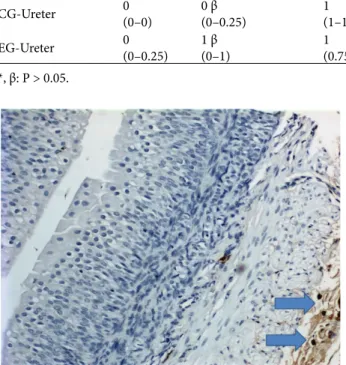
CD117 staining was detected in the renal pelvis (n = 2) and ureter (n = 2) of the CG. These cells were considered as Cajal cells since they were able to be distinguished from mast cells with mast cell tryptase staining (Figures 1 and 2). However, none of the samples obtained from the EG showed CD117 staining. There was no statistical difference between groups regarding CD117-positive cells (P > 0.05) (Figure 3).
4. Discussion
The treatment of choice for UUT stones has been ESWL since the 1980s, when it was first introduced (1,7). The first report regarding ESWL in children revealed that the complications and safety were similar to those in adults (7). Further studies were performed demonstrating the effectiveness of ESWL in children, and then it became a treatment of choice for the stones located in calyces or renal pelvises with a diameter of up to 2 cm (1,2).
223 BOYBEYİ et al. / Turk J Med Sci
The effectiveness of ESWL is measured by the stone-free rate, which has been reported from 50% to 95% in children (1–3,8). Some cases require more than 1 session of ESWL (up to 3 sessions) for a better stone-free rate (1,2,8,9). Each session consists of 1800 to 2000 shock waves (and up to 4000) between 14 and 21 kV (2,3). The usage of ultrasound for localization will provide less radiation exposure. In the present study, we performed ESWL on rabbits with a similar dosage in 3 sessions, just as reported in the literature (8–10).
Although ESWL is an efficient treatment modality in the management of urolithiasis in children, it has some adverse effects (3–5). Some of these are renal and extrarenal
tissue damages, skin bruising, hematuria, transient tubular function deterioration, incomplete fragmentation, residual stone fragments, and obstruction of urinary flow (2–5). Besides the several clinical reports in humans about complications of ESWL, some studies in animals have also reported on the acute and chronic effects of ESWL (4,5,11). Nevertheless, contractility, which has an effect on stone clearance, has not been demonstrated before. There is one study revealing that ureteric contraction frequency was increased by in vitro prostaglandin stimulation as detected in ESWL-treated patients (12). Some of the adverse effects of ESWL on urinary flow are thought to be transient and would resolve several days after ESWL (2–5). We detected alteration in the distribution of ICCs in the EG in the present study. Although there was no statistical difference between groups, this decrease in ICC distribution in the EG might be an indicator of altered ureteric peristalsis, which could be transient or permanent.
Ureteral peristalsis is important in propelling the urine downwards and also stone fragments after ESWL treatment. However, the exact mechanism of Table. The median values of the histopathological grades (interquartile ranges within parentheses).
Groups Edema Inflammation Congestion Hemorrhage Fibrosis Vascularization
CG-Renal pelvis 0 *(0–1) 1(0.75–1) 1(1–1) 0(0–0.25) 0(0–0) 0(0–0)
EG-Renal pelvis 1 *(0–1.5) 1(0.75–3) 1(1–1) 0(0–0.25) 0(0–0.25) 0(0–0)
CG-Ureter 0(0–0) 0 β(0–0.25) 1(1–1) 0(0–0.25) 0(0–0) 0(0–0)
EG-Ureter 0(0–0.25) 1 β(0–1) 1(0.75–1) 0(0–0) 0(0–0) 0(0–0)
*, β: P > 0.05.
Figure 1. Mast cell tryptase expression on samples (DAB, 20×).
Figure 2. CD117-positive, spindle-shaped cells that are
considered Cajal-like cells within the muscular layer of the renal pelvis and proximal ureter in the CG (DAB, 40×).
2.5 N= 6 6 6 6 CONTROL IC C nu mb er ESWL CAJAL-P CAJAL-U 2.0 1.5 1.0 –5 .5 0.0
Figure 3. The comparison of ICC distribution in the pelvis and
224
BOYBEYİ et al. / Turk J Med Sci this propelling movement and the factors deteriorating
it are not clear (6,13,14). This peristalsis occurs even after surgical denervation of ureter or isolation of the pyeloureteric system, indicating autonomous pacemaker activity (6,13,14). Some pacemaker potentials were able to be shown by electrophysiological studies in the renal pelvis in animal studies (6,14). After recognition of ICCs in the gut (15,16), several studies were performed to investigate the presence and distribution of pacemaker cells in the UUT. Some spindle-shaped c-kit-positive cells were detected in the UUT. They were differentiated from other c-kit-positive cells, such as mast cells, by additional staining with mast cell markers, and were called Cajal-like cells (6,13,14). These cells are usually found within smooth muscle layers of the UUT. The number of them is highest in the proximal UUT and decreases gradually in distal parts of ureter. However, the distribution of these cells differs between species. They are found less frequently in pig, for example. On the other hand, differentiation of them requires species-specific antibodies (6,13,14). Therefore, further studies are needed to examine the long-term effects of ESWL on ureteric peristalsis.
In the present study, we could detect only a few Cajal-like cells. Difficulty in double immunohistochemical
staining of ICCs in rabbit tissue was also experienced in previous studies (6). We could detect ICCs in only 2 pelvis and 2 ureter samples in the CG. Although there was no statistical difference between groups, we could not detect any ICCs in the EG. This result may be obtained not only due to the deleterious effect of shock waves to the ICCs and ureteric peristalsis, but also due to the difficulty in staining with rabbit-specific antibodies against ICCs. Therefore, future studies are needed to prove the adverse effect of ESWL on ICC distribution in the UUT with different animal models.
In conclusion, rabbit does not appear a good model for investigating ICCs. ESWL may cause histopathological alterations in the renal pelvis and ureter. Since it has not been statistically proven, the reduced contractility of the ureter after ESWL may not be attributed to altered distribution of ICCs in renal pelvis and ureter.
Acknowledgments
This study was supported by the Kırıkkale University Scientific Research Council (2011/59). It was presented at the 24th Congress of the European Society of Pediatric Urology, Genoa, Italy, in 2013.
References
1. Minevich E. Management of ureteric stone in pediatric patients. Indian J Urol 2010; 26: 564–567.
2. Gnessin E, Chertin L, Chertin B. Current management of paediatric urolithiasis. Pediatr Surg Int 2010; 28: 659–665. 3. Badawy AA, Saleem MD, Abolyosr A, Aldahshoury M,
Elbadry MS, Abdalla MA, Abuzeid AM. Extracorporeal shock wave lithotripsy as first line treatment for urinary tract stones in children: outcome of 500 cases. Int Urol Nephrol 2012; 44: 661–666.
4. Skolarikos A, Alivizatos G, Rosette J. Extracorporeal shock-wave lithotripsy 25 years later: complications and their prevention. Eur Urol 2006; 50: 981–990.
5. Kelley JM. Extracorporeal shock wave lithotripsy of urinary calculi. Theory, efficacy and adverse effects. West J Med 1990; 153: 65–69.
6. Metzger R, Schuster T, Till H, Franke FE, Dietz HG. Cajal-like cells in the upper urinary tract: comparative study in various species. Pediatr Surg Int 2005; 21: 169–174.
7. Newman DM, Coury T, Lingeman JE, Mertz JH, Mosbaugh PG, Steele RE, Knapp PM. Extracorporeal shock wave lithotripsy experience in children. J Urol 1986; 136: 238–240.
8. McAdams S, Shulka AR. Pediatric extracorporeal shock wave lithotripsy: predicting successful outcomes. Indian J Urol 2010; 26: 544–548.
9. Ghalayini IF, Al-Ghazo MA, Khader YS. Evaluation of emergency extracorporeal shock wave lithotripsy for obstructing ureteral stones. Int Braz J Urol, Clin Urol 2008; 34: 433–442.
10. Gillitzer R, Neisius A, Wöllner J, Hampel C, Brenner W, Bonilla AA, Thüroff J. Low-frequency extracorporeal shock wave lithotripsy improves renal pelvic stone disintegration in a pig model. BJU Int 2009; 103: 1284–1288.
11. Karalezli G, Gögüs O, Bedük Y, Köküuslu C, Sarica K, Kutsal O. Histopathologic effects of extracorporeal shock wave lithotripsy on rabbit kidney. Urol Res 1993; 21: 67–70. 12. Horgan PG, Hanley D, Burke J, Couse NF, Fitzpatrick JM.
Extracorporeal shock wave lithotripsy induces the release of prostaglandins which increase ureteric peristalsis. Br J Urol 1993; 71: 648–652.
13. Lang RJ, Tonta MA, Zoltkowski BZ, Meeker WF, Wendt I, Parkington HC. Pyeloureteric peristalsis: role of atypical smooth muscle cells and interstitial cells of Cajal-like cells as pacemakers. J Physiol 2006; 576: 695–705.
14. Lang RJ, Klemm MF. Interstitial cells of Cajal-like cells in the upper urinary tract. J Cell Mol Med 2005; 9: 543–556. 15. Won K, Sanders KM, Ward SM. Interstitial cells of Cajal
mediate mechanosensitive responses in the stomach. P Natl Acad Sci USA 2005; 102: 14913–14918.
16. Soyer T, Ayva Ş, Somuncu S, Atasoy P, Kanmaz T, Çakmak M. Caustic esophageal injury decreases the number of interstitial cells of Cajal in the rat esophagus. Turk J Med Sci 2010; 40: 599–604.