Case Report
95 ©Copyright 2018 by Turkish Ophthalmological Association
Turkish Journal of Ophthalmology, published by Galenos Publishing House.
Introduction
Stickler syndrome is an autosomal-dominant hereditary clinical condition caused by structural abnormalities in collagen types 2, 9, and 11. It can manifest with widely varying clinical signs affecting the ocular, orofacial, musculoskeletal, and/or auditory systems.1,2,3,4 Ophthalmologic complications of the
syndrome are progressive and can lead to blindness.5 In this
report, we present a case of bilateral asymmetric rhegmatogenous retinal detachment associated with Stickler syndrome and the outcomes of treatment.
Case Report
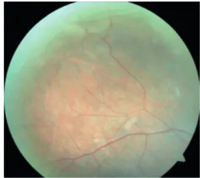
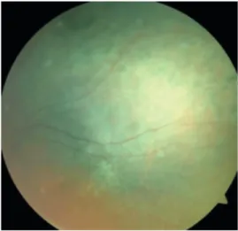
A 17-year-old female patient presented to our clinic with complaints of low vision in both eyes for approximately 1 year. On ophthalmologic examination, her best corrected
visual acuity (BCVA) was 0.1 in the right eye and 0.05 in the left eye. Bilateral subcapsular cataract was detected in anterior segment examination. Fundus examination revealed bilateral rhegmatogenous retinal detachment (Figures 1 and 2), membranous vitreous in the periphery, and chronic detachment with multiple holes and tears. There was proliferative vitreoretinopathy (PVR) grade C2 in the left eye. The patient exhibited the craniofacial structural features of Stickler syndrome (she did not consent to en face photography). Scleral buckling was performed in both eyes as an initial intervention and resulted in postoperative retinal attachment in the right eye (Figures 3, 4, 5). However, the procedure was insufficient for the left eye having PVR C2 (Figure 6). Phacoemulsification, intraocular lens implantation, and pars plana vitrectomy with silicone injection were performed in the left eye. Postoperatively the retina was reattached (Figure 7). Silicone extraction was done 6 months
Abstract
Address for Correspondence: İmren Akkoyun MD, Başkent University Faculty of Medicine, Department of Ophthalmology, Ankara, Turkey
Phone: +90 537 613 38 48 E-mail: [email protected] ORCID-ID: orcid.org/0000-0002-2860-7424
Received: 19.09.2017 Accepted: 29.11.2017
*Başkent University Faculty of Medicine, Department of Ophthalmology, Ankara, Turkey **Çankırı State Hospital, Ophthalmology Clinic, Ankara, Turkey
Bilateral Asymmetric Rhegmatogenous Retinal
Detachment in a Patient with Stickler Syndrome
DOI: 10.4274/tjo.60430 Turk J Ophthalmol 2018;48:95-98
Caner Öztürk*, Almila Sarıgül Sezenöz**, Gürsel Yılmaz*, İmren Akkoyun*
Here we present the long-term anatomical and visual outcomes of bilateral asymmetric rhegmatogenous retinal detachment repair in a patient with Stickler syndrome. A 17-year-old girl presented with decreased visual acuity in both eyes for more than one year. Her best-corrected visual acuity (BCVA) was 0.1 in the right eye and 0.05 in the left eye. Slit-lamp anterior segment examination revealed subcapsular cataract in both eyes. Fundus examination showed bilateral rhegmatogenous retinal detachment, chronic retinal detachment accompanied by multiple retinal holes, tears and membranous fibrillary vitreous in the peripheral retina. Grade C2 proliferative vitreoretinopathy was observed in the left eye. Scleral buckling surgery was performed initially for both eyes. After the primary surgical procedure, retinal reattachment was achieved in the right eye and the left eye underwent phacoemulsification, intraocular lens implantation, pars plana vitrectomy (PPV), and silicone oil injection. After these surgical procedures retinal reattachment was achieved in the left eye. Silicone oil removal was performed six months after PPV surgery. After surgical treatment, BCVA was 0.6 in the right eye at the end of the 3.5-year follow-up period. After silicone oil removal, BCVA reached 0.2 in the left eye after 36 months of follow-up and retinal reattachment was achieved in both eyes. Scleral buckling surgery and PPV are effective and confidential methods for the treatment of chronic retinal detachment cases in Stickler syndrome.
Turk J Ophthalmol 48; 2: 2018
96
after pars plana vitrectomy. All procedures were performed by the same surgeon (I.A.).
In postoperative follow-up at about 3.5 years, the right eye had a BCVA of 0.6 and the retina was further attached (Figures 8, 9). In the left eye, the retina was attached and BCVA was 0.2 at 36-month follow-up after silicone extraction (Figures 10, 11, 12).
Discussion
Stickler syndrome is a disease spectrum that has been investigated since Stickler et al.6 first described it in 1965. Though
rare, it can involve serious complications. Ophthalmologic
Figure 3. Right retinal appearance after scleral buckling in fundus photograph Figure 2. Preoperative appearance of the left retina in fundus photograph Figure 1. Preoperative appearance of the right retina in fundus photograph
Figure 7. Optical cohorence tomography image of left eye after scleral buckling,
pars plana vitrectomy, and silicone injection
Figure 6. Optical cohorence tomography image of left eye after scleral buckling Figure 5. Optical cohorence tomography image of right eye after scleral buckling Figure 4. Right peripheral retinal appearance after scleral buckling in fundus
97
complications of the disease include high myopia, open-angle glaucoma due to dysgenesis at the drainage angle of the anterior chamber, cortical and subcapsular cataract, perivascular retinal lattice degeneration, and adhesive vitreous bands. These vitreoretinal changes cause giant retinal tears and subsequent rhegmatogenous retinal detachment.7,8 Although it is a rare
condition, Stickler syndrome is the most common hereditary cause of rhegmatogenous retinal detachment.9 In a study from
Turkey by Yararcan et al.10 including 6 individuals with Stickler
syndrome in a single family, all of the patients had myopia of 10 diopters or greater, early onset cataract, chorioretinal atrophy, and vitreous liquefaction. Three of the 6 patients in that study had total retinal detachment, one had glaucoma, and one had phthisis bulbi.10 Abeysiri et al.11 presented the 17-year
follow-up results of 30 eyes of 23 patients. In their study, the overall re-attachment rate was 78.4%, with rates of 67% for scleral buckling and 82.4% for pars plana vitrectomy. BCVA improved by 0.33 logarithm of minimum angle of resolution (logMAR) in patients that underwent scleral buckling and 0.32 logMAR in those that underwent vitrectomy.11 In a study by Reddy et al.12
including 16 eyes of 13 patients, the average age of patients with retinal detachment was 10.4 years and patients were followed for 94 months. Scleral buckling was performed in 5 patients (31%), pars plana vitrectomy in 7 patients (44%) and combined surgery in 4 patients (25%). The patients underwent a mean of
Öztürk et al, Bilateral Retinal Detachment in Stickler Syndrome
Figure 12. Optical cohorence tomography image of left eye 36 months after
silicone extraction
Figure 11. Fundus photograph showing appearance of peripheral retina of the left
eye at 36 months after silicone extraction
Figure 10. Fundus photograph showing left retinal appearance at 36 months
after silicone extraction
Figure 9. Fundus photograph showing appearance of the peripheral retinal of the
right eye 3.5 years after scleral buckling
Figure 8. Fundus photograph showing right retinal appearance 3.5 years after
Turk J Ophthalmol 48; 2: 2018
98
3.1 surgical interventions. In long-term follow-up, the retinal re-attachment rate was 100%, but 12 eyes (75%) developed PPV. The patient discussed in the present case had bilateral subcapsular cataract. Her fundus findings were consistent with those described in Stickler syndrome: fibrillary vitreous, lattice degenerations and adhesive vitreous bands, multiple retinal tears and holes, and PPV. The long-term outcomes of treatment were retinal re-attachment with BCVA of 0.6 in the right eye and 0.2 in the left eye. Scleral buckling and pars plana vitrectomy may be an effective and reliable treatment option for severe chronic retinal detachment due to Stickler syndrome.
Ethics
Informed Consent: It was taken.
Peer-review: Externally and internally peer-reviewed. Authorship Contributions
Surgical and Medical Practices: İmren Akkoyun, Concept: İmren Akkoyun, Design: İmren Akkoyun, Data Collection or Processing: Caner Öztürk, Almila Sarıgül Sezenöz, Gürsel Yılmaz, İmren Akkoyun, Analysis or Interpretation: Caner Öztürk, Almila Sarıgül Sezenöz, Gürsel Yılmaz, İmren Akkoyun, Literature Search: Caner Öztürk, Almila Sarıgül Sezenöz, Gürsel Yılmaz, İmren Akkoyun, Writing: Caner Öztürk, Almila Sarıgül Sezenöz, İmren Akkoyun.
Conflict of Interest: No conflict of interest was declared by
the authors.
Financial Disclosure: The authors declared that this study
received no financial support.
References
1. Snead MP, Yates JR. Clinical and molecular genetics of Stickler syndrome. J Med Genet. 1999;36:353-359.
2. Stickler GB, Hughes W, Houchin P. Clinical features of hereditary progressive arthro-ophthalmopathy (Stickler syndrome): a survey. Genet Med. 2001;3:192-196.
3. Snead MP, McNinch AM, Poulson AV, Bearcroft P, Silverman B, Gomersall P, Parfect V, Richards AJ. Stickler syndrome, ocular-only variants and a key diagnostic role for the ophthalmologist. Eye (Lond). 2011;25:1389-1400. 4. Robin NH, Moran RT. Ala-KokkoLStickler Syndrome Synonym: Arthro
ophthalmopathy; In: Pagon RA, Adam MP, Ardinger HH, eds. Gene Reviews. 2000:1993-2015.
5. Antunes RB, Alonso N, Paula RG. Importance of early diagnosis of Stickler syndrome in newborns. J Plast Reconstr Aesthet Surg. 2012;65:1029-1034. 6. Stickler GB, Belau PG, Farrell FJ, Jones JD, Pugh DG, Steinberg AG,
Ward LE. Hereditary progressive arthro-ophthalmopathy. Mayo Clin Proc. 1965;40:433-455.
7. Parma ES, Korkko J, Hagler WS, Ala-Kokko L. Radial perivascular retinal degeneration: a key to the clinical diagnosis of an ocularvariant of stickler syndrome with minimal orthosystemic manifestations. Am J Ophthalmol. 2002;134:728-734.
8. Shuler M, Sullivan J, Hurley B. McNamara Vitreoretinal dystrophies. J Pediatric Retina. 2011:315-319.
9. Richards AJ, Scott JD, Snead MP. Molecular genetics of rhegmatogenous retinal detachment. Eye (Lond). 2002;16:388-392.
10. Yararcan M, Boz U, Derebaşı H, Akın I, Ulkucu N, Cakmaklı Z. Stickler Sendromlu Olgularımız. Ret-Vit. 1998;6:140-147.
11. Abeysiri P, Bunce C, da Cruz L. Outcomes of surgery for retinal detachment in patients with Stickler syndrome: a comparison of two sequential 20-year cohorts. Graefes Arch Clin Exp Ophthalmol. 2007;245:1633-1638. 12. Reddy DN, Yonekawa Y, Thomas BJ, Nudleman ED, Williams GA.
Long-term surgical outcomes of retinal detachment in patients with Stickler syndrome. Clin Ophthalmol. 2016;16:1531-1534.