DAHİLİ BİLİMLER / MEDICAL SCIENCES
Olgu Bildirisi / Case Report
161
Ankara Üniversitesi Tıp Fakültesi Mecmuası 2006; 59:161-163
Ankara Numune Training and Research Hospital, Ankara
Corresponding author
Ankara Numune Training and Research Hospital, Department of Radiology, Talatpaşa Bulvarı, Sıhhıye/Ankara
Phone : +90 (312) 508 44 49 E-mail adress : [email protected]
Received: 31.08.2006 • Accepted: 19. 10.2006
B
rain tumors and intracranial aneurysms rarely coexist. Until now, 180 cases have been reported (1-3), accounting for an incidence rate of nearly 0.3-4 % (1). The coexistence of meningioma and intracranial aneurysms is even rarer with only 94 cases reported in the literature (1-3).Meningioma has been the most common intracranial tumor along with an-eurysm (1). While the anan-eurysm of other arteries along with meningioma is relatively more common, only 3 cases with basilar artery accompanied by men-ingioma have been reported (4). A case of coexisting basilar artery aneurysm and multiple meningiomas has been reported in the light of the relevant literature.
Case report
A 52-year-old male patient presented with headache of 5 months and vertigo attacks. The history was non-specific. He had no family history. The results of neurological examination were normal. No hypertension was detected.
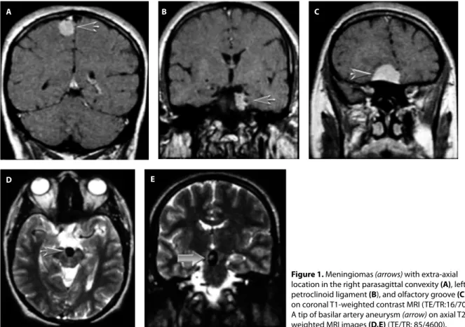
The patient underwent magnetic resonance imaging (MRI). The evaluation revealed meningioma like lesions with dural tails located in the right parasagittal convexity, left petroclinoid ligament, and the olfactory groove. The extraaxi-ally located lesions were isotense with the gray matter on T1-weighted images and slightly hyperintense on T2-weighted images and showed homogenous contrast enhancement after Gd-DTPA injection (Figure 1 a,b,c). A peritumoral edema was detected in the brain parenchyma around the lesion in the olfactory groove.
MRI also showed aneurysm in the basilary tip (Figure 1 d,e). A solitary rysm was detected (with cerebral angiography). It was decided to treat to aneu-rysm via the endovascular route using aproximal occlusion technique.The lesion in the olfactory groove was operated and pathological evaluation confirmed the diagnosis of meningioma.
Brain tumors and intracranial aneurysms rarely coexist. A case of association between multiple intracranial meningiomas and cerebral artery aneurysm is reported.
Key words: Cerebral aneurysm, meningioma, magnetic resonance
Beyin tümörlerine eşlik eden intrakranial anevrizmalar oldukça nadirdir. Çok sayıda menenjioma eşlik eden, baziler arter anevrizması bulunan 52 yaşındaki erkek olgu sunulmaktadır.
Anahtar sözcükler: Serebral anevrizma,menenjiom, manyetik rezonans
Multiple intracranial meningiomas and
accompanying basilar artery aneurysm
Multipl intrakraniyal menenjioma eşlik eden baziler arter anevrizması
Ankara Üniversitesi Tıp Fakültesi Mecmuası 2006; 59(4)
162 Multiple intracranial meningiomas and accompanying basilar artery aneurysm
Discussion
The coexistence of meningioma and intracranial aneu-rysms is a rare occurrence. Only 94 cases have been re-ported up to date (1-3). Three of these cases were rere-ported to be aneurysm in the basilar artery (4).
The aneurysm incidence for the whole population is 0.5 %, while it is 0.3-0.7 % among brain tumor patients. Basi-lar artery aneurysms are rare. About 10% of all intracranial aneurysms arise on the vertebrobasilar circulation. Five per cent arise from the basilary artery bifurcation, and the re-maining 1% to 5% arise from other posterior fossa vessels (1). In one study, aneurysms were detected at a higher rate among the brain tumor patients than in the normal popula-tion (5). However also, another study reported much higher aneurysm incidence in brain tumor patients (4%), but since not all brain tumor patients could undergo four-vessel angi-ography, the actual incidence rate for this group of patients was considered to be even higher than the reported (1).
The advent of MR and CT angiography as well as CT scanning has raised expectations that aneurysm screening may soon become feasible. These technologies can provide detailed noninvasive images of the major cerebral arter-ies and thus avoid the use of the more expensive invasive intra-arterial DS angiography. Several recent studies have shown that MRA reliably detects aneurysms greater than
3 mm in diameter, an important consideration, as risk of rupture may correlate with aneurysm size. Although a neg-ative MRA at the time of screening does not guarantee that an aneurysm is not present or will not form, the likelihood of these events is remote. Accepting these caveats, one may conclude that MRA may be a reliable screen for clinically significant cerebral aneurysm (6-8).
Literature review revealed that in order of incidence, the brain tumors accompanying aneurysm were meningi-oma (29.3%), glimeningi-oma (27.5%), and hypophysis adenmeningi-oma (20.6%) (4). In another study, the rate of meningioma was relatively much higher (44%) (5).
Meningiomas are common tumors (1). The menin-gioma associated symptoms observed in this coexistence are no different than those of the other meningioma cases. Furthermore, the locations preferred by meningiomas are similar (1).In all of the series, incidence rate of ipsilateral aneurysms and meningioma coexistence has been found to be higher (1). Accompanying aneurysm has been encoun-tered in the internal (ICA), middle (MCA) and anterior (ACA) cerebral artery in order of incidence and relatively fewer in more than one artery (4). Some authors have re-ported marked incidence rates in MCA for convexity men-ingiomas and in ICA and anterior communicating artery for basal meningiomas (1).
Figure 1. Meningiomas (arrows) with extra-axial
location in the right parasagittal convexity (A), left petroclinoid ligament (B), and olfactory groove (C) on coronal T1-weighted contrast MRI (TE/TR:16/700). A tip of basilar artery aneurysm (arrow) on axial T2-weighted MRI images (D,E) (TE/TR: 85/4600).
A B C
Journal of Ankara University Faculty of Medicine 2006; 59(4)
163
U. Toprak
In two of the three cases with meningioma accompanied with basilar artery aneurysm, aneurysms were detected in posterior communicating artery, MCA, and other arteries. However, none of these cases had meningiomas located in the basilar artery region (4).
These tumors with high vascularity may increase the regional blood flow, which supports the dominance of ip-silateral aneurysms. However, numerous authors interpret this finding as merely coincidental (1). The authors de-scribe a previously published case report of a patient with an enlarging ACA aneurysm after the posterior fossa men-ingioma removal (9).
None of the meningiomas in our case was located in the area supplied by the basilar artery, supporting previous
claims that multiple meningiomas do not result from hy-pervascularity. Ipsilateral location is coincidental; no sec-ondary etiology for aneurysm was detected.
Other authors have claimed that meningiomas and an-eurysms may be affected by hormonal factors and empha-sized that surgical trauma to the arterial wall or invasion of the artery by the tumor and genetic factors might lead to aneurysms (1).
A probability of incidental aneurysm in the same loca-tion or a distant region in a case with brain tumor should be kept in mind and radiological evaluation (CT and/or MRI) should be performed with great scrutiny with re-spect to aneurysms.
References
1. Scamoni C, Dorizzi A, Dario A, et al. Intracranial meningioma associated with cerebral artery aneurysm. Report of two cases and review of the literature. J Neurosurg Sci 1997;41:273-281. 2. Javadpour M, Khan AD, Jenkinson MD, et al. Cerebral aneurysm
associated with an intracranial tumour: staged endovascular and surgical treatment in two cases. Br J Neurosurg 2004;18:280-284. 3. Lama M, Mottolese C. Middle meningeal artery aneurysm
associated with meningioma. J Neurosurg Sci 2000;44:39-41. 4. Pia HW, Obrador S, Martin JG. Association of brain tumours and
arterial intracranial aneurysms. Acta Neurochir 1972;27:189-204. 5. Licata C, Pasqualin A, Freschini A, et al. Management of associated
primary cerebral neoplasms and vascular malformations. Acta Neurochir 1986;82:28-38.
6. Lindley JG, Wirth FP: Epidemiology of cerebral aneurysms. In Ratcheson RA, Wirth FP (eds): Ruptured Cerebral Aneurysms: Perioperative Management. Baltimore, Williams & Wilkins, 1994, p15.
7. Maeder PP, Meull RA, de Tribolet N: Th ree-dimensional volume rendering for maganetic resonance angiography in the screening and preoperative workup of intracranial aneurysms. J Neurosurg 1996:85;1050-1055.
8. Jager HR, Ellamushi H, Moore EA, et al. Contrast-enhanced MR angiography of intracranial giant aneurysms. AJNR Am J Neuroradiol 2000;21:1900-1907.
9. Prandini MN, Lacanna SN, Tella OI, et al. Aneurysm growth after brain tumor removal: case report. Arq Neuropsiquiatr 2004;62:722-724.