Synchronous lung cancer cases
Senkron akciğer kanseri olgularıArif Osman Tokat
1, Ulaş Kumbasar
2, Adem Güngör
21 Ministry of Health, Ankara Training and Research
Hospital, Ankara
2 Department of Thoracic Surgery, Ankara University
School of Medicine, İbn-i Sina Hospital, Ankara
Synchronous tumor is described as two different tumors presenting at the same time in the same organ. Pulmonary tumors existing in different lobes or different sides of the lungs at the same time are defined as pulmonary synchronous tumors. We describe three different cases that were diagnosed by different methods and that had different clinical outcomes. Within the first case, the second tumor was determined while the patient was receiving chemotherapy. In the second case, the tumor was diagnosed by flow cytometric examination. And in the third case, the tumor was diagnosed intraoperatively by frozen section. Synchronous tumors are uncommon, they occur in 1-2 % of all bronchial carcinomas. Concerning the better survival rates of surgical treatment, the possibility of synchronous tumor should be remembered in the cases that have separate lesions. Key words: Lung cancer, pulmonary, synchronous tumor
Senkron tümör tanımı, bir organda aynı anda iki farklı tümör saptanması olarak özetlenebilir. Ak-ciğerlerde aynı anda farklı lokalizasyonlarda pulmoner tümör saptanması durumunda pulmoner senkron tümörden bahsedilir. Bronş karsinomu serisi içinde karşılaşılan üç pulmoner senkron tü-mör olgusunu, tanı aşamasındaki ve klinik seyirlerindeki farklılıklar nedeniyle sunulması amaçlan-dı. Birinci olgu; akciğer kanseri tanısı ile kemoterapi protokolüde iken ikinci tümör saptanamaçlan-dı. İkinci olguda senkron tümör tanısı preoperatif “flow-sitometrik” inceleme ile ortaya konuldu. Üçüncü olguda ise senkron tümör tanısı ameliyat sırasına “frozen” biyopsi ile ispatlandı. Oldukça seyrek olarak görülen senkron tümörler tüm akciğer kanserlerinin yaklaşık % 1-2’sini oluşturmaktadır. Birden fazla kitle ile başvuran olgularda senkron tümör olasılığı akılda tutularak, olgulara cerrahi tedavi şansı verilebilmekte ve cerrahi olmayan tedavi protokollerine göre daha uzun bir sağkalım şansı sağlanabilmektedir.
Anahtar kelimeler: Akciğer kanseri, pulmoner, senkron tümör
L
ung cancer is one of the most common causes of death associated with cancer. Five year survival rate of surgical treatment is better than medical treatment (1,2). Synchronous tumors are described as two different tu-mors presenting at the same time in the same organ. Pulmonary tutu-mors existing in different lobes or different sides of the lungs at the same time are defined as pulmonary synchronous tumors (3).However surgical management of synchronous tumors is still controversial. Synchronous tumors are uncommon, occurring in 1-2 % of all lung cancers (2). In the literature 5-year survival rate of pulmonary synchronous tumors is reported between 0 % and 6 % (1, 2,4).
In this study we report three different non-small cell pulmonary synchronous tumors and try to argue the role of surgical management.
Case 1
A 58-year-old man was admitted with dispnea. Computed tomography (CT) of the chest demonstrated a peripherally located 6x4x2 cm mass in the left lower lobe.
Corresponding author
Prof. Dr. Adem Güngör
Ankara Üniversitesi Tıp Fakültesi Göğüs Cerrahisi Anabilim Dalı, Ankara
Tel : (312) 310 33 33/2227 Faks : (312) 417 3474
E-mail : [email protected]
Transthoracic fine needle aspiration biopsy revealed ad-enocarcinoma. Brain CT revealed 1 cm. hypodense mass in the left posterior parietal region. Abdomen CT showed no abnormality. As the bone scan of the patient demon-strated a metastatic lesion in the seventh thoracal vertebrae chemotherapy was administered.
Control CT of the patient demonstrated another le-sion in the right upper lobe, regresle-sion of the mediastinal lymph nodes and regression of the lesion located in the left lower lobe. The second control CT of the patient demon-strated regression of the lesion located in the left side but progression of the lesion located in the right upper lobe. Indeed a 3x2 cm pretracheal lymph node was detected. Transthoracic fine needle aspiration biopsy of the lesion located in the right upper lobe was reported as epidermoid carcinoma. In conclusion chemotherapy was administered with the diagnosis of synchronous lung cancer.
Case 2
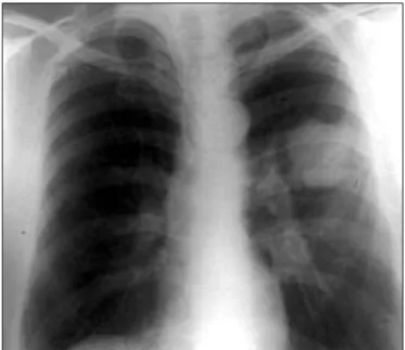
A 63-year-old man was presented to our hospital with dyspnea and chest pain. Physical examination revealed clubbing of the nails. Chest X ray demonstrated two dif-ferent opacities in the left lung (Figure 1).
CT of the chest demonstrated a 5x4x4 cm soft tissue le-sion in the left upper lobe and a 2x2x2 cm lele-sion in the left lower lobe (Figure 2, 3). Respiration function tests were FVC: 2.74 L (%65), FEV1: 1.99 L (%61), arterial blood gas analysis showed pO2: 69.2, pCO2: 37.3, SO2: 95.1 %. Two separate endobronchial lesions were detected one in the left lower lobe bronchus and one in the left upper anterior segment bronchus on bronchoscopy. Specimens
for histopathological examination were taken from each lesion. The histopathologic examination was reported as squamous cell carcinoma. Although these tumors have similar ploidi features, the proliferation of the left lower lobe tumor was found higher than the other tumor at the flow cytometric examination.
Brain-abdomen CT and bone scan revealed normal. Thoracotomy was performed with the diagnosis of pulmo-nary synchronous tumors. On exploration 4x4x5 cm lesion in the upper lobe and 3x3x3 cm lesion located peribron-chially in the lower lobe were detected. Pneumonectomy and systematic nodal dissection were performed.
Histo-pathologic examination of the lesion located in upper lobe was reported as adenosquamous carcinoma and the other tumor was reported as moderately differentiated squamous cell carcinoma. As the pathologic examination showed no nodal involvement each tumor was evaluated as
Figure 1. Chest X-Ray of the patient.
Figure 2. CT of the mass in the left upper lobe.
stage I. However the tumor in the upper lobe was revealed to be T2, N0, M0 and the other was T1, N0, and M0.
During follow up, all control CT’s were normal until the postoperative 21st month. The control abdominal CT revealed a hypodens lesion in the liver, bilaterally adrenal hyperplasia and multiple paraaortic lymphadenopathies. Biopsy of the lesion was reported as metastasis of adenocar-cinoma, and it was thought as a primary tumor originated from the stomach. Furthermore primary gastric tumor was seen on endoscopy. Chemotherapy was planned for the pa-tient. However the patient died because of the complica-tions of gastric carcinoma on 28th month follow up.
Case 3
A 59 year-old-man admitted with a 2 year history of cough. Chest X-Ray showed two different lesions in the right hemithorax (Figure 4).
Thorax CT revealed 3x3x3 cm. lesion located inferiorly in the right upper lobe and another 7-cm. lesion in the laterobasal segment of the right lower lobe (Figure 5,6). Brain-abdomen CT and bone scan showed no abnormal-ity.
On preoperative bronchoscopic examination, an en-dobronchial lesion was detected in the anterior segment bronchus of the right upper lobe. Biopsy of the lesion was reported as squamous cell carcinoma. Respiration function tests of the patient were FVC: 85.2 %, FEV1: 86 % and
arterial blood gas analysis showed PO2: 77, PCO2: 38.6, SO2: 94.4.
Right thoracotomy was performed with the possibility of synchronous tumors. On exploration, a 5-cm. lesion in the left upper lobe and another 8x9x5 cm. lesion in the lower lobe were detected. As the frozen section of the le-sion located in the lower lobe was reported as adenocar-cinoma, right pneumonectomy and systematic nodal dis-section were performed. Histopathological examination was reported as adenosquamous carcinoma for the lower lobe tumor, squamous cell carcinoma for the upper lobe tumor. Resection margins were negative, squamous tumor cells were detected on paraesophageal lymph node. He was asymptomatic on 18th mount follow up.
Figure 4. Chest X-Ray of the patient.
Figure 5. CT of the mass in the right upper lobe.
Discussion
Carcinoma of the lung is the leading cause of cancer death in countries whose statistical analyzes are reliable (3, 5). Synchronous tumors, which are described as multiple primary tumors, are very rare (1, 2, 4, and 6). The most common site for these tumors is breast. Advances in surgi-cal managements and diagnostic methods for carcinoma of the lung lead to an increase in the rate of synchronous tumors (2, 6).
Tobacco smoking and asbestosis are the most common occupational causes of lung cancer (1, 7, and 8). Also it is obvious that similar exposures can cause similar effects on same tissues (8). By the way, detection of multiple lesions of the lungs at the same time should be considered (1, 2, 7, and 8). In some series, detection of a secondary primary tumor for the patients who survives more than 3 years was found to be 10 % (1, 4).
Metastatic disease increases the grade (Grade IV) and decreases overall lifetime in lung cancer. However, a sec-ond lung cancer with different histologic features should be considered as synchronous and should be staged sepa-rately. Differentiation of these clinical entities is important in terms of surgical vs. medical management and prognosis (3, 5).
When more than one lesion was detected in a patient and if there is enough evidence that the second lesion is not metastatic then the possibility of synchronous tumor should be kept in mind. In such cases, separate biopsies should be taken from both lesions. When it is impossible to reach the lesion with bronchoscopy, transthoracic biop-sy might confirm the diagnosis. DNA flow cytometry can be used to differentiate between two synchronous cancers of the same histologic condition (8, 9).
Most authors suggested that after the diagnosis, the two tumors should be staged separately and surgery should not be performed for the tumors having larger grades than grade II (4, 10-12).
In bilaterally lesions wedge resection and segmentec-tomy are the preferred methods. However, for ipsilaterally
lesions surgical methods such as pneumonectomy were ap-plied in some studies (1, 2, 10, 13).
Postoperative survival in synchronous lung cancer hav-ing complete resection has a wide range in different stud-ies. Five year survival was 6 % in Carey (14)’s study and 44 % in Rosengart (7)’s study. However, all cases in every stage were evaluated in these studies. In Pompier (6)’s se-ries, grade I and II cases were evaluated and 5 year survival was found 38 % after complete resection. In this study, 5-year survival was 33 % for the tumors with same histo-logic types while it was 17 % for the tumors with different histologic types (2).
The cases in which more than one tumor is detected radiologically, the possibility of a synchronous tumor or a metastasis of the primary tumor must always be kept in mind and histopathologic diagnosis should be done for both tumors (14). Complete resection should be per-formed if feasible. For synchronous tumors with lower grades, with early surgical intervention, the survival can be as long as solitary lung cancers especially in epidermoid carcinoma (15).
In recent years genetic investigations showed that en-vironmental conditions have an influence on the develop-ment of cancer (16). Besides, smoking and asbestosis are the most common occupational causes of lung cancer. For patients who have similar genetic and environmental con-ditions, there is a possibility of developing tumor in mul-tiple foci.
With the help of developments in treatment of cancer, for the patients who had long overall survival or had cure with the treatment of primary cancer, the possibility of finding a second tumor (metachronous tumor) increases. Simultaneous discovery of tumors, like larynx and bladder tumors which are related to tobacco, with the lung can-cer is possible. Therefore, it is important to keep in mind that, especially for the cases T1-2 N0, the lesions that are accepted as pulmonary metastases may be synchronous tu-mors.
References
1. Adebonojo SA, Moritz DM, Danby CA. The results of modern surgical therapy for multipl primary lung cancers. Chest 1997; 112: 693-701.
2. Pompier RF, Vetto JT, Lee JT et al. Synchronous non-small cell lung cancers. Am J Surg. 1996; 171:521-4.
3. Adebonojo SA, Mortiz DM, Danby CA. The results of Modern Surgical Therapy for Multiple Primary Lung Cancers. Chest 1997; 112: 693-701.
4. Rosengart TK, Martini N, Ghosn P et al. Multipl primary lung carcinomas: prognosis and treatment. Ann Thorac Surg 1991; 52:773-9.
5. Pairolero PC, Williams DE, Bergstrahl EJ et al. Postsurgical stage I bronchogenic carcinoma: morbid implications of recurrent disease. Ann Thorac Surg 1984: 38:331.
6. Kitamura K, Mitsudomi T, Ishidsa T et al. Adenocarcinoma and squamos cell carcinoma in the same lobe of the lung. Respiration 1991; 58: 226-8.
7. Aydıner A, Karadeniz A, Uygun K et al. Multipl primary neoplasms at single instution. Am J Clin Oncol 2000; 23: 364-70. 8. Deschamps C, Pairolero PC, Trastek VF et al. Multipl primary
lung cancer. J Thorac Cardiovasc Surg. 1990; 99:769-78. 9. Hida T, Ariyoshi Y, Sugiura T et al. Synchoronous lung cancer
presenting with small cell carcinoma and adeno carcinoma. Chest 1993; 104:1602-4.
10. Martini N, Melamed MR. Multiple primary lung cancers. J Thorac Cardiovasc Surg 1975; 70:606-12.
11. Ferguson MK. Synchoronous primary lung cancers. Chest 1993; 103:398-400.
12. Ferguson MK, DeMeester TR, DesLauriers J et al. Diagnosis and management of Synchoronous lung cancers. J Thorac Cardiovasc Surg 1985; 89:378-85.
13. Carey FA, Donnely SC, Walker WS et al. Synchronous primary lung cancers: Prevalance in surgical material and clinical implications. Thorax 1993; 48:344-6.
14. Pierard P, Vermylen P, Bosschaerts T et al. Synchronous roentgenographically occult lung carcinoma in patients with resectabl primary lung cancer. Chest 2000; 117:779-85.
15. Colomer EJ, Molina JC, Aguilar X, et al. Synchoronous multiple primary cancer of the lung: A rare association of small cell carcinoma as the main tumor plus epidermoid carcinoma. Arch Bronconeumol 1999; 35:245-7.
16. Hiroshima K, Toyozaki T, Kohno H et al. Synchoronous and metachronous lung carcinomas: molecular evidence for multicentricity. Pathol Int 1998; 48:869-76.