51
7tepeklinik
Closure of a persistant
oroantral fistula with
nasoseptal cartilage as
a complication of
lateral sinus lifting: A
case report
Lateral sinus yükseltme
komplikasyonu olarak
oluşan inatçı
oroantral fistülün
nazoseptal kıkırdak ile
kapatılması: Bir olgu
sunumu
Assist. Prof. Gökhan Gürler
İstanbul Medipol University, Faculty of Dentistry, Department of Oral and Maxillofacial Surgery, İstanbul
Dr. Emrah Dilaver
İstanbul Medipol University, Faculty of Dentistry, Department of Oral and Maxillofacial Surgery, İstanbul
Assist. Prof. Erkan Soylu
İstanbul Medipol University, Faculty of Medicine, Department of Ear Nose and Throat, İstanbul Assist. Prof. Tuba Develi
İstanbul Medipol University, Faculty of Dentistry, Department of Oral and Maxillofacial Surgery, İstanbul
Prof. Çağrı Delilbaşı
İstanbul Medipol University, Faculty of Dentistry, Department of Oral and Maxillofacial Surgery, İstanbul
Received: 27 February 2017 Accepted: 4 April 2017
DOI: 10.5505/yeditepe.2017. 43434
Corresponding author Assist. Prof. Gökhan Gürler
İstanbul Medipol University, Faculty of Dentistry, Department of Oral and Maxillofacial Surgery 34083 Unkapanı-İstanbul
Tel: 212 453 49 40
E-mail: [email protected]
SUMMARY
Oroantral fistula may develop due to tooth extraction, infec-tion or surgical interveninfec-tions in the posterior maxilla. Several surgical techniques have been described for oroantral fistula closure. All these techniques have different advantages and disadvantages. This report presents an oroantral fistula case, which occurred, following lateral sinus lifting procedure. Tra-ditional surgical techniques (buccal advancement flap, pala-tal flap, and Bichat’s buccal fat pad) were performed to close the oroantral fistula, but could not to be managed. Finally, closure was succeeded with septal cartilage graft placed over the bony defect.
Key words: Oroantral fistula, sinus lifting, septal cartilage, complication.
ÖZET
Oroantral fistül, posterior maksillada diş çekimi, enfeksiyon veya cerrahi işlemlere bağlı olarak gelişebilir. Oroantral fistülün kapatılmasına yönelik pek çok cerrahi teknik tanımlanmıştır. Bütün bu tekniklerin kendine özgü avantaj ve dezavantajları vardır. Bu olguda raporunda lateral sinüs yükseltme işlemine bağlı gelişen oroantral fistül sunulmuştur. Geleneksel cerrahi yöntemlerle (bukkal ilerletme flebi, palatal flep, Bichat bukkal yağ dokusu) kapatılamayan defekt, son olarak otojen septal kıkırdak grefti uygulanarak başarıyla kapatılabilmiştir.
Anahtar kelimeler: Oroantral fistül, sinus yükseltme, septal kıkırdak, komplikasyon.
INTRODUCTION
Oroantral fistula (OAF) is an epithelized communication be-tween maxillary sinus and oral cavity.1 It is mostly seen after extraction of maxillary molar and premolar teeth those root apices are anatomically adjacent to the maxillary sinus. The incidence of complication as a result of simple maxillary pre-molar and pre-molar extraction varies from 0.31% to 3.8%.2 Other causative factors for OAF are dentoalveolar infections, cysts or tumors that invade through maxillary sinus, Paget’s dis-ease, trauma and complication of Caldwell-Luc procedure.1,2,3 Commonly used surgical techniques for closure of OAF are described in the literature including buccal advancement flap, buccal fat pad, palatal island flap and buccal sliding flap4,5 This report presents a persistent OAF which occurred following lateral sinus lifting procedure; however, closure could not to be managed performing buccal sliding flap, pal-atal flap and buccal fat pad respectively. Final closure could be accomplished using autogenous septal cartilage graft. CASE REPORT
A 44-year-old male patient referred to our clinic with a complaint of tooth loss in the bilateral posterior maxilla. Panoramic graphy and Cone Beam Computed Tomogra-phy (CBCT) revealed insufficient vertical bone for implant insertion at both sides (Figure 1).
52
7tepeklinik
Figure 1. Preoperative panoramic graphy of the patient.
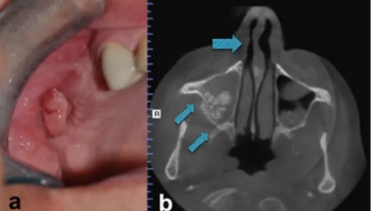
Systemic condition and maxillary sinus examination of the patient were symptomless. Bilateral lateral maxillary sinus lifting and onlay horizontal bone augmentation with otogenous mandibular symphysis block graft for the left posterior region were carried out under conscious sedation and local anesthesia. Patient was symptomless postoperatively but two months later, he referred to our department with a complaint of postnasal flow. At clinical and radiological examination, graft particles migrating from the right augmented site and antral polip formation as well as maxillary sinusitis were observed (Figure 2a-b).
Figure 2 (a-b.) Intraoral appearance of the antral polip (a). Axial view of the CBCT depicting right maxillary sinusitis (double arrow) and nasal septum deviation (ar-row) (b).
He was prescribed antibiotic (1000 mg amoxicillin and clavulanate combination plus 500 mg metronidazole two times a day), and a decongestant (paracetamol 300 mg, phenylyprophanolamin 25 mg and chlorphenramin 2 mg) postoperatively for 10 days. Since the symptoms subsided, infected graft particles were curetted and oroantral fistula was closed with buccal advancement flap. After two weeks of follow-up there was still pus dis-charge from the operation site. He was again prescribed the above-mentioned drugs for another ten days and following regression of the symptoms; the fistula was re-closed with palatal flap. However, in the follow-up period of ten days the fistula recurred, wound dehiscence was observed and pus discharge re-initiated. The patient was again prescribed antibiotic and decongestant and was decided to follow-up for one month. At the end of this period the patients had no symptoms hence, the fistula was attempted to close with Bichat’s buccal fat fad.
Un-fortunately, the same complaints consisted with OAF was observed again. Finally, we consulted patient’s otolar-yngologist. After diagnostic endoscopy and computed tomography, nasal septum deviation that restricts right maxillary sinus drainage was diagnosed (Figure 2-b and Figure 3).
Figure 3. Intranasal endoscopic appearance.
Functional endoscopic sinus surgery (FESS) and na-sal deviation correction i.e septoplasty operation were planned as well as closure of the fistula with septal carti-lage obtained during the septoplasty operation.
Patient was operated under general anesthesia. Local an-esthetic containing epinephrine was injected in the un-cinate process and middle turbinate to control bleeding. Using a rigid 4.0 mm endoscope (Karl Storz, Tutlingen, Germany) the middle turbinate was carefully dislocated toward the septum to visualize the uncinate process and ethmoid bulla. The uncinate process was removed by forceps and natural ostium of the maxillary sinus was en-larged to a size sufficient for clear visualization of the si-nus and its effective drainage after healing. Inflammatory polyps were removed and nasal septal cartilage graft was harvested. OAF epithelium was excised circumferentially. The mucoperiosteal flap was elevated from the buccal side of alveolar crest. Inflamed mucosa was removed un-der fiberoptic endoscope. Nasal septal graft was placed to cover the large opening and sutured to adjacent bone with 4/0 polydioxanone (Figure 4a-b).
Soft tissue was closed with 3/0 silk suture. After one week sutures were removed. Healing was uneventful during the follow-up examination at one year (Figure 5).
53
7tepeklinik
Figure 4 (a-b). Intraoperative appearance of the bony defect on the anterior wall of the maxillary sinus (a). Stabilization of the nasal septal cartilage over the bony defect (b).
Figure 5. Postperative view showing complete healing.
DISCUSSION
Maxillary sinus is one of the paranasal sinuses, which has a close proximity to maxillary premolar and molar teeth. The factors disrupting the continuity of sinus membrane may include; tooth extraction, pathologic lesions, trauma and surgical interventions in the posterior maxilla. Oro-antral fistula is defined as the communication between maxillary sinus and oral cavity. It usually occurs after pre-molar and pre-molar extractions. While the mucosal perfora-tions smaller than 5mm heal spontaneously, larger perfo-rations need surgical interventions.4,5
Several techniques are used in oroantral fistula closure. Although buccal advancement flap and buccal fat pad are commonly used and relatively easy methods, lack of high blood perfusions may lead to recurrent fistulas.4-10 Palatal rotation flap is preferred for oroantral fistula clo-sure with large defects, having 76.9% success rate thanks to its vascular pedicle.5,10 Additionally autogenous and alloplastic graft materials are used to separate the sinus membrane and oral mucosa to facilitate the closure and subsequent implant operation or sinus lifting procedure. The inflammation and infection risk of alloplastic materi-als and high cost limit the use of these materimateri-als in OAF closure.5 The use of autogenous bone grafts harvested from mandibular symphysis has successful results. Au-togenous bone grafts permit defect closure and future implant insertion.
The autogenous cartilage grafts are used to close tym-panic membrane perforations and cerebrospinal leakage while it is easy to adapt, well tolerated, easily manipulat-ed, easily accessible and cost effective as well as it is re-sistant to resorption and infection. Autogenous cartilage grafts have less vascularity than other bone grafts. Due to low vascularity of cartilage grafts, resorption of the graft is limited.6,7 It can be easily trimmed and harvested from the same surgical site with minimal morbidity. Because nasoseptal cartilage is near to the field of repair and can be harvested without additional time, it is a suitable graft in OAF closure. Furthermore, there is no need to change the position of the patient and the surgeon during the operation. This technique causes no esthetic problems, there is minimal donor site complication, and also it is easy to handle.6
In this case we reported a persistent oroantral fistula, which recurred after several closure attempts, because of impaired sinus drainage. The closure was succeeded with FESS and simultaneous application of nasoseptal cartilage. We also emphasized the need for consultation with an ENT specialist in case of severe and persistent OAF patients.
54
7tepeklinik
REFERENCES
1.Lazow SK. Surgical management of the oroantral fistula flap procedures. Oper Tech Otolaryngol Head Neck Surg 1999; 10: 148-152.
2.Punwutukorn C, Waikakul A, Pairuchvej V. Clinically significant oroantral communications – a study of inci-dence and site. Int J Oral Maxillofac Surg 1994; 23: 19-21. 3.Kiran Kumar Krishanappa S, Prashanti E, Sumanth KN, Naresh S, Moe S, Aggarwal H, Mathew RJ. Interventions for treating oro-antral communications and fistulae due to dental procedures. Cochrane Database Syst Rev 2016; 27: CD011784.
4. Procacci P, Alfonsi F, Tonelli P, Selvaggi F, Menchini Fabris GB, Borgia V, De Santis D, Bertossi D, Nocini PF. Surgical Treatment of Oroantral Communications. J Cra-niofac Surg 2016; 27: 1190-1196.
5.Dergin G, Emes Y, Delilbasi C, Gurler G. Management of Oroantral Fistula. In: A Texbook of Advanced Oral and Maxillofacial Surgery. Volume 3. Chapter: Management of Oroantral Fistula, 367-385 pp, Intecch Publisher, 2016. 6.Mohanna PN, Kangesu L, Sommerlad BC. The use of conchal-cartilage grafts in the closure of recurrent palatal fistulae. Br J Plast Surg 2001; 54: 274.
7.Al-Sibahi A, Shanoon A. The use of soft polymethyl-methacry- late in the closure of oro-antral fistula. J Oral Maxillofac Surg 1982; 40: 165-166.
8.Kansu L, Akman H, Uckan S. Closure of oroantral fistula with the septal cartilage graft. Eur Arch Otorhinolaryngol 2010; 267: 1805-1806.
9.Saleh EA, Issa IA. Closure of large oroantral fistulas using septal cartilage. Otolaryngol Head Neck Surg 2013;148:1048-1050.
10.Nezafati S, Vafaii A, Ghojazadeh M. Comparison of pedicled buccal fat pad flap with buccal flap for closure of oro-antral communication. Int J Oral Maxillofac Surg 2012; 41: 624-628.