REVIEW
Kemal Dolay, Department of Surgery, Medipol University Fac-ulty of Medicine, 34214 Istanbul, Turkey
Sami Akbulut, Department of Surgery and Liver Transplant Institute, Inonu University Faculty of Medicine, Turgut Ozal Medical Center, 44280 Malatya, Turkey
Author contributions: Dolay K and Akbulut S contributed to the final review and submission of the manuscript, writing the paper and review of the literature; Dolay K collected endoscop-ic images.
Correspondence to: Sami Akbulut, Assist Prof, FICS, FACS, Department of Surgery and Liver Transplant Institute, Inonu University Faculty of Medicine, Turgut Ozal Medical Center, 44280 Malatya, Turkey. [email protected]
Telephone: +90-422-3410660 Fax: +90-422-3410036 Received: March 17, 2014 Revised: April 30, 2014 Accepted: June 12, 2014
Published online: November 7, 2014
Abstract
Most cases of hepatic hydatid disease exhibit uncom-plicated clinical course and management. However, the diagnosis and management of complicated hepatic hydatid disease is a special issue. One of the most common and serious complications of hepatic hydatid disease is the rupture of the cyst into intrahepatic bile ducts. The clinical appearance of intrabiliary rupture can range from asymptomatic to jaundice, cholecysti-tis, cholangicholecysti-tis, liver abscess, pancreatitis and septice-mia. Current treatments for major ruptures can result in high morbidity and mortality rates. Furthermore, ruptures that cannot be diagnosed preoperatively can induce complications such as biliary fistulae, biloma, cavitary infection and obstructive jaundice. In the past, these complications were diagnosed and treated by surgical methods. Currently, complications in both the pre- and postoperative periods are diagnosed and treated by non-invasive or minimally invasive methods. In clinical practice, endoscopic retrograde cholangio-pancreatography (ERCP) is indicated for patients with preoperative frank intrabiliary rupture in which hydatid
elements are clearly seen in the bile ducts, or for bili-ary adverse events after surgery, including persistent biliary fistulae and jaundice. However, controversy concerning routine preoperative ERCP and prophylactic endoscopic sphincterotomy in patients suspected of having minor cystobiliary communications still remains. In this article, the role of ERCP in the diagnosis and management of hepatic hydatid disease during the pre- and postoperative periods is reviewed.
© 2014 Baishideng Publishing Group Inc. All rights reserved.
Key words: Hydatid cyst; Complications; Intrabiliary
rup-ture; Endoscopic retrograde cholangiopancreatography
Core tip: Intrabiliary rupture is the most common and
serious complication of hepatic hydatid disease. In the past, all the complications due to the intrabiliary rup-ture were diagnosed and treated by surgical methods, whereas nowadays these complications in both the pre- and the postoperative periods can be diagnosed and treated by non-invasive or minimally invasive methods such as endoscopic retrograde cholangiopan-creatography (ERCP). The primary aim of this review was to analyze the efficacy of ERCP in diagnosis and treatment of hepatic hydatid disease during the pre- and postoperative periods.
Dolay K, Akbulut S. Role of endoscopic retrograde chola-ngiopancreatography in the management of hepatic hydatid disease. World J Gastroenterol 2014; 20(41): 15253-15261 Available from: URL: http://www.wjgnet.com/1007-9327/full/ v20/i41/15253.htm DOI: http://dx.doi.org/10.3748/wjg.v20. i41.15253
INTRODUCTION
Hydatid disease results from infestation of a parasite
DOI: 10.3748/wjg.v20.i41.15253 © 2014 Baishideng Publishing Group Inc. All rights reserved.
Role of endoscopic retrograde cholangiopancreatography in
the management of hepatic hydatid disease
originating from a Mediterranean strain of Echinococcus
seen mostly in South America, North Africa, the Middle East and Eastern Europe[1,2]. Hydatid disease is endemic
in most parts of the world, especially in the Mediter-ranean where sheep husbandry is common, and is an important medical health problem in these regions. Fur-thermore, increasing migration raises the incidence of the disease in areas where it was once rare[3,4].
Approxi-mately 4000 patients per year are diagnosed with hydatid disease in Turkey[5].
The clinical presentation of hydatid disease varies depending on the location, growth rate and size of the cysts, and organ affected. Symptoms typically develop as a result of the compression of adjacent structures or viscera as a result of surrounding inflammation, or from the rupture of the cyst into the bile duct, pleural space, or peritoneal cavity. The liver is most commonly affect-ed, with involvement of the right lobe in 55%-80% of patients. Most patients with hepatic hydatid cysts (HHC) present with an uncomplicated course at diagnosis and treatment planning is not problematic. However, the diagnosis and management of complicated courses of HHC depend on the experience levels of the surgeon, interventional radiologist and therapeutic endoscopist.
Common complications of HHC include rupture into intrahepatic bile ducts or the peritoneal cavity, inva-sion of other organs, pressure on the biliary tree and other neighboring structures, and infection[6,7]. Of these,
intrabiliary rupture (IBR) is the most common and seri-ous complication, occurring in 2%-42% of cases[7-11].
According to Dew[12], who was the first to report this
complication, IBR usually occurs in the biliary ducts of the right lobe (55%-60%), less commonly in the left lobe biliary ducts (30%-35%), and rarely in the common bile duct. The clinical presentation of IBR can range from asymptomatic to jaundice, cholecystitis, cholangitis, liver abscess, pancreatitis and septicemia, depending on the size of the cystobiliary communication. However, un-diagnosed ruptures can result in biliary leakage, biloma, cavitary infection and obstructive jaundice after HHC surgery[7,13-15].
In the past, IBR complications were diagnosed and treated by surgical methods, accompanied by 50% mor-bidity and 4.5% mortality rates. More recently, non-invasive or minimally invasive methods have been used to diagnose and treat these complications during both the pre- and the postoperative periods. In clinical practice, it is generally agreed that endoscopic retrograde cholangiopancreatog-raphy (ERCP) is indicated for patients with biliary adverse events after surgery, such as persistent biliary fistulae and jaundice, and preoperative frank IBR that is suspected clinically (because of jaundice), biochemically (because of overt cholestasis), or radiologically (as a dilated main biliary ductal system or hydatid elements evident in the bile ducts). However, the use of routine preoperative ERCP and prophylactic endoscopic sphincterotomy (ES) in patients with suspected minor cystobiliary communica-tions remains controversial. Furthermore, the increasing
use of magnetic resonance cholangiopancreatography (MRCP) fuels the debate on the routine use of preopera-tive diagnostic ERCP. The goal of this article is to review the efficacy of ERCP for the diagnosis and treatment of hepatic hydatid disease during the pre- and postoperative periods.
HYDATID CYSTS AND INTRABILIARY
RUPTURE
The parasite that causes the hydatid cyst becomes ap-parent after three weeks and can attain a bulk of 3 cm in the liver parenchyma after three months. Atrophy and fibrosis occur as a result of the pressure of the growing cyst. During this growing period, the intracystic pressure may rise to 80 cm H2O within a live cyst and can cause
compression and obstruction of the biliary tree if cen-trally located. Even if there is no rupture into the bile ducts, cholestasis and partial dilatation of the proximal part of affected ducts can be detected biochemically and radiologically. Continued growth can result in rupture into the bile ducts due to the increased intracystic pres-sure and fragility of the cyst wall[16-18].
Rupture into the bile ducts is the most common complication of hepatic hydatid disease and its incidence is reported to be as high as 42% in some clinical se-ries[8-11,19,20]. There are two theories describing the
patho-genesis of IBR. According to the first theory, progressive necrosis and communication between the cyst and the biliary tree are caused by the compression of the HHC wall. The second theory states that the trapping of small biliary radicals in the pericystic wall causes high intracys-tic pressure and results in atrophy, followed by the rup-ture of biliary radicals[21,22].
The communication between the ruptured cyst and bile ducts can be classified as either major or minor/ simple based on size. Simple communications are rela-tively small communications between the cyst wall and small biliary radicals, which are seen in 10%-37% of HHC patients. In these cases, bile ducts are in com-munication with the cyst, but neither daughter cysts nor germinative membrane is found within the bile ducts. Cystic fluid, scolices or very tiny hydatid elements may enter the bile ducts, but do not cause obstruction due to their size. Simple communications are difficult to dem-onstrate preoperatively as they do not exhibit indirect symptoms, such as dilatation of bile ducts, seldom cause biliary colic, and patients are generally asymptomatic. Although simple communications can be demonstrated intraoperatively, they are most often recognized by post-operative biliary leakage. In contrast, major, open wide communications between the cyst wall and biliary tree, seen with frank IBR in 3%-17% of HHC patients, can be detected preoperatively. In this type of cystobiliary communication, cystic content empties into the biliary tree causing intermittent or total obstruction[23,24]. Major
communications most commonly present as biliary colic, jaundice and cholangitis, which can worsen, resulting in
liver abscess, septicemia or anaphylaxis. Although rare, complications such as acute cholecystitis and pancreati-tis can also occur due to hydatid remnants within bile ducts[7,12-15].
PREOPERATIVE ERCP
In clinical practice, there are four recommendations concerning preoperative ERCP for diagnosing and treat-ing cystobiliary communications: (1) perform elective surgery to overcome acute conditions such as cholangitis and biliary obstruction; (2) reduce the risk of postopera-tive persistent biliary fistulae and the duration of hos-pitalization by preoperative ES in patients with minor communications; (3) remove hydatid elements within the bile ducts and schedule hydatid cyst surgery; and (4) in cases of major rupture, employ a therapeutic approach by emptying hydatid contents within the cyst and bile ducts[10,16,18-20,25,26].
Diagnostic ERCP
The first management approach for hepatic hydatid disease consists of a clinical history, detailed physical examination, routine blood tests (complete blood count, sedimentation, liver biochemistry), serologic examination and imaging methods such as ultrasonography (US) and computed tomography (CT). These evaluations help to determine whether the hydatid disease has a complicated or uncomplicated course[7,16,17,27,28].
Although routine ERCP is not yet accepted for un-complicated hydatid cysts, some clinical centers advise their use in order to entirely determine bile duct anato-my and describe the communication between the cyst and bile ducts[16,29]. With consideration of cases where
cystobiliary communication is common in liver hydatid cyst surgery (e.g., large or multiple cysts, hilar or caudate
lobe cysts or cysts accompanied with leukocytosis), the routine use of ERCP in uncomplicated hydatid cystic disease is suggested to prevent or decrease postoperative biliary complications[10,11,20,30,31]. However, postoperative
bile leakage has been reported in patients who under-went preoperative ERCP and ES for prevention of fis-tula formation, not all communications can be detected with diagnostic ERCP, and most of the patients with postoperative bile leakage heal spontaneously without a need for ERCP[3,10,11,20,30,31]. Moreover, in a variable
per-centage (0%-30%) of patients who underwent ERCP for suspected frank IBR (i.e., presented with jaundice and/or
dilated bile ducts), only bile duct compression by the cyst was observed[5,10,18].
There are some limitations associated with ERCP for the diagnosis of hydatid disease. Primarily, minor cysto-biliary communications may not be detected with ERCP due to increased intracystic pressure, the minimal com-munication of cyst and biliary tree, and to daughter cysts temporarily obstructing the cystobiliary opening. Intra-hepatic bile ducts may not completely fill with contrast media due to the increased pressure, increasing the false
negativity ratio up to 17%-20%. Also, if contrast media is too intense, cysts within the main bile ducts may not be apparent and daughter cysts or old hydatid particles may mimic gallstones. Finally, hydatid elements that are solely in intrahepatic ducts may be overlooked with ERCP. Therefore, surgeons should remain vigilant for preoperative minor cystobiliary communications despite their apparent absence[3,10,16,32-35]. Some of these
limita-tions can be minimized with correct pressure injection from different points or by applying occlusion cholangi-ography. Moreover, ERCP can reveal cystobiliary fistulae and filling defects due to hydatid elements in bile ducts, as well as indirect signs of minor cystobiliary communi-cation, such as cystic pressure, dilatation and erratic fill-ing of neighborfill-ing bile ducts.
Studies have suggested that MRCP is comparable to ERCP as a diagnostic tool because of its high sensitiv-ity in bile duct pathologies, and advise its use in place of ERCP as it is a non-invasive method[10,36,37]. In cases
of cystobiliary communication, ERCP can then be used as a therapeutic means. In our clinical practice, we do not perform preoperative diagnostic ERCP if there is no history of cystobiliary pancreatic symptoms, overt cholestasis in blood studies or biliary ductal dilatation distal to the cyst in US and CT images. In cases of mi-nor cholestasis or partial dilatation of intrahepatic bile ducts proximal to the cyst, MRCP in addition to US and CT prove to be more useful than ERCP for diagnosing minor communications in uncomplicated cases.
Although frank IBR can be easily diagnosed with US, CT or MRCP, ERCP is usually the first modality chosen in cases of complicated HHC where frank IBR is sus-pected by obstructive jaundice, cholangitis or biliary sep-sis, abnormal biochemical liver function tests, presence of cholestatic enzymes or hyperbilirubinemia, if hydatid elements are present within the bile ducts, or for any kind of dilatation of main bile ducts[16,19,31,35,38]. Hence,
preoperative ERCP is more commonly used therapeuti-cally than diagnostitherapeuti-cally[3,7,23].
In cases of frank IBR, white, shiny hydatid mem-branes are mostly seen within the duodenum or protrud-ing from the papilla Vateri durprotrud-ing ERCP. On cholangi-ography, linear wavy filling defects of laminated hydatid membranes can be easily seen within the main bile duct. Daughter cysts can be seen as rounded or oval lucent filled defects. In late phases, membrane particles lose their shiny characteristic and mimic bile stones as they become colorized by bile acids. In patients with uncom-plicated HHC, both diagnostic and therapeutic ERCP should be performed if cystic fluid is colorized by bile when using the PAIR (puncture, aspiration, injection and re-aspiration) protocol[7,16].
Therapeutic ERCP
Preoperative therapeutic ERCP has a reported success rate as high as 80%-100%[4,20,39-47], and can be used when
cystobiliary communication or hydatid membranes and/or daughter cysts that cause biliary obstruction are
demonstrated by cholangiography. ES, extraction by bal-loon or basket catheter, nasobiliary drainage (NBD) and biliary stenting are some therapeutic methods that can be performed by ERCP. As described above, preopera-tive ES reduces the risk of postoperapreopera-tive external biliary fistulae, morbidity and the length of hospitalization in patients diagnosed with cystobiliary communication by ERCP. However, in the series reported by Rodriguez
et al[39], patients undergoing preoperative ERCP
devel-oped uncontrollable fistulae. In another series reported by Galati et al[23] ES was found to reduce postoperative
fistulae and cavitary infection due to cystobiliary rup-ture. Additional advantages of endoscopic treatment for HHC patients are that it allows for elective surgery and significantly decreases morbidity and mortality associated with the procedure. However, further studies are needed to evaluate the prophylactic use of ES for the prevention of biliary fistulae[13,20,47,48].
In major ruptures, hydatid membranes or daughter cysts encountered in bile ducts can be emptied out by ES, and a Dormia basket and biliary occlusion balloon can be used to clean out the common and main bile ducts. If hydatid membranes are sufficiently large, they can fill the duodenal lumen and hide the already sphinc-terotomized papilla orifice. In our ERCP procedure, guidewire placement is routinely used before ES when large amounts of hydatid elements are observed in bile ducts. These techniques can be performed successively many times under C-arm fluoroscopy even when en-doscopic manipulation is made difficult by the hydatid
materials around the papilla. After emptying hydatid membranes and big daughter vesicles, saline irrigation of the bile duct may be necessary to flush out the hydatid material and small daughter cysts (Figure 1).
As bile ducts cannot be purified from hydatid ele-ments using ES alone, NBD and biliary stenting can be added. NBD may be the first choice of temporary treatment, particularly when life-threatening sepsis from acute cholangitis occurs. Once the obstructed bile ducts are drained, ERCP can be re-performed to completely clean out bile ducts of remaining hydatid elements. If there is an obstruction or stricture in the biliary tree, en-doscopic balloon dilatation and biliary stenting may be needed[10,18,19,43,49-51].
There are numerous studies demonstrating that the use of ERCP to clean out bile ducts and cystic materials in cases of frank IRB significantly decreases cyst vol-ume during successive follow-ups and 25% of patients are cured and do not need any further surgical treat-ment[4,16,35,39,42-46,52,53]. An endoscopic transpapillary
treat-ment for frank IBR in which hydatid eletreat-ments within the bile ducts were cleaned out using a biliary occlusion balloon and basket catheter, and a saline solution was infused in the cystic cavity by an endoscopically placed NBD catheter was first described in 1987 by al Karawi
et al[54]. Five of the 6 patients treated by these authors
were cured completely without needing any further sur-gical treatment[55]. Similar successes were reported in five
patients described by Akkiz et al[42] in 1996 and seven
pa-tients described by Signh et al[56] in 2006, demonstrating
A B
C
Figure 1 Endoscopic retrograde cholangiopancreatography. A: Magnetic resonance image of a hydatid cyst with major rupture; B: Emptying of hydatid mem-branes with endoscopic sphincterotomy and a biliary occlusion balloon; C: Extraction of hydatid materials.
that therapeutic ERCP is both effective and safe for the treatment of ruptured hydatid cysts.
This technique has been performed in our clinic with some modifications, which we refer to as nasocystic-biliary drainage (NCBD). In this procedure, hydatid ele-ments are emptied into the duodenum, and a hydrophilic guidewire is negotiated into the cyst. Hydatid materials are then completely emptied out over the guidewire by balloon and basket catheter, and a redesigned 10F NBD catheter is inserted into the cystic cavity to facilitate full emptying of the cyst contents. The cystic cavity and common bile duct are periodically irrigated using a saline solution through the nasobiliary catheter for seven days to ensure total clearance of the germinal layers and the remaining small daughter cysts. Small additional holes are placed in the choledochal and duodenal portion of the NCBD catheter to avoid the risk of cholangitis due to the continuous drainage of hydatid materials into bile ducts and obstruction. This redesign allows the NCBD cath-eter to act as a stent between the common bile duct and duodenum, and for periodic irrigation of the cystic cavity. Furthermore, the NCBD decreases the external biliary loss[26]. At present, 12 of the 14 patients that have
under-gone this procedure have been completely cured (Figure 2). The procedure failed in one patient who underwent a pre-vious major cystobiliary rupture operation (duodenotomy and open sphincteroplasty), and complete extraction of hydatid remnants in the common bile duct could not be
achieved due to the lack of dilatation of extrahepatic bile ducts. The procedure could not be completed in another patient who refused treatment on the fourth day[26]. As a
result, we suggest that ERCP and NCBD should be the first choice of treatment in patients with major cystobili-ary rupture in whom bile ducts are dilated.
POSTOPERATIVE ROLE OF ERCP
In the postoperative period, ERCP can be used to clarify the causes of already present or recurrent symptoms or biochemical abnormalities, overcome obstruction or chol-angitis due to residual materials within bile ducts, treat postoperative external biliary fistulae, and resolve second-ary bilisecond-ary strictures[5,16,19,52]. Early postoperative
compli-cations include persistent biliary fistulae and obstructive jaundice due to hydatid cyst surgery, whereas as scleros-ing cholangitis and stenosis of the sphincter of Oddi are considered late postoperative complications[19,50,57].
Biliary stricture is a rare but problematic postopera-tive complication of hydatid cyst disease. It is reported that 27% of all postoperative biliary strictures are sec-ondary to hydatid cyst surgery[58]. Predisposing factors
for caustic sclerosing cholangitis and biliary strictures include scolicidal agents, cystobiliary communications, hydatid cystic elements, secondary biliary infections, distal bile duct obstruction due to gallstones, papillary stenosis and centrally located hydatid cysts[16,58]. A few
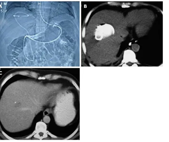
A B
C
Figure 2 Nasocystic-biliary drainage procedure. A: Topographic images of hydatid cyst with major rupture by nasocystic drainage; B: Drainage catheter within the cystic cavity; C: Computed tomography of the same patient taken at the three-month follow-up.
studies claim that endoscopic or percutaneous methods should be used first for the management of postopera-tive strictures. Biliary strictures affect mostly long and multiple segments, are located proximally (Bismuth type
Ⅲ and Ⅳ), and cholangiographic signs show diffuse involvement as primary sclerosing cholangitis. Although ERCP can be an appropriate method for the treatment of complications due to stricture, such as jaundice, chol-angitis and sepsis, the repairs are costly and lengthy and can result in cholangitis and liver abscess formation. However, therapeutic ERCP should be preferred for bili-ary strictures distally located and involving single short segments, whereas percutaneous transhepatic cholangi-ography should be used for proximally located, multiple and long segment biliary strictures[16,35,39,58].
The formation of biliary fistulae is the most com-mon postoperative complication, with a postoperative incidence as high a 50%-63%[53,59]. Although biliary
leak-age risk is higher in the early postoperative period, it begins to decrease from the tenth day after surgery. Per-sistent biliary fistulae are defined by a high amount of biliary drainage lasting more than ten days after surgery, and ERCP use is advised in these cases[5,15,60]. It is
dif-ficult to determine whether postoperative biliary fistulae will subside spontaneously or will turn into a persistent form. However, the presence of a large amount of leak-ing bile in the early postoperative period, or drainage that does not decrease day by day suggests that the fis-tula will not spontaneously subside. In such cases, there is no need to wait ten days, and ERCP can be performed early in the postoperative period, as any delay will cause the chronicity of fistulae and infective complications. In contrast, small or decreasing amounts of drainage may be followed conservatively[61,62].
When biliary fistulae are observed, it is important to evaluate the clinical effectiveness of biliary drainage. Effective biliary drainage is displayed by controlled fis-tulae, which show no signs of localized or generalized peritonitis, and no pathophysiologic signs of cholestasis. In uncontrolled biliary fistulae, there is generally insuf-ficient biliary drainage and this condition results in intra-abdominal bile collection. The condition requires urgent hemodynamic stabilization of the patient, and drainage of the collection percutaneously, laparoscopically or sur-gically. Undrained bile will become infected, resulting in subphrenic, suphepatic abscess formation or generalized peritonitis. In some cases, ineffective biliary draining can lead to life-threatening cholangitis, intrahepatic abscesses and septicemia, which require urgent manipulations[51].
To treat biliary fistulae, ERCP with endoscopic treat-ments, such as ES, NBD and biliary stenting, are used to bypass the sphincter of Oddi, reduce duodenobiliary pressure differences, and to divert the bile flow into the duodenum. As pressure within the biliary tree decreases, bile will flow into the duodenum by way of the papilla Vateri instead of flowing into the cystic cavity. Although the advantages of these techniques are debated, the suc-cess rate has been reported as high as 90% accompanied
by a low mortality rate[16]. In the patients who
under-went ES in the study by Rodriguez et al[39] postoperative
fistulae were closed successfully in approximately 25 d. In the study by Tekant et al[63] postoperative persistent
biliary fistulae subsided spontaneously in seven days in all but one patient who underwent ES. The same study suggested that late ES may delay fistula closing, and thus ERCP should be performed at an early period. Reports in the literature show that postoperative ES fistulae close within 3-43 d, yet hospitalization typically lasts only 3-9 d for ERCP procedures[7,43,49,52,64].
In a study by Bilsel et al[50] ES was considered to be the
appropriate treatment modality as stent usage was not cost-effective and required a second ERCP for removal. How-ever, the same study reported that ES should be combined with biliary stenting for chronic and high-output fistulae in major cystobiliary communications, which was also in-dicated in a study by Adas et al[65]. Akcakaya et al[66] suggest
that ES should be the first method selected, but in spe-cific cases where there is a defect in emptying of hydatid materials, or a stricture or gallstone is present in the com-mon bile duct, biliary stenting should be used directly. However, Cicek et al[5] recommend using NBD, rather
than ES, with biliary stenting, as NBD is more effective in decreasing intrabiliary pressure and permits follow-up by cholangiography. Moreover, preoperative NBD aids in the intraoperative localization of cystobiliary communi-cations[67-69]. Despite these apparent advantages, ES may
be needed in some cases where hydatid membranes are within the bile ducts, and because of the low tolerability of lengthy hospitalization for NBD.
In our experience, ES is the primary choice as it is a safe and effective treatment for biliary fistulae resulting from hydatid cyst surgery. In addition, early ERCP is limited to patients with fistula output that is high or does not decrease within the first week. Because small hydatid particles cannot be visualized clearly with cholangiogra-phy, all of our patients undergo ES, bile ducts are con-trolled routinely with balloons and basket catheters, and hydatid elements are cleaned out if present. If biliary strictures accompany biliary fistulae, ES is supplemented with biliary stenting. Finally, NCBD is performed fol-lowing ES in cases of biliary sepsis, particularly when the bile ducts are filled with hydatid elements.
CONCLUSION
Preoperative ERCP is the primary diagnostic and thera-peutic method for frank IBR. Following a diagnosis of frank IBR, cystic material can be emptied by NBD, with-out a need for further surgical intervention. However, preoperative ERCP is not indicated for cases involving large or multiple cysts, hilar or caudate lobe cysts, or if there is minimal dilatation of intrahepatic bile ducts proximal to the cyst. Moreover, ERCP can fail to detect simple communications and may promote the formation of postoperative biliary fistulae. Even in the most ex-perienced hands, serious complications can result from
ERCP, including pancreatitis, bleeding, infection or per-foration. Therefore, suspected minor communications should be investigated by non-invasive methods such as MRCP, US and CT.
Therapeutic ERCP is indicated for early (persistent biliary fistula and obstructive jaundice) and late (scleros-ing cholangitis and sphincter of Oddi stenosis) postop-erative biliary adverse events, of which biliary fistulae are the most common. Although some fistulae spontane-ously resolve, high-output or persistent fistulae require ERCP. If biliary fistulae are accompanied by strictures or do not close after ES, biliary stenting should be added. However, biliary stenting and NBD should be confined to rare cases.
ACKNOWLEDGMENTS
Authors would like to thank to Dr Pelin Basim for wrote the English language of this review article.
REFERENCES
1 Eckert J, Deplazes P. Biological, epidemiological, and clini-cal aspects of echinococcosis, a zoonosis of increasing con-cern. Clin Microbiol Rev 2004; 17: 107-135 [PMID: 14726458 DOI: 10.1128/CMR.17.1.107-135.2004]
2 McManus DP, Zhang W, Li J, Bartley PB. Echinococcosis. Lancet 2003; 362: 1295-1304 [PMID: 14575976 DOI: 10.1016/ S0140-6736(03)14573-4]
3 Ponchon T, Bory R, Chavaillon A. Endoscopic retrograde cholangiography and sphincterotomy for complicated hepatic hydatid cyst. Endoscopy 1987; 19: 174-177 [PMID: 3622400 DOI: 10.1055/s-2007-1018274]
4 Becker K, Frieling T, Saleh A, Häussinger D. Resolution of hydatid liver cyst by spontaneous rupture into the biliary tract. J Hepatol 1997; 26: 1408-1412 [PMID: 9210631 DOI: 10.1016/S0168-8278(97)80479-5]
5 Cicek B, Parlak E, Disibeyaz S, Oguz D, Cengiz C, Sahin B. Endoscopic therapy of hepatic hydatid cyst disease in pre-operative and postpre-operative settings. Dig Dis Sci 2007; 52: 931-935 [PMID: 17333353 DOI: 10.1007/s10620-006-9426-4] 6 Dadoukis J, Gamvros O, Aletras H. Intrabiliary rupture of
the hydatid cyst of the liver. World J Surg 1984; 8: 786-790 [PMID: 6506741 DOI: 10.1007/BF01655782]
7 Paksoy M, Karahasanoglu T, Carkman S, Giray S, Senturk H, Ozcelik F, Erguney S. Rupture of the hydatid disease of the liver into the biliary tracts. Dig Surg 1998; 15: 25-29 [PMID: 9845559 DOI: 10.1159/000018582]
8 Ramia JM, Figueras J, De la Plaza R, García-Parreño J. Cys-to-biliary communication in liver hydatidosis. Langenbecks Arch Surg 2012; 397: 881-887 [PMID: 22374106 DOI: 10.1007/ s00423-012-0926-8]
9 Akcan A, Sozuer E, Akyildiz H, Ozturk A, Atalay A, Yilmaz Z. Predisposing factors and surgical outcome of complicated liver hydatid cysts. World J Gastroenterol 2010; 16: 3040-3048 [PMID: 20572308 DOI: 10.3748/wjg.v16.i24.3040]
10 Murty TV, Sood KC, Rakas FS. Biliary obstruction due to ruptured hydatid cyst. J Pediatr Surg 1989; 24: 401-403 [PMID: 2732886 DOI: 10.1016/S0022-3468(89)80281-7]
11 Zaouche A, Haouet K, Jouini M, El Hachaichi A, Dziri C. Management of liver hydatid cysts with a large biliocystic fistula: multicenter retrospective study. Tunisian Surgical Association. World J Surg 2001; 25: 28-39 [PMID: 11213153 DOI: 10.1007/s002680020005]
12 Dew H. Some complications of hydatid disease. Br J Surg 1930; 18: 275-293 [DOI: 10.1002/bjs.1800187010]
13 Kayaalp C. Hydatid cyst of the liver. 4th ed. In: Surgery of the liver, biliary tract, and pancreas. In: Blumgart LH, edi-tor. Philadelphia, PA: Saunders Elsevier, 2007; 952-970 14 Bektaş M, Dökmeci A, Cinar K, Halici I, Oztas E,
Karayal-cin S, Idilman R, Sarioglu M, Ustun Y, Nazligul Y, Ormeci N, Ozkan H, Bozkaya H, Yurdaydin C. Endoscopic man-agement of biliary parasitic diseases. Dig Dis Sci 2010; 55: 1472-1478 [PMID: 19513838 DOI: 10.1007/s10620-009-0850-0] 15 Manouras A, Genetzakis M, Antonakis PT, Lagoudianakis
E, Pattas M, Papadima A, Giannopoulos P, Menenakos E. Endoscopic management of a relapsing hepatic hydatid cyst with intrabiliary rupture: a case report and review of the literature. Can J Gastroenterol 2007; 21: 249-253 [PMID: 17431515]
16 El Malki HO, El Mejdoubi Y, Souadka A, Mohsine R, Ifrine L, Abouqal R, Belkouchi A. Predictive model of biliocystic communication in liver hydatid cysts using classification and regression tree analysis. BMC Surg 2010; 10: 16 [PMID: 20398342 DOI: 10.1186/1471-2482-10-16]
17 Reedy N, Rao GV, Ong WC, Rupa B. Parasitic disease: En-doscopic diagnosis and management of tropical parasitic infestations. 1st ed. ERCP. In: Baron TH, Kozarek R, Carr-Locke DL, editors. Philadelphia, PA: Saunders Elsevier, 2008; 393-398
18 Kayaalp C, Bostanci B, Yol S, Akoglu M. Distribution of hyda-tid cysts into the liver with reference to cystobiliary communi-cations and cavity-related complicommuni-cations. Am J Surg 2003; 185: 175-179 [PMID: 12559452 DOI: 10.1016/S0002-9610(02)01202-3] 19 Lewall DB, McCorkell SJ. Rupture of echinococcal cysts:
diagnosis, classification, and clinical implications. AJR Am J Roentgenol 1986; 146: 391-394 [PMID: 3510519]
20 Dolay K, Soylu A, Yanar H, Aygun E, Ertekin C. Manage-ment of intrabiliary rupture of hepatic hydatid cysts with endoscopic nasocystic drainage”. Paris: 15th United Euro-pean Gastroenterlogy Week (UEGW), 2007: 27-31
21 Hankins JR. Management of complicated hepatic hydatid cysts. Ann Surg 1963; 158: 1020-1034 [PMID: 14081536 DOI: 10.1097/00000658-196312000-00014]
22 Alper A, Ariogul O, Emre A, Uras A, Okten A. Choledo-choduodenostomy for intrabiliary rupture of hydatid cysts of liver. Br J Surg 1987; 74: 243-245 [PMID: 3580792 DOI: 10.1002/bjs.1800740405]
23 Galati G, Sterpetti AV, Caputo M, Adduci M, Lucandri G, Brozzetti S, Bolognese A, Cavallaro A. Endoscopic retro-grade cholangiography for intrabiliary rupture of hydatid cyst. Am J Surg 2006; 191: 206-210 [PMID: 16442947 DOI: 10.1016/j.amjsurg.2005.09.014]
24 el-Tahir MI, Omojola MF, Malatani T, al-Saigh AH, Ogun-biyi OA. Hydatid disease of the liver: evaluation of ultra-sound and computed tomography. Br J Radiol 1992; 65: 390-392 [PMID: 1611417 DOI: 10.1259/0007-1285-65-773-390] 25 Ozaslan E, Bayraktar Y. Endoscopic therapy in the manage-ment of hepatobiliary hydatid disease. J Clin Gastroenterol 2002; 35: 160-174 [PMID: 12172363 DOI: 10.1097/00004836-2 00208000-00009]
26 Ascenti G, Scribano E, Loria G, Vallone A, Pandolfo I, Gaeta M. [Computerized tomography in the assessment of ob-structive jaundice caused by hepatic hydatid cysts]. Radiol Med 1995; 89: 804-808 [PMID: 7644732]
27 Pawlik TM. Echinococcal disease of the liver. 9th edition. Current Surgical Therapy. In: Cameron JL, editor. Philadel-phia, PA: Elsevier-Mosby, 2008: 331-35
28 Unalp HR, Baydar B, Kamer E, Yilmaz Y, Issever H, Tarcan E. Asymptomatic occult cysto-biliary communication without bile into cavity of the liver hydatid cyst: a pitfall in conservative surgery. Int J Surg 2009; 7: 387-391 [PMID: 19573629 DOI: 10.1016/j.ijsu.2009.06.012]
29 Lygidakis NJ. Diagnosis and treatment of intrabiliary rupture of hydatid cyst of the liver. Arch Surg 1983; 118: 1186-1189 [PMID: 6615202 DOI: 10.1001/archsurg.1983.01390100056014]
30 Pitt HA, Korzelius J, Tompkins RK. Management of hepatic echinococcosis in Southern California. Am J Surg 1986; 152: 110-115 [PMID: 3728803 DOI: 10.1016/0002-9610(86)90159-5] 31 Kilic M, Yoldas O, Koc M, Keskek M, Karakose N, Ertan T,
Gocmen E, Tez M. Can biliary-cyst communication be pre-dicted before surgery for hepatic hydatid disease: does size matter? Am J Surg 2008; 196: 732-735 [PMID: 18513700 DOI: 10.1016/j.amjsurg.2007.07.034]
32 Kuntz C, Manner M. [Hepatic echinococcosis with gallstones of the echinococcal cavity]. Dtsch Med Wochenschr 1995; 120: 1699-1702 [PMID: 7497895 DOI: 10.1055/s-2008-1055531] 33 Regan JK, Brown RD, Marrero JA, Malik P, Rosenberg F,
Venu RP. Chronic pancreatitis resulting from primary hy-datid disease of the pancreas: a case report and review of the literature. Gastrointest Endosc 1999; 49: 791-793 [PMID: 10343231 DOI: 10.1016/S0016-5107(99)70304-49]
34 Uccheddu A, Murgia C, Licheri S, Dazzi C, Cagetti M. [En-doscopic retrograde cholangiography in the diagnosis of hepatic hydatidosis]. G Chir 1989; 10: 46-50 [PMID: 2518529] 35 Magistrelli P, Masetti R, Coppola R, Costamagna G,
Duras-tante V, Nuzzo G, Picciocchi A. Value of ERCP in the diagno-sis and management of pre- and postoperative biliary com-plications in hydatid disease of the liver. Gastrointest Radiol 1989; 14: 315-320 [PMID: 2680737 DOI: 10.1007/BF01889226] 36 Kaltenthaler EC, Walters SJ, Chilcott J, Blakeborough A,
Ver-gel YB, Thomas S. MRCP compared to diagnostic ERCP for diagnosis when biliary obstruction is suspected: a systematic review. BMC Med Imaging 2006; 6: 9 [PMID: 16907974 DOI: 10.1186/1471-2342-6-9]
37 Kumar R, Reddy SN, Thulkar S. Intrabiliary rupture of hyda-tid cyst: diagnosis with MRI and hepatobiliary isotope study. Br J Radiol 2002; 75: 271-274 [PMID: 11932222]
38 Zargar SA, Khuroo MS, Khan BA, Dar MY, Alai MS, Koul P. Intrabiliary rupture of hepatic hydatid cyst: sonographic and cholangiographic appearances. Gastrointest Radiol 1992; 17: 41-45 [PMID: 1544557 DOI: 10.1007/BF01888506] 39 Rodriguez AN, Sánchez del Río AL, Alguacil LV, De Dios
Vega JF, Fugarolas GM. Effectiveness of endoscopic sphinc-terotomy in complicated hepatic hydatid disease. Gastroin-test Endosc 1998; 48: 593-597 [PMID: 9852449 DOI: 10.1016/ S0016-5107(98)70041-0]
40 Dumas R, Le Gall P, Hastier P, Buckley MJ, Conio M, Del-mont JP. The role of endoscopic retrograde cholangiopan-creatography in the management of hepatic hydatid disease. Endoscopy 1999; 31: 242-247 [PMID: 10344429 DOI: 10.1055/ s-1999-14209]
41 Akoglu M, Hilmioglu F, Balay AR, Sahin B, Davidson BR. Endoscopic sphincterotomy in hepatic hydatid disease open to the biliary tree. Br J Surg 1990; 77: 1073 [PMID: 2207579 DOI: 10.1002/bjs.1800770940]
42 Akkiz H, Akinoglu A, Colakoglu S, Demiryürek H, Yagmur O. Endoscopic management of biliary hydatid disease. Can J Surg 1996; 39: 287-292 [PMID: 8697318]
43 Spiliadis C, Georgopoulos S, Dailianas A, Konstantinidis A, Rimikis M, Skandalis N. The use of ERCP in the study of patients with hepatic echinococcosis before and after surgi-cal intervention. Gastrointest Endosc 1996; 43: 575-579 [PMID: 8781936 DOI: 10.1016/S0016-5107(96)70194-3]
44 de Aretxabala X, Perez OL. The use of endoprostheses in bili-ary fistula of hydatid cyst. Gastrointest Endosc 1999; 49: 797-799 [PMID: 10343233 DOI: 10.1016/S0016-5107(99)70306-8] 45 Sáez-Royuela F, Yuguero L, López-Morante A,
Pérez-Alva-rez JC, Martín-Lorente JL, Ojeda C. Acute pancreatitis caused by hydatid membranes in the biliary tract: treatment with en-doscopic sphincterotomy. Gastrointest Endosc 1999; 49: 793-796 [PMID: 10343232 DOI: 10.1016/S0016-5107(99)70305-6] 46 Hilmioglu F, Karincaoglu M, Yilmaz S, Yildirim B,
Kirimlio-glu V, Aladag M, Onmus H. Complete treatment of ruptured hepatic cyst into biliary tree by ERCP. Dig Dis Sci 2001; 46: 463-467 [PMID: 11318516]
47 Kattan YB. Intrabiliary rupture of hydatid cyst of the liver. Br J Surg 1975; 62: 885-890 [PMID: 1191949 DOI: 10.1002/ bjs.18006211089]
48 Ulualp KM, Aydemir I, Senturk H, Eyuboğlu E, Cebeci H, Unal G, Unal H. Management of intrabiliary rupture of hy-datid cyst of the liver. World J Surg 1995; 19: 720-724; discus-sion 728 [PMID: 7571669 DOI: 10.1007/BF00295913] 49 Saritas U E, Akoglu M, Sahin B. Effectiveness of endoscopic
treatment modalities in complicated hepatic hydatid dis-ease after surgical intervention. Endoscopy 2001; 33: 858-863 [PMID: 11571682 DOI: 10.1055/s-2001-17342]
50 Bilsel Y, Bulut T, Yamaner S, Buyukuncu Y, Bugra D, Aky-uz A, Sokucu N. ERCP in the diagnosis and management of complications after surgery for hepatic echinococcosis. Gastrointest Endosc 2003; 57: 210-213 [PMID: 12556786 DOI: 10.1067/mge.2003.64]
51 Kornaros SE, Aboul-Nour TA. Frank intrabiliary rupture of hydatid hepatic cyst: diagnosis and treatment. J Am Coll Surg 1996; 183: 466-470 [PMID: 8912615]
52 Giouleme O, Nikolaidis N, Zezos P, Budas K, Katsinelos P, Vasiliadis T, Eugenidis N. Treatment of complications of he-patic hydatid disease by ERCP. Gastrointest Endosc 2001; 54: 508-510 [PMID: 11577320 DOI: 10.1067/mge.2001.118256] 53 Somani SK, Srivastava AP. Resolution of hepatic hydatid
cyst with biliary communication with ERCP. J Gastroint Dig Syst 2012; 2: 114 [DOI: 10.4172/2161-069X.1000114]
54 al Karawi MA, Yasawy MI, el Shiekh Mohamed AR. En-doscopic management of biliary hydatid disease: report on six cases. Endoscopy 1991; 23: 278-281 [PMID: 1743129 DOI: 10.1055/s-2007-1010686]
55 Al Karawi MA, Mohamed AR, Yasawy I, Haleem A. Non-surgical endoscopic trans-papillary treatment of ruptured echinococcus liver cyst obstructing the biliary tree. Endoscopy 1987; 19: 81-83 [PMID: 3569154 DOI: 10.1055/s-2007-1013021] 56 Singh V, Reddy DC, Verma GR, Singh G. Endoscopic
management of intrabiliary-ruptured hepatic hydatid cyst. Liver Int 2006; 26: 621-624 [PMID: 16762008 DOI: 10.1111/ j.1478-3231.2006.01259.x]
57 Agarwal S, Sikora SS, Kumar A, Saxena R, Kapoor VK. Bile leaks following surgery for hepatic hydatid disease. Indian J Gastroenterol 2005; 24: 55-58 [PMID: 15879650]
58 Yilmaz U, Sakin B, Boyacioglu S, Saritas U, Cumhar T, Ako-glu M. Management of postoperative biliary strictures sec-ondary to hepatic hydatid disease by endoscopic stenting. Hepatogastroenterology 1998; 45: 65-69 [PMID: 9496489] 59 Simşek H, Ozaslan E, Sayek I, Savaş C, Abbasoğlu O, Soylu
AR, Balaban Y, Tatar G. Diagnostic and therapeutic ERCP in hepatic hydatid disease. Gastrointest Endosc 2003; 58: 384-389 [PMID: 14528213 DOI: 10.1067/S0016-5107(03)00013-0] 60 Kayaalp C, Bzeizi K, Demirbag AE, Akoglu M. Biliary
com-plications after hydatid liver surgery: incidence and risk factors. J Gastrointest Surg 2002; 6: 706-712 [PMID: 12399060 DOI: 10.1016/S1091-255X(02)00046-X]
61 Dolay K, Akçakaya A, Soybir G, Cabioğlu N, Müslümanoğlu M, Iğci A, Topuzlu C. Endoscopic sphincterotomy in the management of postoperative biliary fistula A complication of hepatic hydatid disease. Surg Endosc 2002; 16: 985-988 [PMID: 12163969 DOI: 10.1007/s00464-001-9020-y]
62 Zeybek N, Dede H, Balci D, Coskun AK, Ozerhan IH, Peker S, Peker Y. Biliary fistula after treatment for hydatid disease of the liver: when to intervene. World J Gastroenterol 2013; 19: 355-361 [PMID: 23372357 DOI: 10.3748/wjg.v19.i3.355] 63 Tekant Y, Bilge O, Acarli K, Alper A, Emre A, Arioğul O.
Endoscopic sphincterotomy in the treatment of postopera-tive biliary fistulas of hepatic hydatid disease. Surg Endosc 1996; 10: 909-911 [PMID: 8703149 DOI: 10.1007/BF00188481] 64 Vignote ML, Miño G, de la Mata M, de Dios JF, Gomez F.
Endoscopic sphincterotomy in hepatic hydatid disease open to the biliary tree. Br J Surg 1990; 77: 30-31 [PMID: 2302509 DOI: 10.1002/bjs.1800770110]
65 Adas G, Arikan S, Gurbuz E, Karahan S, Eryasar B, Kara-tepe O, Tekant Y. Comparison of endoscopic therapeutic modalities for postoperative biliary fistula of liver hydatid cyst: a retrospective multicentric study. Surg Laparosc En-dosc Percutan Tech 2010; 20: 223-227 [PMID: 20729689 DOI: 10.1097/SLE.0b013e3181e12ee6]
66 Akcakaya A, Sahin M, Karakelleoglu A, Okan I. Endoscopic stenting for selected cases of biliary fistula after hepatic hydatid surgery. Surg Endosc 2006; 20: 1415-1418 [PMID: 16736309 DOI: 10.1007/s00464-005-0572-0]
67 Marks JM, Ponsky JL, Shillingstad RB, Singh J. Biliary
stent-ing is more effective than sphincterotomy in the resolu-tion of biliary leaks. Surg Endosc 1998; 12: 327-330 [PMID: 9543522 DOI: 10.1007/s004649900663]
68 Youngelman DF, Marks JM, Ponsky T, Ponsky JL. Compari-son of bile duct pressures following sphincterotomy and endobiliary stenting in a canine model. Surg Endosc 1997; 11: 126-128 [PMID: 9069142 DOI: 10.1007/s004649900313] 69 Sharma BC, Reddy RS, Garg V. Endoscopic management
of hepatic hydatid cyst with biliary communication. Dig Endosc 2012; 24: 267-270 [PMID: 22725113 DOI: 10.1111/ j.1443-1661.2011.01225.x]
P- Reviewer: Moralioglu S S- Editor: Qi Y L- Editor: A E- Editor: Liu XM