138
Turk J Gastroenterol 2014; 25 (Suppl.-1): 138-41
Fatty liver disease might increase the risk of abdominal operation
in patients with fatty liver and the prevalence of cancer in
first-degree relatives
Metin Başaranoğlu1, Billur Canbakan2, Kemal Yıldız3, Bahadır Ceylan4, Birol Baysal3, Ömer Uysal5, Hakan Şentürk3
1Department of Gastroenterology, Türkiye Yüksek İhtisas Hospital, Ankara, Turkey
2Department of Gastroenterology, İstanbul University Cerrahpaşa Faculty of Medicine, İstanbul, Turkey 3Department of Gastroenterology, Bezmialem Vakif University Faculty of Medicine, İstanbul, Turkey 4Department of Clinical Microbiology and Infectious Diseases, Medipol University Hospital, İstanbul, Turkey 5Department of Biostatistics, Bezmialem Vakif University Faculty of Medicine, İstanbul, Turkey
ABSTRACT
Background/Aims: We investigated the risk of abdominal operation in patients with fatty liver and the risk of any cancer in first-degree relatives of patients with fatty liver.
Materials and Methods: We evaluated 105 patients with nonalcoholic fatty liver disease (NAFLD), 121 patients with biopsy-proven hepatitis C (61 patients with fatty liver and 60 patients without fatty liver), 50 patients with inflammatory bowel disease (IBD), and 109 patients with dyspepsia.
Results: There was no difference in sex, mean age, and marital status among the groups except that patients with IBD were younger than the others (p<0.001). The frequency of cancer among family members was 18% in IBD, 9% in dyspepsia, 28% in hepatitis C with steatosis, 21.5% in hepatitis C without steatosis, and 27% in NAFLD (p=0.006). Then, we divided the study group into two groups as follows: group 1: (IBD + dyspepsia + hepatitis C without ste-atosis) and group 2: (hepatitis C with steatosis + NAFLD). We found that the frequency of cancer was 16% in group 1 versus 24.4 % in group 2 (p=0.037). We also investigated the risk of abdominal operation in patients with fatty liver. The results were as follows: 33% in group without fatty liver versus 43% in group with fatty liver (p=0.043).
Conclusion: Understanding the underlying causes of fatty liver forms might decrease the cancer frequency in the population and number of operations in patients with fatty liver.
Keywords: Abdominal operation, cancer, dyspepsia, fatty liver, hepatitis C, nonalcoholic
INTRODUCTION
Nonalcoholic fatty liver disease (NAFLD) is one of the most prevalent forms of chronic liver diseases in gas-troenterology and hepatology practices (1,2). Contrib-uting factors to this include the increasingly sedentary lifestyle of the population, besides the increased con-sumption of a high-fat (HF) diet and high-fructose corn syrup (HFCS) (3-5). It is now accepted that in the setting of excessive central adiposity, insulin resistance is the major underlying cause of fat accumulation in the liver (6-10). Insulin resistance and the metabolic syndrome are growing threats to the health of people in devel-oped nations (1,2). NAFLD has the potential to progress to cirrhosis, hepatocellular carcinoma (HCC), end-stage
liver disease, and liver transplantation (11-16). However, one of the most important and unresolved problems is the pathogenesis and underlying mechanisms in the development of NAFLD, despite the growing body of literature (17-22). In this study, we investigated the risk of abdominal operation in patients with fatty liver. Ad-ditionally, the risk of cancer in the first-degree relatives of patients with fatty liver was evaluated.
MATERIALS AND METHODS
A randomized prospective study was performed at the Department of Gastroenterology, Bezmialem Vakif Uni-versity, İstanbul, Turkey. The local ethic committee ap-proved this study.
xxxxxxxxxxxxx
This study was presented by Dr. Hakan Sentürk as a poster at the 63rd Annual Meeting of the American Association for the Study of Liver Disease (AASLD), 9-13 November 2012, Boston, USA.
Address for Correspondence: Hakan Şentürk, Department of Gastroenterology, Bezmialem Vakif University Faculty of Medicine, İstanbul, Turkey E-mail: [email protected]
Received: March 03, 2014 Accepted: July 23, 2014
© Copyright 2014 by The Turkish Society of Gastroenterology • Available online at www.turkjgastroenterol.org • DOI: 10.5152/tjg.2014.7674
Or
iginal Ar
ticle
Retraction Notice
The following article is retracted from publication in Turkish Journal of Gastroenterology by decision of editors of the journal since it is a duplicate publication, which has been previously published in Wien Klin Wochenschr. 2015 Apr 14 DOI: 10.1007/s00508-015-0744-4.
139
Alcohol history was excluded from each patient by self-report, and an alcohol abuse history was taken from family members for each patient. Any drug history or history of chronic hepatitis, hy-pertension, and diabetes were all questioned. Ultrasonographic evaluation of the hepatobiliary system was performed. A fatty liver was diagnosed by increased echogenicity or increased liv-er-kidney contrast. Serum serology of HBsAg (hepatitis B surface antigen), anti-HBs hepatitis B surface), anti-HBc-total (anti-hepatitis B core-total), and anti-HCV (anti-(anti-hepatitis C virus) was measured. If necessary, liver biopsies were processed and read by a single pathologist. NAFLD was diagnosed by experienced pathologists according to standard criteria (23-25).
We first performed a power analysis/sample size determination (a single-sample z-test) for the proportion. We evaluated 105 pa-tients with NAFLD, 121 papa-tients with hepatitis C (61 with hepatic steatosis, and 60 without steatosis), 50 patients with inflamma-tory bowel disease (IBD), and 109 patients with dyspepsia. Statistical analysis
The data were presented as mean±SD and were analyzed by SPSS 13 (SPSS Inc, Chicago, IL, USA). Features of groups were
compared with each other by t-test and chi-square test. Prob-ability (p) values <0.05 were considered statistically significant. RESULTS
There was no difference in sex, mean age, and marital status among the groups except that patients with IBD were younger than others (p<0.001). The frequency of cancer in their first-de-gree relatives was 18% in IBD, 9% in dyspepsia, 28% in hepatitis C with steatosis, 21.5% in hepatitis C without steatosis, and 27% in NAFLD (p=0.006 among the groups), as shown in Figure 1. After the exclusion of patients with dyspepsia, there was no signifi-cant difference among the groups. Then, we divided the study group into two groups-group 1: IBD + dyspepsia + hepatitis C without steatosis; and group 2: hepatitis C with fatty liver steato-sis + NAFLD-and performed the analysteato-sis. We found that the fre-quency of cancer was 16% in group 1 without fatty liver versus 24.4% in group 2 with fatty liver (p=0.037), as shown in Figure 2. In the second part of the study, we investigated the frequency of operation in patients with fatty liver. We found that the frequen-cy of operation in patients with fatty liver was 4% in IBD (after Figure 1. The frequency of cancer in family members was 18% in IBD
(gr1), 9% in dyspepsia (gr2), 28% in hepatitis C with steatosis (gr3), 21.5% in hepatitis C without steatosis (gr4), and 27.9% in NAFLD (gr5) (p=0.006 among the groups). There was no difference among the groups in cancer frequency after the exclusion of patients with dyspepsia (p>0.05).
18.0%
9.2% p=0.006
Frequency of cancer in family
60.0% 50.0% 40.0% 30.0% 20.0% 10.0% .0% -10.0% 28.3% 21.5% 27.9% gr1 gr2 gr3 gr4 gr5 *
Figure 2. After dividing the study group into two groups as group 1: IBD + dyspepsia + hepatitis C without steatosis and group 2: hepatitis C with steatosis + NAFLD, we performed the analysis: 16% in group 1 without fatty liver vs. 24.4 % in group 2 with fatty liver (p=0.037).
p=0.037 group 1 group 2 24.40% 16% 30% 25% 20% 15% 10% 5% 0%
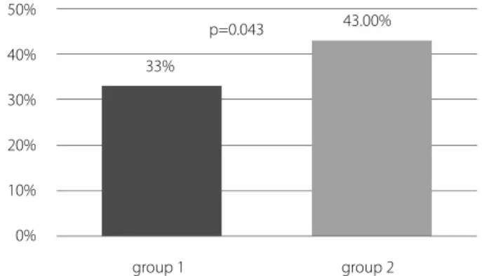
Figure 4. The frequency of operation in patients with fatty liver was 33% in group 1 without fatty liver vs. 43% in group 2 with fatty liver (p=0.043). Group 1: IBD + dyspepsia + hep C without fatty liver. Group 2: hep C with steatosis + NAFLD. p=0.043 group 1 group 2 43.00% 33% 50% 40% 30% 20% 10% 0%
Figure 3. The frequency of operation was 4% in patients with IBD (after the exclusion of intestinal surgeries due to IBD), 40% in dyspepsia, 45% in hepatitis C with steatosis, 41% in hepatitis C without steatosis, and 44% in NAFLD, respectively (p=0.001 among the groups). There was no differ-ence among the groups in the operation frequency after the exclusion of patients with IBD (p>0.05).
The frequency of previous operations in patients 60.0% 40.0% 20.0% .0% 40.4% 45.0% 41.3% 44.2% 4.0% gr1 gr2 gr3 gr4 gr5
Başaranoğlu et al. Fatty liver disease
Turk J Gastroenterol 2014; 25 (Suppl.-1): 138-41
Or
iginal Ar
140
the exclusion of intestinal surgeries due to IBD), 40% in dyspep-sia, 45% in hepatitis C with steatosis, 41% in hepatitis C without steatosis, and 44% in NAFLD (p=0.001 among the groups), as shown in Figure 3. On further analysis, the prevalence of opera-tions was 33% in group 1 without fatty liver versus 43% in group 2 with fatty liver (p=0.043), as shown in Figure 4.
DISCUSSION
In this study, we showed an increase in both the frequency of operations in patients with fatty liver and the frequency of can-cer in their first-degree relatives, independent of the underly-ing chronic disease. All of these results were obtained indepen-dently of sex, mean age, and marital status of the patients. We showed that the frequency of cancer among the family mem-bers was 9% in dyspepsia, 21.5% in hepatitis C without steato-sis, 28% in hepatitis C with steatosteato-sis, and 27% in NAFLD, respec-tively. These results indicated that fat accumulation in the liver might increase the cancer rate among family members. We consider that obesity-induced fatty liver has a more detrimen-tal effect on the body system than hepatitis C-induced fatty liver. Then, we divided the study group into two big groups by the occurrence of fat to see whether there were any changes in the statistical results: group 1: (IBD + dyspepsia + hepatitis C without steatosis) and group 2: (hepatitis C with steatosis + NAFLD). As expected, although the frequency of cancer was 16% in group means none fat group, the frequency of cancer reached 24.4% in the fatty liver group, with statistical signifi-cance (p=0.037).
In this study, we also investigated the risk of abdominal op-eration in patients with fatty liver. The abdominal opop-eration rate was 33% in patients without fatty liver. On the other hand, 43% of patients with fatty liver underwent abdominal opera-tion previously. These frequencies were statistically meaningful (p=0.043).
A growing body of literature has indicated that there is an association between obesity, type 2 diabetes mellitus, and particular cancer types, such as enhanced risk of colorectal, esophageal, and kidney cancer, by large epidemiological stud-ies (12-15). This risk was maintained, even after adjusting for factors, such as BMI, family history, physical activity, smoking, red meat consumption, hormone, and aspirin use. The cause of these findings was not investigated in this study. However, shared mechanisms in the development of fatty liver disease and cancer might be a reason of the increased frequency of cancer rates. Some genes, ligands, and various factor receptors (insulin, IGF1 axis, ectopic E synthesis, adipokines, PTEN, PI3K, mTOR, MAPK, HIF-1α, COX-2, MIF), might increase the risk of cancer, which might be dealing with the pathogenesis of fatty liver.
On the other hand, an impaired immune system is one of the underlying mechanisms in the development of fatty liver (1-4,7,18,19). Patients with fatty liver have chronic low-grade
in-flammation. Thus, increased systemic body inflammation and its consequences might lead to increased frequency of opera-tions in patients with fatty liver.
In conclusion, independent of the underlying chronic disease, the occurrence of fat in the liver increases both the frequency of operations in patients with fatty liver and the frequency of cancer in their first-degree relatives. These findings indicate that we should carefully focus on the prevention and therapy of fatty liver. Understanding the underlying causes of fatty liver forms the basis for rational preventive and treatment strategies. These attempts might decrease the cancer frequency in the popula-tion and number of operapopula-tions in patients with fatty liver.
Conflict of Interest: No conflict of interest was declared by the authors.
REFERENCES
1. Basaranoğlu M, Neuschwander-Tetri Brent A. Nonalcoholic fatty liver disease: Clinical features and pathogenesis. Gastroenterol-ogy & HepatolGastroenterol-ogy. 2006;2:282-291.
2. Basaranoglu M, Ormeci N. Nonalcoholic fatty liver disease: Diag-nosis, pathogenesis, and management. Turk J Gastroenterol 2014; 25: 127-32.
3. Tetri LH, Basaranoglu M, Brunt EM, Yerian LM, Neuschwander-Tetri BA. Severe NAFLD with hepatic necroinflammatory changes in mice fed trans fats and a high-fructose corn syrup equivalent. Am J Physiol Gastrointest Liver Physiol 2008; 295: 987-95.
4. Neuschwander-Tetri BA, Ford DA, Acharya S, et al. Dietary fatty acid induced NASH is normalized following loss of trans-fatty acids from hepatic lipid pools. Lipids 2012; 47: 941-50. 5. Basaranoglu M, Basaranoglu G, Sabuncu T, Sentürk H. Fructose
as a key player in the development of fatty liver disease. World J Gastroenterol 2013; 19: 1166-72.
6. Basaranoglu M, Sonsuz A, Senturk H, Akin P. The low incidence of primary liver disease in patients with nonalcoholic steatohepati-tis. J Hepatol 2001; 35: 684-5.
7. Basaranoglu M, Kayacetin S, Yilmaz N, Kayacetin E, Tarcin O, Son-suz A. Understanding mechanisms of the pathogenesis of nonal-coholic fatty liver disease. World J Gastroenterol 2010; 16: 2223-6. 8. Basaranoglu M, Basaranoglu G. Pathophysiology of insulin
resis-tance and steatosis in patients with chronic viral hepatitis. World J Gastroenterol 2011; 17: 4055-62.
9. Canbakan B, Tahan V, Balci H, et al. Leptin in nonalcoholic fatty liver disease. Ann Hepatol 2008; 7: 249-54.
10. Basaranoglu M. Serum levels of triglyceride and insulin resistance in patients with NASH. Can J Gastroenterol 2002; 16: 813-4. 11. Caldwell SH, Oelsner DH, Iezzoni JC, Hespenheide EE, Battle EH,
Driscoll CJ. Cryptogenic cirrhosis: Clinical characterization and risk factors for underlying disease. Hepatology 1999; 29: 664-9. 12. Calle EE, Rodriguez C, Walker-Thurmond K, Thun MJ. Overweight,
obesity, and mortality from cancer in a prospectively studied co-hort of U.S. adults. N Engl J Med 2003; 348: 1625-38.
13. Jemal A, Siegel R, Ward E, Murray T, Xu J, Thun MJ. Cancer statis-tics, 2007. CA Cancer J Clin 2007; 57: 43-66.
14. Gallagher EJ, LeRoith D. Insulin, insulin resistance, obesity, and cancer. Curr Diab Rep 2010; 10: 93-100.
15. Braun S, Bitton-Worms K, LeRoith D. The link between the meta-bolic syndrome and cancer. Int J Biol Sci 2011; 7: 1003-15.
Başaranoğlu et al. Fatty liver disease Turk J Gastroenterol 2014; 25 (Suppl.-1): 138-41
Or
iginal Ar
141
16. Basaranoglu M, Turhan N, Sonsuz A, Basaranoglu G. Mallory-DenkBodies in chronic hepatitis. World J Gastroenterol 2011; 17: 2172-7. 17. Canbakan B, Senturk H, Canbakan M, Toptas T, Tuncer M. Reliabil-ity of caspase activReliabil-ity as a biomarker of hepatic apoptosis in non-alcoholic fatty liver disease. Biomark Med 2011; 5: 813-5. 18. Tarcin O, Basaranoglu M, Tahan V, et al. Time course of collagen
peak in bile duct-ligated rats. BMC Gastroenterology. 2011; 11: 45. 19. Canbakan B, Senturk H, Canbakan M, et al. Is alanine aminotrans-ferase level a surrogate biomarker of hepatic apoptosis in nonal-coholic fatty liver disease? Biomark Med 2010; 4: 205-14. 20. Ipekci SH, Basaranoglu M, Sonsuz A. The fluctuation of serum
lev-els of aminotransferase in patients with nonalcoholic steatohepa-titis. J Clin Gastroenterol 2003; 4: 371.
21. Tahan V, Canbakan B, Balci H, et al. Serum gamma-glutamyltrans-peptidase distinguishes non-alcoholic fatty liver disease at high risk. Hepatogastroenterology 2008; 55: 1433-8.
22. Canbakan B, Senturk H, Tahan V, et al. Clinical, biochemical and his-tological correlations in a group of drinker subjects with non-alcoholic fatty liver disease. Acta Gastroenterol Belg 2007; 70: 277-84. 23. Basaranoglu M, Turhan N, Sonsuz A, Basaranoglu G. Mallory Denk Bodies in chronic liver diseases. World J Gastroenterol 2011; 17: 2172-7.
24. Brunt EM. Nonalcoholic fatty liver disease: what the pathologist can tell the clinician. Dig Dis 2012; 30 (Suppl 1): S61-8.
25. Basaranoglu M, Basaranoglu G, Sentürk H. From fatty liver to fibro-sis: A tale of “second hit”. World J Gastroenterol 2013; 19: 1158-65.
Başaranoğlu et al. Fatty liver disease
Turk J Gastroenterol 2014; 25 (Suppl.-1): 138-41
Or
iginal Ar