ContentslistsavailableatScienceDirect
Pathology
–
Research
and
Practice
j o ur na l h o me p a g e :w w w . e l s e v i e r . c o m / l o c a t e / p r p
Clinicopathologic
and
prognostic
significance
of
immunohistochemical
expression
of
HIF-1
␣,
CXCR4
and
CA9
in
colorectal
carcinoma
夽
Burcu
Saka
a,∗,
Ozgur
Ekinci
b,
Ayse
Dursun
b,
Nalan
Akyurek
baDepartmentofPathologyIstanbulMedipolUniversity,Bagcilar,Istanbul,Turkey bDepartmentofPathology,GaziUniversitySchoolofMedicine,Ankara,Turkey
a
r
t
i
c
l
e
i
n
f
o
Articlehistory: Received6November2016 Accepted10April2017 Keywords: Colorectalcarcinoma Prognosis Survival HIF-1␣ CA9 CXCR4a
b
s
t
r
a
c
t
Objective:ToinvestigatetheimmunohistochemicalexpressionsofHIF-1␣,CA9andCXCR4inresected humanCRCspecimensinrelationtoclinicopathologicandprognosticvariables.
Methods:Atotalof186patients(mean(SD)age:56.7(12.6)years,54.0%weremales)withcolorectal ade-nocarcinomawereincludedinthisretrospectivestudy.Resectionspecimensoftheprimarytumorwere reviewedtoconfirmthediagnosesandthestageofthedisease.Dataonage,gender,tumor characteris-tics(localization,size,macroscopicgrowthpattern,histologictype,grade,angiolymphaticinvasion,TNM stage),appliedtreatmentsandclinicaloutcome(overallsurvival,localrecurrenceanddistant metasta-sis)wereobtainedfromthehospitalrecords.Immunohistochemicalanalysisoftissuespecimenswas performedtodetermineHIF-1␣,CA9andCXCR4expressions.
Results:Overall,94.0%ofcasesshowedHIF-1␣immunoreactivity,89%showedCXCR4immunoreactivity, and15.6%showedCA9immunoreactivity,whileweakexpressionofimmunohistochemicalmarkerswas notedin51.1%,93.0%and50.5%ofcases,respectively.
HIF-1␣expressionwashigheramongmalesthaninfemales(median(min-max)finalscoreof6(0–9) vs.3(0–9),p=0.013).CA9expressedathigherlevelsinulcerovegetativeanddepressedtumorsthan inpolypoidones[0(0–9)vs.0(0–6),p=0.039].CXCR4expressionwassignificantlyhigherintumors <5cmthan≥5cm[6(0–9)vs.3(0–9),p=0.028]andingrade1–2thangrade3tumors[4(0–9)vs.3(0–9), p=0.030].NosignificantdifferencewasnotedinsurvivalwithrespecttostrengthofHIF-1␣,CA9and CXCR4immunoreactivity.
Conclusion:Inconclusion,ourfindingsrevealedweak-to-moderateHIF-1␣andCXCR4immunoreactivity inmajorityofresectionsamples,andweakCA9immunoreactivityinmajorityofCA9positivecases.Other thangender(HIF-1␣),macroscopicgrowthpattern(CA9)andtumorsizeandhistologicgrade(forCXCR4), noneoftheclinicopathologicandprognosticfactorsinvestigatedwereassociatedwithexpressionof immunohistochemicalmarkersandlevelofimmunoreactivityhadnoimpactonsurvival.
©2017ElsevierGmbH.Allrightsreserved.
1. Introduction
Colorectalcancer(CRC)isthethird mostcommoncancerin malesandthesecondinfemales [1,2].CRCremainsthesecond leadingcausesofcancer-relatedmortalityworldwide,inrelation
夽 Thisstudywaspresentedasaposteratthe21stNationalCongressofPathology, November2011,Izmir,Turkey.
∗ Correspondingauthor.
E-mailaddresses:[email protected](B.Saka),[email protected] (O.Ekinci),[email protected](A.Dursun),[email protected](N.Akyurek).
tohighratesofrecurrenceandmetastasisandthefactthatalmost halfofthecasesarediagnosedattheadvancedstage[3–5].
Hypoxia-signaling pathway is a common hallmark of solid tumors,suchasCRC,andresultsinincreasedexpressionof hypoxia-inducible factors(HIFs) within thetumormicroenvironment to compensate forinsufficientblood supply tothegrowingtumor [6–9].Hypoxia-induciblefactor1␣(HIF-1␣)isaheterodimeric transcriptionfactorthatshowsincreasedexpressioninresponse to hypoxia during tumor growth which leads to upregulation ofseveralgenesinvolved incellproliferationapoptosis,glucose metabolism, pH regulation and angiogenesis [10–12]. Overex-pression of HIF-1␣ plays an important role in regulating cell survival, metabolic changes, angiogenesis and associated with http://dx.doi.org/10.1016/j.prp.2017.04.001
784 B.Sakaetal./Pathology–ResearchandPractice213(2017)783–792 tumorgrowth,metastasis,resistancetochemotherapyand
radio-therapyandthusconsideredtobeapoorprognosticfactorseveral typesofcancersincludingCRC[7,13–18].
Low oxygen concentration is accompanied with a decrease inextracellularpHinmicroenvironmentofhypoxic tumorcells [19,20].CarbonicanhydraseIX(CA9)isamembraneassociatedzinc metallo-enzymewhichplaysakeyroleintumoracid-basebalance bycatalyzingthereversiblehydrationofcarbondioxideto bicar-bonateandaproton[21–24].Thus,CA9enableshypoxictumorcells tosurviveintheacidicmicroenvironment[25,26]andshownto beover-expressedinvarioushumantumors,includingcolorectal carcinomas[22,24,27–29].
CA9isoneofthegenesupregulatedbyHIF-1␣andthus expres-sionofCA9isdirectlylinkedtoanincreaseofHIF-1[23,26,30,31], whilecorrelatestocellsurvival,proliferation,migration,growth, adhesion,pHvalue,andcell-signalingpathways[29–31].
‘Signaling/homing’iscontemporarymetastasistheory includ-ingreleaseofchemokines(signals)fromtargetorgans(home)and theirinteractionwithtumorcellsbychemokinereceptorsleading tometastasis[32].TheC-X-Cchemokinereceptortype4(CXCR4) isoneofthekeyfactorsinthecrosstalkbetweencancercellsand theirmicroenvironment[35,36].CXCR4expressionwasshownto promoteangiogenesisandassociatedwithapoorprognosisin sev-eralhumantumortypes,whileCXCR4expressioninprimaryCRC wasassociatedwithrecurrenceandsurvivalandsuggestedasarisk factorforthedevelopmentofcolorectallivermetastases[33–37].
HIF-1␣ and CA-9 are potential intrinsic markers of tumor hypoxiaandpredictorofanadversediseaseprognosis[7,22,24,38]. ThefactthatHIF-1␣isalsoinducedbynon-hypoxicstimuli[39] raisestheconcernabouttheconsiderationofHIF-1␣asaproper markerofhypoxia[40]withlikelihoodofproteinsunderthe con-trolofHIF-1␣suchasCA9tobemorerelevantmarkersoftumor hypoxia[41].HIF-1␣isalsooneofinducersforCXCR4 transcrip-tion[42,43],andacorrelationreportedbetweenHIF-1␣andCXCR4 immunohistochemicalexpressions[44].
AlthoughHIF-1␣,CA9andCXCR4expressionarepotential can-didatesfortargetedtherapyinCRCpatients,mostoftheprevious studieswerebasedoninvitrotechniquesorfocusedon tumorige-nesiswithonlyafewstudiesontheprobableclinicalrelevanceof thesepotentialmarkers,particularlyamongCRCpatients.Giventhe potentialprognosticroleandpossibleinteractionofHIF-1␣,CA9 andCXCR4inCRC,thepresentstudywasdesignedtoinvestigate theimmunohistochemicalexpressionsofHIF-1␣,CA9andCXCR4 inresectedhumanCRCspecimensinrelationtoclinicopathologic andprognosticvariables
2. Materialsandmethods 2.1. Studypopulation
Atotalof186patients(mean(SD)age:56.7(12.6)years,54.0% weremales)withcolorectaladenocarcinomatreatedatGazi Uni-versity Medical School Hospitalbetween 1993and 2010,were includedinthisretrospectivestudy.
Thestudy was conductedin full accordance withlocal GCP guideline and current legislations, while the permission was obtainedfromMedicalEthicsCommitteeofGaziUniversitySchool ofMedicinefortheuseofpatientdataforpublicationpurposes. 2.2. Assessments
Resectionspecimensoftheprimarytumorwerereviewedbythe samepathologisttoconfirmthediagnosesandthestageofthe dis-easeaccordingtotheAmericanJointCommittee(AJCC-7)onColon and Rectum Cancer tumor-node-metastasis (TNM) staging
sys-tem[45].Dataonage,gender,tumorcharacteristics(localization, size,macroscopicgrowthpattern,histologictype,grade, angiolym-phaticinvasion andTNMstage), appliedtreatmentsandclinical outcome(overallsurvival,localrecurrenceanddistantmetastasis) wereobtainedfromthehospital records.Immunohistochemical analysisoftissuespecimenswasperformedtodetermineHIF-1␣, CA9andCXCR4expressions.
2.3. Immunohistochemistry
Tissue microarray blocks, which contains tumor of 4 cores in 1mm diameterfor each case, wereprepared withVeridiam VTA-100tissuearrayer.Fourm-thickparaffinsectionswerecut. Sectionswere mounted on silanizedslides and allowedto dry overnightat56◦C.Afterdeparaffinizationandrehydratation,slides wereincubatedwith3%hydrogenperoxidesolutionfor10min. Afterwashingwithdistilledwater, tissuesectionsweretreated for20minwithbufferinamicrowaveovenathightemperature. ForHIF-1␣citrate(pH 6.0),forCA9ethylenediaminetetraacetic acid(EDTA)(pH9.0)andforCXCR4Tris-EDTA(pH9.0)wereused asbuffers.Theslideswerethenincubatedwiththeprimary anti-body for60minat roomtemperaturefor CA9andovernightat 4◦CforHIF-1␣andCXCR4.Theprimaryantibodieswerediluted includingHIF-1␣(monoclonalmouseantibody,SantaCruz)1:50, CXCR4 (polyclonalgoatantibody, ABCAM)1:50and CA9 (poly-clonalrabbitantibody,SantaCruz,H-120)at1:100.Theslideswere stainedusingthestreptavidin-biotinindirectimmunoperoxidase technique.TissuestainingwasvisualizedbyDABsubstrate chro-mogen solution(Lab-Vision, Neomarkers,USA).Counterstaining wasdoneusingHarrishematoxylin.Finally,theslideswere dehy-drated,andmounted.Positivecontrolsweretorsionedtestistissue, clearcellrenalcellcarcinomaand hepatocellularcarcinomafor HIF-1␣,CA9andCXCR4respectively(Fig.1).
2.4. AssessmentofHIF-1˛,CA9andCXCR4staining
Immunohistochemicalpositivitywasdefinedasnuclear stain-ing for HIF-1␣, membranous staining for CA9 and cytoplasmic stainingforCXCR4.Foreachmarker,extentandintensitywas eval-uatedandscoredseparately.Theextentofstainingwasscoredas:0, nocellsstained;1,lessthan30%oftumorcellsstained;2,31%–60% ofcellsstained;and3,morethan60%ofcellsstained.Intensitywas alsoscoredas0-none,1-minimal,2-moderate,3-strongby com-paringtothepositivecontrols.Thesetwoscoresweremultipliedto obtain‘finalscore’.Accordingtothefinalscores,immunoreactivity wasregardedasweak(0–3),moderate(4–6)andstrong(7–9).
2.5. Statisticalanalysis
Statisticalanalysiswas madeusingIBM SPSSStatistics (IBM Corp. Released 2012.IBM SPSS Statistics for Windows,Version 21.0.,Armonk,NY:IBMCorp).Chi-square(2)testforthe com-parisonofcategoricaldataandMann-WhitneyUtestandKruskal Wallistestfornumericaldata.Correlationbetween immunohisto-chemicalmarkerswereanalyzedviaSpearman’scorrelationtest. Gender,tumorsiteandmacroscopicgrowthpatternweretested byvarianceanalysisandsignedranktests.Datawereexpressedas “mean(standarddeviation;SD)”, median(minimum-maximum) andpercent(%)whereappropriate.p<0.05wasconsidered statis-ticallysignificant.
Fig.1. Immunohistochemistrystainingcharacteristicsofcolorectalcancercellswithrespecttointensityofexpressionandpositivecontrolsoftorsionedtestistissue,clear cellrenalcellcarcinomaandhepatocellularcarcinomaforHIF-1␣,CA9andCXCR4,respectively.DABx400.
3. Results
3.1. Patientandtumorcharacteristicsandclinicaloutcome
Mosttumorswerelocatedtocolon(68.0%),had ulcerovegeta-tive+depressedgrowthpattern(91.0%)andsized≥5cm(53.0%). Conventionaladenocarcinomawasthemostcommontype(82.8%), and most tumors were grade1-2 (74.0%), lymph node positive (N1+N2 in59.0%), StageIII+IV(67.0%) tumors with angiolym-phaticinvasion(53.0%),andinvasionbeyondmuscularis(T3+T4 in96.0%)(Table1).
Afteramedian23.0monthsoffollow-up,distantmetastasiswas notedin27.4%andlocalrecurrencein7.0%ofthepatients,while survivalratewas71.0%(Table1).
AllstageIIIandIVpatientsreceivedadjuvantchemotherapy. The adjuvant chemotherapy for patients with stage III CRC in ourinstitution wasan oxaliplatin-basedregimen.In metastatic CRC,patientsreceivedirinotecan-basedregimencombinedwith bevacizumab.Threepatientshad additionalneoadjuvantand22 patientshadadjuvantradiotherapy.
3.2. IntensityandpatternsofHIF-1˛,CA9andCXCR4expression Overall, 94.0% of cases showed HIF-1␣ immunoreactivity, 89% showed CXCR4 immunoreactivity, and 15.6% showed CA9 immunoreactivity,whileweakexpressionof immunohistochem-ical markers was noted in 51.1%, 93.0% and 50.5% of cases, respectively(Table2).
ThepatternofHIF-1␣expressionwasmostlynuclear,onlyafew tumorsshowedweakcytoplasmicstaininginadditiontonuclear positivity.CA9expressionwasassociatedwithmembranous
stain-ing in 89.3% of cases with/without accompanying cytoplasmic reaction.CXCR4waspositiveonlyinthecytoplasm(Fig.1).
3.3. HIF-1˛,CA9andCXCR4expressionwithrespectto clinicopathologicfeatures
HIF-1␣expressionwashigher(median(min-max)finalscoreof 6(0–9)vs.3(0–9),p=0.013)amongmalesthaninfemales.No sig-nificantdifferencewasnotedinfinalHIF-1␣expressionscoreswith respecttoclinicopathologicfeaturesotherthangender(Table3).No significantdifferencewasnotedinstrengthofHIF-1␣ immunore-activitywithrespecttoclinicopathologicparameters(Table3).
CA9expressedathigherlevels(median(min-max)finalscore of0(0–9)vs.0(0–6),p=0.039)inulcerovegetativeanddepressed tumorsthanin polypoidones(median(min-max)finalscoreof 0(0–9)vs.0(0–6),p=0.039).Nosignificantdifferencewasnoted in finalCA9expression scoreswithrespecttoclinicopathologic featuresotherthanmacroscopicgrowthpattern(Table4).No sig-nificantdifferencewasnotedinstrengthofCA9immunoreactivity withrespecttoclinicopathologicparameters(Table4).
CXCR4expressionwassignificantlyhigherintumors<5cmthan those≥5cm(median(min-max)finalscoreof6(0–9)vs.3(0–9), p=0.028) and alsoin grade 1–2than grade 3 tumors (median (min-max)finalscoreof4(0–9)vs.3(0–9),p=0.030).No signifi-cantdifferencewasnotedinfinalCXCR4expressionscoreswith respecttoclinicopathologicalfeaturesotherthantumorsizeand histologicgrade(Table5).Nosignificantdifferencewasnotedin strengthofCXCR4immunoreactivitywithrespectto clinicopatho-logicparameters(Table5).
786 B.Sakaetal./Pathology–ResearchandPractice213(2017)783–792
Table1
ClinicopathologiccharacteristicsofCRC.
n(%)
Age(year),mean(SD) 56.7(12.6)
<50 53(28.0) ≥50 133(72.0) Gender Female 86(46.0) Male 100(54.0) Tumortype Adenocarcinoma 154(82.0)
Signetringcellcarcinoma 8(4.3)
Mucinouscarcinoma 24(12.9)
Tumorlocalization
Rectumandrectosigmoid 59(32.0)
Colon 127(68.0)
Macroscopicgrowthpattern
Ulcerovegetative+Depressed 170(91.0) Polypoid 14(8.0) Tumorsize <5cm 87(47.0) ≥5cm 99(53.0) Histologicgrade Grade1and2 138(74.0) Grade3 48(26.0) Angiolymphaticinvasion Negative 87(47.0) Positive 99(53.0) Invasiondepth T1+T2 8(4.0) T3+T4 178(96.0)
Lymphnodestatus
N0 77(41.0)
N1+N2 109(59.0)
Metastasisstatusatthetimeofdiagnosis
M0 135(73.0)
M1 51(27.0)
Stage
StageI+II 62(33.0)
StageIII+IV 124(67.0)
Distantmetastasisatthetimeofdiagnosisorfollowup
Nometastasis 90(48.0)
Livermetastasisa 59(32.0)
Non-livermetastasis 37(20.0)
Distantmetastasisatfollowup 51(27.4) Localrecurrence
No 173(93.0)
Yes 13(7.0)
Followupduration(months),median(min-max) 23.0(2–109) Survivaloutcome
Survived 132(71.0)
Died 54(29.0)
a
aIsolatedorwithotherdistantorganmetastasis.
Table2
Expressionof immunohistochemical markers inthe overall study population (n=186).
Positiven(%) Strengthofimmunoreactivity,n(%)
weak moderate strong
Immunohistochemicalmarkers
HIF-1␣expression 175(94.0) 95(51.1) 73(39.2) 18(9.7)
CA9expression 165(89.0) 173(93.0) 10(5.4) 3(1.6)
CXCR4expression 29(15.6) 94(50.5) 60(32.3) 32(17.2)
3.4. HIF-1˛,CA9andCXCR4expressionandsurvival
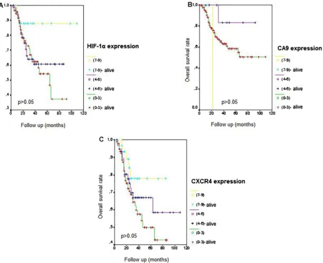
KaplanMeieroverallsurvivalcurvesrevealednosignificant dif-ferencein survivalwithrespecttostrengthofHIF-1␣(Fig.2A), CA9(Fig.2B)andCXCR4(Fig.2C)immunoreactivitybasedonfinal scores.
3.5. CorrelationbetweenHIF-1˛,CXCR4,andCA9expressions NosignificantcorrelationwasnotedbetweenHIF-1␣,CA9and CXCR4 expression in terms of extent,intensity and final score (Table6).
4. Discussion
OurfindingsinaretrospectivecohortofCRCpatientsrevealed immunoreactivityforHIF-1␣,CA9andCXCR4in94.0%,15.0%and 89.0%oftumorspecimens andweakexpressionin 51.1%,93.0% and50.5%ofcaseswithpositiveimmunoreactivity,respectively. IncreasedexpressionofHIF-1␣among males,increased expres-sionofCA9intumorswithulcerovegetativegrowthpatternand increasedexpressionofCXCR4insmallersizeandlower histolog-icalgradetumorswerenoted.Noneoftheimmunohistochemical markersshowedanassociationwithotherclinicopathologic fac-torsaswellaswithprognosticfactors,andoverallsurvivalrates werealsosimilarwithrespecttolevelofHIF-1␣,CA9andCXCR4 immunoreactivity.
HIF-1␣overexpressionwasreportedtobeevidentinnearly55% ofCRCpatients[46],associatedwithpoorprognosisresistanceto chemotherapyand increasedmortality[14,17,47,48]in patients withCRC.HIF-1␣overexpressionwasalsoshowntobean inde-pendentdeterminantofincreaseddiseaserecurrenceinpatients withcolorectallivermetastases(CRLM)[49].
Alargestudyof731CRCspecimensdemonstratedthatHIF-1␣ overexpressionwasindependentlyassociatedwithpoorprognosis [14],whiledatafromameta-analysisof23studiescomprising2984 CRCpatientsalsorevealedasignificantassociationofHIF overex-pressionwithincreasedmortalityriskincludingoverallsurvival anddiseasefreesurvival(DFS)[50].
Although94.0%ofcasesshowedHIF-1␣immunoreactivityin our cohort, weak-to-moderate nuclear expression occurred in majorityofcases,despitestrongnuclearstaininghas characteris-ticallybeenreportedforHIF-1␣expressioninCRCcases[51].This differencemayberelatedtoourmethodofsampling,since microar-raycoreswereselectedfromtumorareasawayfromnecrosisinour study.
Notably,noassociationofthepresenceofHIF-1␣ immunore-activitywasshownwithclinicopathologicandprognosticsfactors, andthelevelofHIF-1␣immunoreactivityhadnoimpactonpatient survivalinourcohort.
Similarly,inapaststudyfromTurkeyconductedamongpatients withmetastaticCRCreceivingchemotherapy, overexpressionof HIF-1␣wasreportedin55%intissuesampleswithnosignificant impactofHIF-1␣expressionrateonsurvival[38].Authorsalso notedthatHIF-1␣expressionwassimilarintheprimarytumorsof thepatientswithvs.withoutmetastasisorinpatientswithsingle vs.multiplemetastasesatthetimeofdiagnosis[38].
PreoperativeHIF-1␣expressionassessedviaIHCwasshown to have a negative predictive and prognostic value in terms of pathologic response and response to chemotherapy in clin-ical stage II/IIIrectal cancerpatients treated with neoadjuvant chemoradiotherapy[52–54].Howeverotherstudiesusingreverse transcription-polymerase chain reaction to measure HIF-1␣ expressionrevealed inconsistentresultsquestioningtherole of HIF-1␣ as a predictive marker in the preoperative treatment
Table3
HIF-1␣expressionwithrespecttoclinicopathologicfeatures.
Immunoreactivity
Finalscore Strength,n(%)
median(min-max) pvaluea weak moderate strong pvalueb
Age <50 3(0–9) 0.197 30(56.6) 20(37.7) 3(5.6) 0.423 ≥50 4(0–9) 65(48.8) 53(39.8) 15(11.2) Sex Female 3(0–9) 0.013 52(60.4) 28(32.5) 6(6.9) 0.055 Male 6(0–9) 43(43.0) 45(45.0) 12(12.0) Tumorlocalization
Rectumandrectosigmoid 4(0–9) 0.482 29(49.1) 25(42.3) 5(8.4) 0.816
Colon 3(0–9) 66(51.9) 48(37.8) 13(77.1)
Macroscopicgrowthpattern
Ulcerovegetative+Depressed 3(0–9) 0.501 88(51.7) 69(40.5) 13(7.6) 0.192 Polypoid 4.5(1–9) 7(50.0) 4(28.5) 3(21.4) TumorSize <5cm 6(0–9) 0.081 40(45.9) 40(45.9) 7(8.0) 0.275 ≥5cm 3(0–9) 55(55.5) 33(33.3) 11(11.1) Histologicgrade Grade1and2 4(0–9) 0.194 68(49.2) 56(40.5) 14(10.1) 0.704 Grade3 3(0–9) 27(56.2) 17(35.4) 4(8.3) Angiolymphaticinvasion Negative 3(0–9) 0.836 44(50.5) 29(33.3) 12(13.8) 0.070 Positive 3(0–9) 50(50.5) 44(44.4) 5(5.0) InvasionDepth T1+T2 3(3–6) 0.841 6(75) 2(25) 0(0.0) 0.143 T3+T4 3.5(0–9) 89(0.5) 71(39.8) 18(10.1)
Lymphnodestatus
N0 3(0–9) 0.755 39(50.6) 29(37.6) 9(11.6) 0.763
N1+N2 3(0–9) 56(51.3) 44(40.3) 9(8.2)
Metastasisstatusatthetimeofdiagnosis
M0 3(0–9) 0.228 71(52.5) 54(40) 10(7.4) 0.289
M1 6(0–6) 24(47.0) 19(37.2) 8(15.7)
Stage
StageI+II 3(0–9) 0.803 33(53.2) 22(35.4) 7(11.3) 0.715
StageIII+IV 3.5(0–9) 62(50.0) 51(41.1) 11(8.8)
Distantmetastasisatdiagnosisorfollowup
Nometastasis 4(0–9) 0.814 43(47.7) 40(44.4) 7(7.7) 0.573 Livermetastasisc 3(0–9) 30(50.8) 22(37.2) 7(11.9) Non-livermetastasis 3(0–9) 22(59.4) 11(29.7) 4(10.8) Localrecurrence No 3(0–9) 0.633 88(50.8) 68(39.3) 17(9.8) 0.961 Yes 3(0–9) 7(53.8) 5(38.4) 1(7.7) aMann-WhiteyUtest. b2test.
c isolatedorwithotherdistantorganmetastasis.
[55,56].Accordingly, besidesdifferenttumor down-staging and tumorregressionsystems,useofdifferentassessmentmethodhas alsobeensuggestedtoberesponsibleforthediscrepancybetween studiesintermsofutilityofHIF-1␣asapredictiveand prognos-ticmarkerinCRC[54].GeneticvariationsinHIF-1␣genehasalso beensuggested tomodulatetheefficacyof postoperative adju-vantchemotherapyandthustoaffectthepredictiveroleofHIF-1␣ expressionofclinicaloutcomesinCRCpatients[54–56].
IncreasedHIF-1␣expressionwasnotedinCRCsamplesobtained frommale than femalepatientsin our cohort.No suchgender differencewasreportedinpaststudiesregardingHIF-1␣ expres-sioninCRCsamples.Nonetheless,genderinfluenceonsensitivity tooxygendeprivationandthusHIF1-dependentcardiacadaptive responsestohypoxiawasreportedintheliterature,withsmaller changesingeneexpressionunderhypoxiaamongfemales[57].
CA9 is suggested to have a potential role as a diagnostic biomarker,prognosticindicatoraswellasatumortherapeutic tar-getinseveralcancertypes[58,59].Beingoverexpressedinhypoxic CRCtumorascomparedwithnormoxiccondition,CA9is consid-eredanimportantbiomarkerforhypoxicCRCtumordiagnosis[23]. Indeed,acorrelationofserumCA9levelswithtumortissueCA9 lev-elswasreportedinclinicalCRCpatientsindicatingnotonlytumor
CA9butalsoserumCA9tobeconsideredasabiomarkerforhypoxic tumordiagnosis[23].
CA9expressionwasshowntooccurparticularlyinareasofhigh proliferationincolorectaltumors[60],athigherlevelinlatethan earlystagetumors[23],andwithmorediffusestainingcompared withbenignlesions[29].
Broad expression of CA9 was shown in adenoma and T1 colorectal cancer specimens based on immunofluorescent and immunohistochemicalstaining,flowcytometry,andquantitative real-time-polymerasechainreactioninapaststudy,indicatingthe stronglikelihoodofCA9tobeinvolvedinthecarcinogenesisofCRC [61].
AlthoughexpressionofCA9isregulatedbyanddirectlylinkedto anincreaseofHIF-1[23,30,31],CA9immunoreactivitywasevident onlyin15.6%ofresectionsampleswithweakimmunoreactivity inmajorityofpositivecasesinourcohort,despiteidentification ofHIF-1␣positivityin94%ofcases.Besides,nocorrelationwas evidentbetweenHIF-1␣andCA9immunoreactivityinourcohort. Inapaststudyontheimpactofsamplingmethodofcolon car-cinomatissuesamples(colonbiopsyvs.surgicalresection)onthe expressionlevelsofpotentialcancerbiomarkergenes,significantly increased expressionof hypoxia markers GLUT-1 and CA9 was
788 B.Sakaetal./Pathology–ResearchandPractice213(2017)783–792
Table4
CA9expressionwithrespecttoclinicopathologicfeatures.
Immunoreactivity
Finalscore Strength,n(%)
median(min-max) pvaluea weak moderate strong pvalueb
Age <50 0(0–9) 0.295 46(86.7) 5(9.4) 2(3.7) 0.121 ≥50 0(0–9) 127(95.4) 5(3.7) 1(0.7) Sex Female 0(0–6) 0.697 83(96.5) 3(3.4) 0(0.0) 0.081 Male 0(0–9) 90(90) 7(7.0) 3(3.0) Tumorlocalization
Rectumandrectosigmoid 0(0–9) 0.416 54(91.5) 4(6.7) 1(1.7) 0.849
Colon 0(0–9) 119(94.0) 6(4.7) 2(1.6)
Macroscopicgrowthpattern
Ulcerovegetative+Depressed 0(0–9) 0.039 159(93.5) 8(4.7) 3(1.7) 0.346 Polypoid 0(0–6) 12(85.7) 2(14.2) (0.0) TumorSize <5cm 0(0–9) 0.436 81(93.1) 5(5.7) 1(1.1) 0.878 ≥5cm 0(0–9) 92(92.9) 5(5) 2(2.0) Histologicgrade Grade1and2 0(0–9) 0.844 128(92.7) 8(5.7) 2(1.4) 0.872 Grade3 0(0–9) 45(93.7) 2(4.2) 1(2.0) Angiolymphaticinvasion Negative 0(0–9) 0.880 80(91.9) 3(3.4) 2(2.3) 0.443 Positive 0(0–9) 91(91.9) 7(7.0) 1(1.0) InvasionDepth T1+T2 0(0-0) 0.217 8(100) 0(0.0) 0(0.0) 0.429 T3+T4 0(0–9) 165(92.7) 10(5.6) 3(1.7)
Lymphnodestatus
N0 0(0–9) 0.937 71(92.2) 4(5.2) 2(2.6) 0.700
N1+N2 0(0–9) 102(93.8) 6(5.5) 1(0.9)
Metastasisstatusatthetimeofdiagnosis
M0 0(0–9) 0.170 126(93.3) 7(5.2) 2(1.5) 0.777
M1 0(0–9) 47(92.2) 3(5.9) 1(1.9)
Stage
StageI+II 0(0–9) 0.796 58(93.5) 2(3.2) 2(3.2) 0.323
StageIII+IV 0(0–9) 115(92.7) 8(6.5) 1(0.8)
Distantmetastasisatdiagnosisorfollowup
Nometastasis 0(0–6) 0.094 87(96.6) 3(3.3) 0(0.0) 0.20 Livermetastasisc 0(0–9) 52(88.1) 5(8.5) 2(3.4) Non-livermetastasis 0(0–9) 34(91.9) 2(5.4) 1(2.7) Localrecurrence No 0(0–9) 0.990 161(93.0) 10(5.8) 2(1.2) 0.198 Yes 0(0–9) 12(92.3) 0(0.0) 1(7.7) aMann-WhiteyUtest. b 2test.
c isolatedorwithotherdistantorganmetastasis.
showninresectionsamples[62].Authorsconcludedthisincrease tobeasaconsequenceofinductionofhypoxicstresssignalbythe clampingofpartofthecolonduringsurgicalresection[62].This seemsnotablegiventhatCA9specificantibodiescouldnot discrim-inatecellsthatcurrentlyorpreviouslyexistinhypoxicconditions sincetransmembraneCA9proteinsremainstableforarelatively longtimeafterre-oxygenationwithahalf-lifeofalmost40h[23]. In a past study in rectal cancer tumor samples from 166 patients,CA9stainingintensitywasshowntodiffersignificantly withrespecttotreatmentcategoriesandresponsetotherapy[29]. Tumorstreatedwithlong-courseRTwithoutchemotherapywere showntobeCA9positivewithmoderate/strongstainingintensity, whiletumorstreatedwithchemoradiotherapywerereportedto bemostly CA9negative[29].Only3 patientsinourcohort had neoadjuvantradiotherapy,whilenonehadsystemicneoadjuvant chemotherapy.Therefore,low/weakCA9immunoreactivityinour CRCsamplesseemsnottobeassociatedwithchemoradiotherapy relatedalterationintumoroxygenationandfinalimmunoreactivity outcome[29,63].Also,lackofpreoperativeradiotherapyin major-ityofourpatientsexcludesthelikelihoodofradiotherapyrelated alterationin tumorsize,stageand grade,nodalstatusand thus HIF-1␣,CA9andCXCR4stainingintheresectionsamples[29].
Nonetheless,higher final scoreswerenoted for CA9 expres-sioninulcerovegetative-depressedthaninpolypoidmacroscopic growthpatterninourcohort.Thisseemsnotable,giventhehigher rateofsubmucosalmassiveinvasionandlymphnodemetastasis anda higherdegreedysplasiareportedin non-polypoidgrowth (NPG)than polypoidgrowth(PG)submucosalCRC[64,65].CA9 expression was known to correlate to cell survival, prolifera-tion,migration,growthandadhesion[30,31].Accordingly,higher expressionofCA9inNPGthanPGgrowthtumorspecimensinour cohortseemsconsistentwithconsiderationofNPGtobeamore aggressiveCRCgrowthpatternthanPG[64].
InapastanalysisoftumorsectionsofCRCpatients,alow CA-9expressioninthetumorwascorrelatedwithbetterdisease-free survival(DFS)andoverallsurvival[66].Consideringdisease out-come in rectal cancer, moderate/strong expression of CA9 and positive HIF-1␣expression inresectionsampleswasassociated with47.5-foldriskofdiseasespecificmortality[67],while neg-ative/weak CA9 staining intensity per se was shown to be an independentpredictoroflongerDFSanddiseasespecificsurvival (DSS)inrectalcancer[29].
Overallsurvivalratewas71.0%atamedian23-monthfollow upwithnoimpactofintensityofHIF-1␣,CA9andCXCR4
expres-Table5
CXCR4expressionwithrespecttoclinicopathologicfeatures.
Immunoreactivity
Finalscore Strength,n(%)
median(min-max) pvaluea weak moderate strong pvalueb
Age <50 4(0–9) 0.537 25(47.2) 17(32.0) 11(20.7) 0.701 ≥50 3(0–9) 69(51.8) 43(32.3) 21(15.8) Sex Female 3(0–9) 0.188 48(55.8) 23(26.7) 15(17.4) 0.302 Male 4(0–9) 46(46.0) 37(37.0) 17(17) Tumorlocalization
Rectumandrectosigmoid 4(0–9) 0.740 29(49.2) 22(37.3) 8(13.6) 0.502
Colon 3(0–9) 65(51.2) 38(29.9) 24(18.9)
Macroscopicgrowthpattern
Ulcerovegetative+Depressed 4(0–9) 0.836 84(49.4) 56(33) 30(17.6) 0.854 Polypoid 3(0–9) 8(57.2) 4(28.6) 2(14.3) TumorSize <5cm 6(0–9) 0.028 40(45.9) 26(29.8) 21(24.2) 0.100 ≥5cm 3(0–9) 54(54.5) 33(33.3) 12(12.1) Histologicgrade Grade1and2 4(0–9) 0.030 65(47.1) 47(34.0) 26(18.8) 0.270 Grade3 3(0–9) 29(60.4) 13(27.1) 6(12.5) Angiolymphaticinvasion Negative 3(0–9) 0.549 47(54.0) 26(29.8) 12(13.8) 0.409 Positive 4(0–9) 46(46.7) 33(33.3) 20(20.2) InvasionDepth T1+T2 3(0–9) 0.793 5(62.5) 1(12.5) 2(25.0) 0.741 T3+T4 3.5(0–9) 89(50.0) 59(33.2) 30(16.8)
Lymphnodestatus
N0 3(0–9) 0.777 39(50.6) 28(36.4) 10(13.0) 0.636
N1+N2 3(0–9) 55(50.5) 32(29.4) 22(20.2)
Metastasisstatusatthetimeofdiagnosis
M0 4(0–9) 0.386 66(48.8) 51(37.7) 18(13.3) 0.817
M1 3(0–9) 28(55.0) 9(17.6) 14(27.5)
Stage
StageI+II 4(0–9) 0.820 30(48.4) 25(40.3) 7(11.3) 0.146
StageIII+IV 3(0–9) 64(51.6) 35(28.2) 25(20.2)
Distantmetastasisatdiagnosisorfollowup
Nometastasis 4(0–9) 0.467 43(47.7) 33(36.6) 14(15.5) 0.317 Livermetastasisc 3(0–9) 31(52.5) 14(23.7) 14(23.7) Non-livermetastasis 3(0–9) 20(54.0) 13(35.1) 4(10.8) Localrecurrence No 4(0–9) 0.061 85(49.2) 57(32.9) 31(17.9) 0.335 Yes 2(0–9) 9(69.0) 3(23.1) 1(7.7) aMann-WhiteyUtest. b2test.
c isolatedorwithotherdistantorganmetastasis.
sionsonsurvivalinourcohortofCRCpatients,whilepatientswith rectumandrectosigmoidtumorscomposedonethirdofstudy pop-ulation.
Althoughnoneofimmunohistochemicalmarkersstudiedwas associatedwithprognosticfactorsorsurvival inourcohortand despitepositiveHIF-1␣expressioninmajorityofsamples,presence ofCA9immunoreactivityonlyin15.6%ofresectionsamplesand weakexpressionin93.0%ofpositivesamplesseemsconsistentwith thefavorablesurvivaloutcome.
Favorablesurvivaloutcomeinourcohortseemsalsoinlinewith thefactthatmostofourpatientshadgrade1–2tumorswithno metastasisat thetime ofdiagnosis,andallstageIII–IVpatients weretreatedwithadjuvantchemotherapy.Nonetheless,giventhe distantmetastasisrate(52.0%,32.0%totheliver)during23-month followup,itshouldbenotedthat5-yearsurvivalrateforpatients withdistantmetastaticCRCis13%[3]andthemortalityiscaused bylivermetastasisinmostcases[33].
Besides, albeit strength of immunoreactivity had noimpact onsurvivaland both HIF-1␣and CXCR4expressions were sim-ilarin patientswith vs. withoutdistantmetastasis atthe time ofdiagnosisor followupin ourcohort,theassociationof each immunomarkerwithpoorsurvivalratesandincreasedmetastatic
potential has consistently been reported in the past studies [14,18,23,35,36,48,61,66,68].
High expression of CXCR4 in early stage CRC tumor speci-mens was reported to be associated with an increased risk of loco-regionalrecurrenceand/orlivermetastasis,andpooroverall survival[68].SerumCXCR4levelswerealsoshowntobestrongly associatedwiththenumberofmetastaticsitesandlivermetastasis inadvancedCRC[35],whileCXCR4expressionwasassociatedwith theoutgrowthofcoloncarcinomamicro-metastasesandthe devel-opmentofCRLM[36,37,69].Hence,CXCR4expressionisconsidered likelytopromotetumormetastasesandthusserumCXCR4levels maybeapotentialprognosticmarkerinmetastatic/recurrentCRC [33].
Increasedriskofrecurrenceandpoorsurvivalinpatientswith CXCR4overexpressioninprimarytumorswithCRCisconsistent withtherole ofCXCR4 receptor inintravasationand enhanced lymphnodemetastases,asstronglyrelatedtotumorprogression andpoorprognosisinadvancedCRC[68,70–76].
IncreasedCXCR4expressioningrade1–2thaningrade3CRC resectionsamplesin ourcohort is consistentwitha significant correlationbetweenCXCR4expressionandtumorgradinginCRC resectionspecimensreportedinapaststudy[37].
790 B.Sakaetal./Pathology–ResearchandPractice213(2017)783–792
Fig.2.KaplanMeieroverallsurvivalcurvesbasedonA)HIF-1␣B)CA9andC)CXCR4expressionfinalscoresindicatingstrengthofimmunoreactivity.
Table6
Correlationbetweentheextent,intensityandfinalscoresofimmunohistochemical markers.
CA9-CXCR4 CA9-HIF1␣ HIF1␣-CXCR4
r p r p r p
Extent −0.027 0.711 −0.066 0.374 −0.038 0.607
Intensity −0.030 0.687 0.003 0.968 −0.023 0.755
Finalscore −0.040 0.584 −0.019 0.798 −0.020 0.790
r:correlationcoefficient.Spearmancorrelationanalysis.
Analysisoftumortissuesamplesrevealedpositiveexpression ratesofCXCR4tobe61.2%insamplesfromstageII–IIIcolon can-cerpatients[77]andtobe38.6%instageI/IIcoloncancerpatients withoutchemotherapy[76].Nosignificantassociationsbetween theexpressionofCXCR4andclinicopathologicandprognostic fac-torsincludinggender,age,tumorlocation,tumorsize,TNMstaging histologicaltype,lymphovascularinvasioninbothstageII–III[77] stageI/II[76]coloncancer.StageII–IIIcoloncancerpatientswith thanwithoutCXCR4hadsignificantlylower3-yearsurvivalrate (27.3%vs.76.3%)[77],whilenosignificantdifferencewasnoted betweenlow-expression and high-expression groups of CXCR4 intermsof5-yeardisease-freesurvivalinstageI/IIcoloncancer patients[76].
Accordingly,otherthangender(HIF-1␣),macroscopicgrowth pattern(CA9)andtumorsizeandhistologicgrade(CXCR4),none oftheclinicopathologicandprognosticfactorsstudiedincluding tumorlocalization, histologictype, tumorstage,angiolymphatic invasion,depthofinfiltration,lymphnodestatus,localrecurrence, metastasisandoverallsurvivalwasassociatedwithexpressionof
immunohistochemicalmarkersinourcohort.Albeitlackofimpact ofHIF-1␣,CA9and CXCR4expressiononsurvival inourcohort isagainstthepaststudies,ourfindingssupporttheirfindingsin termsofnocorrelationofHIF-1␣,CA9andCXCR4overexpression withclinicalorhistopathologicalandprognosticfactorsotherthan survival[29,38,76,77].
Certainlimitationstothisstudyshouldbeconsidered.First,due toretrospectivesinglecenterdesignofthepresentstudy, establish-ingthetemporalitybetweencauseandeffectaswellasgeneralizing ourfindingstooverallCRCpopulationseemsdifficult.Second, HIF-1␣,CA9andCXCR4expressionwasbasedonimmunohistochemical analysisnotonwereevaluatedbasedonproteinexpressionlevels, notongeneticlevelsandresultswerenotconfirmedbywestern blottinganalysis.Third,differencesinneoadjuvantandadjuvant treatmentand prognosis of tumors locatedto rectum or colon perseislikelytoinfluenceclinicaloutcome.Nevertheless,despite thesecertainlimitations,giventhelargesamplesizeandlong-term followup,ourfindingsrepresenta valuablecontributiontothe literature.
5. Conclusions
Inconclusion,ourfindingsrevealedweak-to-moderateHIF-1␣ and CXCR4 immunoreactivity in majority of resection samples, andweakCA9immunoreactivityinmajorityofCA9positivecases. Otherthan gender(HIF-1␣), macroscopicgrowthpattern (CA9) andtumorsizeandhistologicgrade(forCXCR4),noneofthe clini-copathologicandprognosticfactorsinvestigatedwereassociated withexpression of immunohistochemicalmarkers and level of immunoreactivity had noimpact onsurvival. Besidesinterplay
betweenpotentialtumorhypoxiamarkersandcellularsignal trans-ductionpathways,prognosispersedepends ona multifactorial processofinteractingandcorrelatedparameters.Thus,ourfindings emphasizetheneedforfurtherlargerscaleandlongerterm stud-ieswithstandardizedandvalidatedassaymethodologytoclarify thepredictiveandprognosticroleofimmunohistochemical mark-ersinCRCintermsoftumorigenesis,clinicaloutcomeandtumor responsetotherapy.
References
[1]L.A.Torre,F.Bray,R.L.Siegel,J.Ferlay,J.Lortet-Tieulent,A.Jemal,Global cancerstatistics,CACancerJ.Clin.65(2015)(2012)87–108.
[2]R.Siegel,C.Desantis,A.Jemal,Colorectalcancerstatistics,CACancerJ.Clin.64 (2014)(2014)104–117.
[3]R.Siegel,J.Ma,Z.Zou,A.Jemal,Cancerstatistics,CACancerJ.Clin.64(2014) (2014)9–29.
[4]A.Jemal,F.Bray,M.M.Center,J.Ferlay,E.Ward,D.Forman,Globalcancer statistics,CACancerJ.Clin.61(2011)69–90.
[5]R.Siegel,D.Naishadham,A.Jemal,Cancerstatistics,CACancerJ.Clin.63 (2013)11–30.
[6]A.Nishimoto,N.Kugimiya,T.Hosoyama,T.Enoki,T.S.Li,K.Hamano,HIF-1␣ activationunderglucosedeprivationplaysacentralroleintheacquisitionof anti-apoptosisinhumancoloncancercells,Int.J.Oncol.44(2014)2077–2084. [7]Y.Y.Qiu,S.J.Hu,Y.J.Bao,etal.,Anti-angiogenicandanti-proliferativeeffectsof
inhibitionofHIF-1␣byp-HIF-1␣RNAiincolorectalcancer,Int.J.Clin.Exp. Pathol.8(2015)7913–7920.
[8]P.Vaupel,A.Mayer,Hypoxiaincancer:significanceandimpactonclinical outcome,CancerMetastasisRev.26(2007)225–239.
[9]J.A.Bertout,S.A.Patel,M.C.Simon,TheimpactofO2availabilityonhuman cancer,Nat.Rev.Cancer8(2008)967–975.
[10]S.K.Chia,C.C.Wykoff,P.H.Watson,etal.,Prognosticsignificanceofanovel hypoxia-regulatedmarkercarbonicanhydraseIX,ininvasivebreast carcinoma,J.Clin.Oncol.19(2001)3660–3668.
[11]N.J.Mabjeesh,S.Amir,Hypoxia-induciblefactor(HIF)inhuman tumorigenesis,Histol.Histopathol.22(2007)559–572.
[12]A.L.Harris,Hypoxia−akeyregulatoryfactorintumourgrowth,Nat.Rev. Cancer2(2002)38–47.
[13]Y.Xia,H.K.Choi,K.Lee,Recentadvancesinhypoxia-induciblefactor(HIF)-1 inhibitors,Eur.J.Med.Chem.49(2012)24–40.
[14]Y.Baba,K.Nosho,K.Shima,etal.,HIF1Aoverexpressionisassociatedwith poorprognosisinacohortof731colorectalcancers,Am.J.Pathol.176(2010) 2292–2301.
[15]L.Xie,X.Xue,M.Taylor,etal.,Hypoxia-induciblefactor/MAZ-dependent inductionofcaveolin-1regulatescolonpermeabilitythroughsuppressionof occludin,leadingtohypoxia-inducedinflammation,Mol.Cell.Biol.34(2014) 3013–3023.
[16]J.E.Shay,H.Z.Imtiyaz,S.Sivanand,etal.,Inhibitionofhypoxia-inducible factorslimitstumorprogressioninamousemodelofcolorectalcancer, Carcinogenesis35(2014)1067–1077.
[17]S.Rasheed,A.L.Harris,P.P.Tekkis,etal.,Hypoxia-induciblefactor-1alphaand −2alphaareexpressedinmostrectalcancersbutonlyhypoxia-inducible factor-1alphaisassociatedwithprognosis,Br.J.Cancer100(2009) 1666–1673.
[18]A.Wouters,B.Pauwels,F.Lardon,J.B.Vermorken,Review:implicationsof invitroresearchontheeffectofradiotherapyandchemotherapyunder hypoxicconditions,Oncologist12(2007)690–712.
[19]R.A.Gatenby,R.J.Gillies,Amicroenvironmentalmodelofcarcinogenesis,Nat. Rev.Cancer8(2008)56–61.
[20]P.Vaupel,F.O.Kallinowski,P.Kunieff,Bloodflowoxygenandnutrientsupply, andmetabolicmicroenvironmentofhumantumors:areview,CancerRes.49 (1989)6449–6465.
[21]A.J.Majmundar,W.J.Wong,M.C.Simon,Hypoxia-induciblefactorsandthe responsetohypoxicstress,Mol.Cell.40(2010)294–309.
[22]V.Askoxylakis,V.Ehemann,S.Rana,etal.,Bindingofthephagedisplay derivedpeptideCaIX-P1onhumancolorectalcarcinomacellscorrelateswith theexpressionofcarbonicanhydraseIX,Int.J.Mol.Sci.13(2012)
13030–13048.
[23]S.S.Guan,C.C.Cheng,A.S.Ho,etal.,Sulfonamidederivativetargetingcarbonic anhydraseIXasanuclearimagingprobeforcolorectalcancerdetectionin vivo,Oncotarget6(2015)36139–36155.
[24]S.Carlin,N.Khan,T.Ku,V.A.Longo,S.M.Larson,P.M.Smith-Jones,Molecular targetingofcarbonicanhydraseIXinmicewithhypoxicHT29colorectal tumorxenografts,PLoSOne5(2010)e10857.
[25]J.Pastorek,S.Pastorekova,Hypoxia-inducedcarbonicanhydraseIXasatarget forcancertherapy:frombiologytoclinicaluse,Semin.CancerBiol.31C (2015)52–64.
[26]R.Opavsky,S.Pastorekova,V.Zelnik,etal.,HumanMN/CA9gene:anovel memberofthecarbonicanhydrasefamily:structureandexontoprotein domainrelationships,Genomics33(1996)480–487.
[27]J.Y.Winum,M.Rami,A.Scozzafava,J.L.Montero,C.Supuran,Carbonic anhydraseIX:Anewdruggabletargetforthedesignofantitumoragents, Med.Res.Rev.28(2008)445–463.
[28]A.J.Kivela,J.Kivela,J.Saarnio,S.Parkkila,Carbonicanhydrasesinnormal gastrointestinaltractandgastrointestinaltumours,WorldJ.Gastroenterol.11 (2005)155–163.
[29]E.KorkeilaK,P.M.Talvinen,H.Jaakkola,etal.,Expressionofcarbonic anhydraseIXsuggestspooroutcomeinrectalcancer,Br.J.Cancer100(2009) 874–880.
[30]C.C.Wykoff,N.J.Beasley,P.H.Watson,etal.,Hypoxia-inducibleexpressionof tumor-associatedcarbonicanhydrases,CancerRes.60(2000)7075–7083. [31]J.Pouyssegur,F.Dayan,N.M.Mazure,Hypoxiasignalingincancerand
approachestoenforcetumourregression,Nature441(2006)437–443. [32]H.L.McLeod,J.A.McKay,E.S.Collie-Duguid,J.Cassidy,Therapeutic
opportunitiesfromtumourbiologyinmetastaticcoloncancer,Eur.J.Cancer 36(2000)1706–1712.
[33]Y.J.Choi,W.J.Chang,S.W.Shin,K.H.Park,S.T.Kim,Y.H.Kim,Theprognostic roleofserumC-X-Cchemokinereceptortype4inpatientswithmetastaticor recurrentcolorectalcancer,Onco.TargetsTher.9(2016)3307–3312. [34]U.M.Domanska,R.C.Kruizinga,W.B.Nagengast,etal.,Areviewon
CXCR4/CXCL12axisinoncology:noplacetohide,Eur.J.Cancer.49(2013) 219–230.
[35]A.C.Yopp,J.Shia,J.M.Butte,etal.,CXCR4expressionpredictspatientoutcome andrecurrencepatternsafterhepaticresectionforcolorectallivermetastases, Ann.Surg.Oncol.19(2012)S339–S346.
[36]C.Rubie,V.O.Frick,M.Wagner,etal.,Chemokineexpressioninhepatocellular carcinomaversuscolorectallivermetastases,WorldJ.Gastroenterol.41 (2006)6627–6633.
[37]C.Rubie,O.Kollmar,V.O.Frick,etal.,DifferentialCXCreceptorexpressionin colorectalcarcinomas,ScandJ.Immunol.68(2008)635–644.
[38]V.Berk,K.Deniz,O.Bozkurt,etal.,PredictiveSignificanceofVEGFandHIF-1␣ expressioninpatientswithmetastaticcolorectalcancerreceiving
chemotherapycombinationswithbevacizumab,AsianPac.J.CancerPrev.16 (2015)6149–6154.
[39]M.A.Dery,M.D.Michaud,D.E.Richard,Hypoxia-induciblefactor1:regulation byhypoxicandnon-hypoxicactivators,Int.J.BiochemCellBiol.37(2005) 535–540.
[40]U.R.Jewell,I.KvietikovaI,A.Scheid,C.Bauer,R.H.Wenger,M.Gassmann, InductionofHIF-1alphainresponsetohypoxiaisinstantaneous,FASEBJ.15 (2001)1312–1314.
[41]D.Lando,D.J.Peet,D.A.Whelan,J.J.Gorman,M.L.Whitelaw,Asparagine hydroxylationoftheHIFtransactivationdomainahypoxicswitch,Science 295(2002)858–861.
[42]E.Schutyser,Y.Su,Y.Yu,etal.,HypoxiaenhancesCXCR4expressioninhuman microvascularendothelialcellsandhumanmelanomacells,Eur.Cytokine Netw.18(2007)59–70.
[43]P.J.Maxwell,R.Gallagher,A.Seaton,etal.,HIF-1andNF-kappaB-mediated upregulationofCXCR1andCXCR2expressionpromotescellsurvivalin hypoxicprostatecancercells,Oncogene26(2007)7333–7345. [44]R.Marechal,P.Demetter,N.Nagy,etal.,HighexpressionofCXCR4may
predictpoorsurvivalinresectedpancreaticadenocarcinoma,Br.J.Cancer100 (2009)1444–1451.
[45]SeventhEditionoftheAmericanJointCommittee(AJCC-7)onColonand RectumCancerTumor-node-metastasis(TNM)Staging,2009http:// cancerstaging.org/references-tools/quickreferences/documents/ colonmedium.pdf.
[46]D.Cao,M.Hou,Y.S.Guan,M.Jiang,Y.Yang,H.F.Gou,ExpressionofHIF-1␣ andVEGFincolorectalcancer:associationwithclinicaloutcomesand prognosticimplications,BMCCancer9(2009)432.
[47]A.M.Shannon,D.J.Bouchier-Hayes,C.M.Condron,D.Toomey,Tumour hypoxia:chemotherapeuticresistanceandhypoxia-relatedtherapies,Cancer Treat.Rev.29(2003)297–307.
[48]M.Ioannou,E.Paraskeva,K.Baxevanidou,etal.,HIF-1␣incolorectal carcinoma:reviewoftheliterature,J.BUON20(2015)680–689.
[49]M.Shimomura,T.Hinoi,S.Kuroda,etal.,Overexpressionofhypoxiainducible factor-1alphaisanindependentriskfactorforrecurrenceaftercurative resectionofcolorectallivermetastases,Ann.Surg.Oncol.20(2013)S527–536. [50]Z.Chen,X.He,W.Xia,J.Huang,Prognosticvalueandclinicopathological
differencesofHIFsincolorectalcancer:evidencefrommeta-Analysis,PLoS One8(2013)e80337.
[51]N.Simiantonaki,M.Taxeidis,C.Jayasinghe,U.Kurzik-Dumke,C.J.Kirkpatrick, Hypoxia-induciblefactor1alphaexpressionincreasesduringcolorectal carcinogenesisandtumorprogression,BMCCancer8(2008)320. [52]S.A.Lee-Kong,J.A.Ruby,D.B.Chessin,etal.,Hypoxia-relatedproteinsin
patientswithrectalcancerundergoingneoadjuvantcombinedmodality therapy,Dis.ColonRectum55(2012)990–995.
[53]B.M.Havelund,F.B.Sorensen,J.Lindebjerg,K.L.Spindler,A.Jakobsen, PretreatmentHIF-1alphaandGLUT-1expressionsdonotcorrelatewith outcomeafterpreoperativechemoradiotherapyinrectalcancer,Anticancer Res.31(2011)1559–1565.
[54]S.Lin,H.Lai,Y.Qin,J.Chen,Y.Lin,Thymidinephosphorylaseand hypoxia-induciblefactor1-␣expressioninclinicalstageII/IIIrectalcancer: associationwithresponsetoneoadjuvantchemoradiationtherapyand prognosis,Int.J.Clin.Exp.Pathol.8(2015)10680–10688.
[55]Y.Toiyama,Y.Inoue,S.Saigusa,etal.,Geneexpressionprofilesofepidermal growthfactorreceptor:vascularendothelialgrowthfactorand
792 B.Sakaetal./Pathology–ResearchandPractice213(2017)783–792 hypoxia-induciblefactor-1withspecialreferencetolocalresponsivenessto
neoadjuvantchemoradiotherapyanddiseaserecurrenceafterrectalcancer surgery,Clin.Oncol.(R.Coll.Radiol.)22(2010)272–280.
[56]Y.Zhang,P.Wang,X.C.Zhou,etal.,GeneticvariationsintheHIF1Agene modulateresponsetoadjuvantchemotherapyaftersurgeryinpatientswith colorectalcancer,AsianPac.J.CancerPrev.15(2014)4637–4642. [57]R.Bohuslavová,F.Koláˇr,L.Kuthanová,J.Neckáˇr,A.Tichopád,G.Pavlinkova,
GeneexpressionprofilingofsexdifferencesinHIF1-dependentadaptive cardiacresponsestochronichypoxia,J.Appl.Physiol.109(2010)1195–1202. [58]C.Ward,S.P.Langdon,P.Mullen,etal.,Newstrategiesfortargetingthe
hypoxictumourmicroenvironmentinbreastcancer,CancerTreat.Rev.39 (2013)171–179.
[59]A.B.Stillebroer,P.F.Mulders,O.C.Boerman,W.J.Oyen,E.Oosterwijk,Carbonic anhydraseIXinrenalcellcarcinoma:implicationsforprognosisdiagnosis, andtherapy,Eur.Urol.58(2010)75–83.
[60]J.Saarnio,S.Parkkila,A.Parkkila,etal.,Immunohistochemicalstudyof colorectaltumorsforexpressionofanoveltransmembranecarbonic anhydraseMN/CAIX,withpotentialvalueasamarkerofcellproliferation, Am.J.Pathol.153(1998)(1998)279–285.
[61]H.Takahashi,Y.Suzuki,J.Nishimura,etal.,Characteristicsofcarbonic anhydrase9expressingcellsinhumanintestinalcryptbase,Int.J.Oncol.48 (2016)115–122.
[62]S.Pringels,N.VanDamme,B.DeCraene,etal.,Clinicalprocedureforcolon carcinomatissuesamplingdirectlyaffectsthecancermarker-capacityof VEGFfamilymembers,BMCCancer12(2012)515.
[63]L.Rubbia-Brandt,V.Audard,P.Sartoretti,etal.,Severehepaticsinusoidal obstructionassociatedwithoxaliplatin-basedchemotherapyinpatientswith metastaticcolorectalcancer,Ann.Oncol.15(2004)460–466.
[64]I.Hirata,F.Y.Wang,M.Murano,etal.,Histopathologicalandgenetic differencesbetweenpolypoidandnon-polypoidsubmucosalcolorectal carcinoma,WorldJ.Gastroenterol.13(2007)2048–2052.
[65]E.Miranda,A.Destro,A.Malesci,etal.,Geneticandepigeneticchangesin primarymetastaticandnonmetastaticcolorectalcancer,Br.J.Cancer95 (2006)1101–1107.
[66]M.Verstraete,A.Debucquoy,E.Devos,etal.,Investigationofpossible endogenoushypoxiamarkersincolorectalcancer,Int.J.RadiatBiol.89(2013) 9–15.
[67]E.Korkeila,J.Sundström,S.Pyrhönen,K.Syrjänen,Maineffectsand interactionsofcarbonicanhydraseIX,hypoxia-induciblefactor-1ö,ezrinand glucosetransporter-1inmultivariateanalysisfordiseaseoutcomeinrectal cancer,AnticancerRes.32(2012)3299–3303.
[68]J.Kim,H.Takeuchi,S.T.Lam,etal.,ChemokinereceptorCXCR4expressionin colorectalcancerpatientsincreasestheriskforrecurrenceandforpoor survival,J.ClinOncol.23(2005)2744–2753.
[69]I.S.Zeelenberg,L.Ruuls-VanStalle,E.Roos,ThechemokinereceptorCXCR4is requiredforoutgrowthofcoloncarcinomamicrometastases,CancerRes.63 (2003)3833–3839.
[70]H.J.Son,S.Y.Song,W.Y.Lee,etal.,Characteristicsofearlycolorectal carcinomawithlymphnodemetastaticdisease,Hepatogastroenterology55 (2008)1293–1297.
[71]S.B.Lim,C.S.Yu,S.J.Jang,T.W.Kim,J.H.Kim,J.C.Kim,Prognosticsignificance oflymphovascularinvasioninsporadiccolorectalcancer,Dis.ColonRectum 53(2010)377–384.
[72]P.Alamo,A.Gallardo,F.DiNicolantonio,etal.,Highermetastaticefficiencyof KRasG12VthanKRasG13Dinacolorectalcancermodel,FASEBJ.29(2015) 464–476.
[73]Z.Wu,X.Han,J.Yan,etal.,Theprognosticsignificanceofchemokinereceptor CXCR3expressionincolorectalcarcinoma,Biomed.Pharmacother.66(2012) 373–377.
[74]A.Ottaiano,R.Franco,T.A.Aiello,etal.,OverexpressionofbothCXC chemokinereceptor4andvascularendothelialgrowthfactorproteins predictsearlydistantrelapseinstageII-IIIcolorectalcancerpatients,Clin. Cancer.Res.12(2006)2795–2803.
[75]T.Murakami,K.Kawada,M.Iwamoto,etal.,TheroleofCXCR3andCXCR4in colorectalcancermetastasis,Int.J.Cancer132(2013)276–287.
[76]C.Du,Y.Yao,W.Xue,W.G.Zhu,Y.Peng,J.Gu,Theexpressionofchemokine receptorsCXCR3andCXCR4inpredictingpostoperativetumourprogression instagesI-IIcoloncancer:aretrospectivestudy,BMJOpen4(2014)e005012. [77]X.F.Li,X.G.Guo,Y.Y.Yang,A.Y.Liu,EffectofCXCR4andCD133co-expression
ontheprognosisofpatientswithstageII?IIIcoloncancer,AsianPac.J.Cancer Prev.16(2015)1073–1076.