Address for Correspondence / Yazışma Adresi: Dr.Mehmet Düzlü, Gazi University Faculty of Medicine, Department of Otorhinolaryngology, Besevler, Ankara, Turkey. Tel: +90 5052753097. E-mail: [email protected]
©Telif Hakkı 2015 Gazi Üniversitesi Tıp Fakültesi - Makale metnine http://medicaljournal.gazi.edu.tr/ web adresinden ulaşılabilir. ©Copyright 2015 by Gazi University Medical Faculty - Available on-line at web site http://medicaljournal.gazi.edu.tr/
doi:http://dx.doi.org/10.12996/gmj.2015.22
An Atypical Presentation of Renal Cell Carcinoma in a Child: A Neck Mass
Atipik Prezentasyonlu Bir Pediatrik Renal Hücreli Karsinom Vakası: Boyunda Kitle
Raşit Cevizci
1, Mehmet Düzlü
2, Utku Aydil
2, Recep Karamert
2, İbrahim Onur Özen
3, Faruk Güçlü Pınarlı
4, İpek Işık Gönül
5,
Arzu Okur
4, Muammer Melih Şahin
2, Metin Yılmaz
21
Medipol University Faculty of Medicine, Department of Otorhinolaryngology, Istanbul, Turkey
2
Gazi University Faculty of Medicine, Department of Otorhinolaryngology, Ankara, Turkey
3
Gazi University Faculty of Medicine, Department of Pediatric Surgery, Ankara, Turkey
4
Gazi University Faculty of Medicine, Department of Pediatric Oncology, Ankara, Turkey
5 Gazi University Faculty of Medicine, Department of Pathology, Ankara, Turkey
ABSTRACT
The most common malignant diseases of the head and neck in the adolescent period are acute leukemia, Hodgkin’s disease, non-Hodgkin’s lymphoma, nasopharyngeal carcinoma, and thyroid carcinoma. Although renal cell carcinoma (RCC) may metastasize in the cervical lymph nodes, both occurrence and presentation as a neck mass are rare in the childhood period. In this report, we present a sixteen-year-old girl who was admitted with a left neck mass at presentation. The primary tumor was found to be located in the left kidney and both masses were surgically removed, revealing a diagnosis of a translocation renal cell carcinoma with metastases. Distant metastases of abdominal and thoracic malignancies may present with metastatic lymph nodes, and should be included in the differential diagnosis of the neck masses in children.
Key Words: Neck mass, renal cell carcinoma
ÖZET
Adölesan dönemde baş boyun bölgesinde en sık rastlanılan malign hastalıklar; akut lösemi, Hodgkin lenfoma, non-Hodgkin lenfoma, nazofarenks kanseri ve tiroid kanseridir. Renal hücreli karsinom (RHK) servikal lenf nodlarına metastaz yapabilmekle beraber, çocukluk döneminde görülmesi ve boyun kitlesi ile başvurusu çok nadirdir. Bu yazıda ilk başvuru şikayeti sol boyunda kitle olan on altı yaşında bir kız çocuğunu sunuyoruz. Hastanın sol böbreğinde yerleşik olduğu tespit edilen primer tümör ve boyun metastazı eksize edildi ve patolojik tanı translokasyonal renal hücreli karsinom olarak rapor edildi. Abdominal ve torasik malignensiler metastatik lenf nodları ile prezente olabileceğinden çocuklarda boyun kitlelerine yaklaşımda ayırıcı tanıda bulundurulmalıdır.
Anahtar Sözcükler: Boyun kitlesi, renal hücreli karsinom
INTRODUCTION
Neoplastic neck masses are relatively rare in the childhood period and the most common causes of the neck masses are either infectious or congenital conditions (1). The most common malignancies encountered in the childhood are Hodgkin’s lymphoma (HL), non-Hodgkin’s lymphoma (NHL), rhabdomyosarcoma, neuroblastoma, acute lymphoblastic leukemia (ALL), nasopharynx carcinoma (NF) and thyroid cancer (2).
In case of a metastatic neck mass, the primary lesion should be investigated through the upper aerodigestive tract and thyroid initially. On the other hand, a supraclavicular mass should alert the physician for distant primaries such as lung, kidney and stomach. A comprehensive study investigating 845 genitourinary tumors reported that the rate of neck metastases is only 2.5% (3). Neck mass is rarely a presenting symptom for visceral organ malignancies. In this case report, we present a sixteen-year-old girl with a neck mass as the presenting symptom of a translocation renal cell carcinoma. According to our comprehensive literature review, this is the first pediatric case with a renal cancer who presented solely with a neck mass.
CASE REPORT
A sixteen-year-old girl was admitted to our clinic with a complaint of a mass on the left side of her neck for two months. The patient expressed that the mass was enlarging and painful. She did not report dysphagia, hoarseness, shortness of breath, fever and night sweats, but had anorexia and 5% weight lost within the last three months. Physical examination revealed a painful firm and fixed supraclavicular mass, which was measured to be three centimeters in the largest dimension. Fiberoptic examination of the upper aerodigestive tract revealed no pathology. The parameters of the complete blood count were all within normal ranges. Serological studies including serum IgMs for rubella, toxoplasmosis, Epstein-Barr virus (EBV), Cytomegalovirus (CMV) and Herpes-simplex virus (HSV) were all negative.
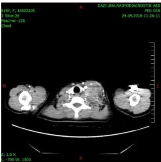
On ultrasound, a neck mass, which was in close relationship with the left lobe of the thyroid gland located behind the sternocleidomastoid muscle was detected. The mass was extending to the left supraclavicular fossa. A computed tomography (CT) was obtained, and a left supraclavicular fossa mass, which was 75 millimeters in largest dimension, was confirmed on CT images (Figure 1).
Magnetic resonance imaging (MRI) of the neck and mediastinum showed that there was a lower neck mass including solid and cystic components; starting from the level of corpus of the fifth cervical vertebra and extending to the left side of the clavicle’s medial edge and to the left side of the thoracic inlet. The dimensions of the mass lesion were measured to be 5x7x9 centimeters. The preliminary diagnosis was lymphoma and an excisional biopsy was performed. Histopathological examination revealed a malignant epithelial tumor metastasis with a differential diagnosis of paraganglioma, which could not be ruled out (Figure 2).
To rule out secondary endocrine neoplasia and a functioning paraganglioma, serum adrenocorticotropic hormone (ACTH), cortisole, prolactine, androstenedione, insulin, and urinary catecholamines were studied, but all were found to be within the normal ranges. Abdomen CT showed a solid mass of 3.5x4.5x4 centimeters located at the superior pole of the left kidney. There was also a left adrenal gland mass which was 17 millimeters in the largest dimension. On positron emission tomography (PET)/CT, maximum standardized uptake values (SUVmax) for the supraclavicular mass, the renal mass and the adrenal mass were 4.0, 5.4, and 6.9, respectively.
Together with the left kidney and the adrenal gland, the neck mass removed surgically. The postoperative period was uneventful. The final histopathological diagnosis was a metastatic translocation cell carcinoma of the kidney (Figure 3). Immunohistochemically, the tumor cells were positive for transcription factor E3 (TFE3). Meanwhile, a paravertebral mass extending from the 8th to 12th thoracic and lumbar vertebrae was also diagnosed. As translocation carcinomas are known to be resistant to chemotherapy and the extended surgical removal of the mass was not possible, external beam radiotherapy with a total dose of 3000cGy was applied to C5-T3 and T7-T12 intervertebral spaces to provide local control. The follow-up evaluation revealed an increase in the size of the residual mass with MRI three months later. Sunitinib was started at a dose of 50mg daily p.o. for 2 weeks every 4 weeks but no response was obtained after three courses. The patient discontinued the treatment thereafter because of its side effects such as fatigue and malaise, and finally the patient deceased due to the progressive disease after the two years of diagnosis.
Figure 1: An axial CT image is showing the supraclavicular mass within the neigborhood of the left thyroid lobe.
Figure 2: Malignant epithelial tumor composed of cells mainly with clear cytoplasm and in part with eosinophylic cytoplasm forming gland-like structures (HEx400).
Figure 3: Lymph node structure with epithelial tumor metastasis (HEx10). Black arrow: residual lymph node tissue.
DISCUSSION
When a neck mass is encountered in adult patients, a physician should consider a malignant tumor, especially a metastatic lymph node related with cancers of the upper aerodigestive tract (4). In contrast to adult population, malignant head and neck tumors are not so frequently seen during the childhood period, congenital masses and infectious lymphadenopathy such as mycobacterial cervical lymphadenitis should be considered primarily in case of a neck mass (2). The most common head and neck malignancy in children is lymphoma which includes mainly HL and NHL. Lymphomas can originate from a lymph node or extranodal sites and often are associated with extensive lesions within the mediastinum or abdomen (5). The most common soft tissue sarcoma in children is rhabdomyosarcoma, and more than one third of cases occur in the head and neck region. Differentiated thyroid cancers are the most common endocrine malignancies in childhood, accounting for the 2% of all thyroid cancers. Nasopharyngeal carcinoma is rare in children but most commonly presents with a painless upper neck mass (6). Other causes of malignant neck masses are neuroblastoma, salivary gland malignancies and metastatic carcinomas (2). While neuroblastoma and embryonal rhabdomyosarcoma are the most frequently seen conditions in young children, lymphomas, thyroid carcinoma and nasopharyngeal carcinoma are also frequently seen in older children and adolescents.
Wilms’ tumor is the most common cause of renal mass in children by far and account for 87% of all pediatric renal masses, occuring in approximately 1:10,000 children (7). The infrequent renal tumors of childhood are congenital mesoblastic nephroma, rhabdoid tumor of the kidney, clear cell sarcoma of the kidney, and renal cell carcinoma. RCC uncommonly presents in the first two decades of life, and it is extremely rarer when compared with Wilms’ tumor. Less than 1% of all cases occur in children, and the mean age of presentation in childhood is nine years (8). RCC can metastasize to reach any area but the most common sites are lungs, regional lymph nodes, bone, and liver (9). Although the head and neck region is a quite rare metastatic site, RCC metastases can also be seen in nose, paranasal sinuses, larynx, tongue, jaws and thyroid gland (4, 10). Only 1% of the patients with RCC have metastases which are confined to the head and neck region (11).
GMJ
2015; 26: 68-70
Akyurek et al.
RCC in a child
69
A comprehensive literature review on cervical lymph node metastasis as the presenting symptom of renal cell carcinoma was done recently by a few authors. Pompeo et al. reported a RCC case who presented with a cervical metastasis as the first sign leading to diagnosis (12). Ozkiris et al. and Chori et al. also reported two RCC cases presented with neck masses (13, 14). Chhabraet and colleagues reported a patient presenting with metastatic renal cell carcinoma to the neck mimicking a carotid body tumor (15). All these cases were in the sixth and the seventh decades. A younger patient was presented by Langille et al., who reported a 29-year-old male case presenting with a metastatic mass located at the supraclavicular fossa at diagnosis (16).
The biology of the pediatric RCC is distinct from its adult counterpart. The most common subtype of RCC in adults is a clear cell histology, whereas the translocation subtype is the most common type of pediatric RCC, as in our patient. Metastases are present in 20% of the patients at presentation (17). In children, RCC associated with Xp11.2 translocations/TFE3 gene fusions is a more advanced-stage disease as compared to the other types of RCC (18,19). Although the translocation RCC is highly malignant and easily invades regional lymph nodes, it was also suggested that local lymph node metastases in the absence of haematogenous disease spread does not indicate a bad prognosis. However, the prognosis of the patients with distant metastases is ominous due to the lack of effective therapeutic modalities (20).
The standard treatment of renal carcinoma is the surgical resection of primary tumor and its metastases. As RCC is known to be resistant to chemotherapy and radiotherapy, these procedures’ role is unclear for residual and widespread diseases. Although multitargeted tyrosine kinase inhibitors such as sorafenib and sunitinib might have an effect on survival in adults , there are only a few case reports concerning these therapies in children, and their efficacy is unknown (21). As sunitinib selectively inhibits several growth factor receptors including VEGF, a rapid and complete tumor necrosis of the widespread metastatic disease after the sunitinib use has been reported (22). However, in our patient, we didn’t have any objective response to the therapy, and she discontinued sunitinib after three courses because of its side effects.
In conclusion, herein we report a sixteen-year-old girl who had a solitary neck mass as a presenting symptom of an RCC. To our knowledge, our case is the first patient in the childhood period that was presented with a cervical mass and diagnosed with renal cell carcinoma. Although quite rare, a neck mass can be the first symptom of RCC even in a child, and the possibility of RCC should be considered in the differential diagnosis.
Conflict of Interest
No conflict of interest was declared by the authors. REFERENCES
1. Boring CC, Squires TS, Tong T. Cancer statistics. CA Cancer J Clin 1993;43:7-26.
2. Showkat SA, Lateef M, Wani AA, Lone SA, Singh K, Yousuf I. Clinicopathological profile of cervicofacial masses in pediatric patients. Indian J Otolaryngol Head Neck Surg 2009;61:141-6. 3. Hessan H, Strauss M, Sharkey FE. Urogenital tract carcinoma
metastatic to the head and neck. Laryngoscope 1986;96:1352–6. 4. Ogunyemi O, Rojas A, Hematpour K, Rogers D, Head C, Bennett C.
Metastasis of genitourinary tumors to the head and neck region. Eur Arch Otorhinolaryngol 2010;267:273-9.
5. Tracy TF Jr, Muratore CS. Management of common head and neck masses. Semin Pediatr Surg 2007;16:3-13.
6. Chadha NK, Forte V. Pediatric head and neck malignancies. Curr Opin Otolaryngol Head Neck Surg 2009;17:471-6.
7. Anand R, Narula MK, Gupta I, Chaudhary V, Choudhury SR, Jain M. Imaging spectrum of primary malignant renal neoplasms in children. Indian J Med Paediatr Oncol 2012;33:242-9.
8. McHugh K. Renal and adrenal tumours in children. Cancer Imaging 2007;7:41-51.
9. Maldazys JD, deKernion JB. Prognostic factors in metastatic renal carcinoma. J Urol 1986;136:376-9.
10. Som PM, Norton KL, Shugar JM, Reede DL, Norton L, Biller HF. Metastatic hypernephroma to the head and neck. AJNR Am J Neuroradiol 1987; 8:1103–6.
11. Pritchyk KM, Schiff BA, Newkirk KA, Krowiak E, Deeb ZE. Metastatic renal cell carcinoma to the head and neck. Laryngoscope 2002;112:1598-602.
12. Pompeo AC, Kanashiro H, Silva MN. Renal cell carcinoma presenting as a cervical mass. Int Braz J Urol 2005;31:151-2. 13. Ozkiriş M, Kubilay U, Sezen OS. Cervical lymph node metastasis in
renal cell carcinoma. J Oral Maxillofac Pathol 2011;15:211-3. 14. Choi YR, Han HS, Lee OJ, Lim SN, Kim MJ, Yeon MH et al.
Metastatic renal cell carcinoma in a supraclavicular lymph node with no known primary: a case report. Cancer Res Treat 2012;44:215-8.
15. Chhabra P, Bhatt V, Brown AM. Metastatic renal cell carcinoma in the neck: an unusual presentation. Dent Update 2009;36:511-3. 16. Langille G, Taylor SM, Bullock MJ. Metastatic renal cell carcinoma
to the head and neck: summary of 21 cases. J Otolaryngol Head Neck Surg 2008;37:515-21.
17. Dome JS, Fernandez CV, Mullen EA, Kalapurakal JA, Geller JI, Huff V et al. COG Renal Tumors Committee. Children's Oncology Group's 2013 blueprint for research: renal tumors. Pediatr Blood Cancer 2013; 60:994-1000.
18. Song HC1, Sun N2, Zhang WP1, He L3, Fu L3, Huang C1. Biological characteristics of pediatric renal cell carcinoma associated with Xp11.2 translocations/TFE3 gene fusions. J Pediatr Surg 2014;49:539-42.
19. Wu A, Kunju LP, Cheng L, Shah RB. Renal cell carcinoma in children and young adults: analysis of clinicopathological, immunohistochemical and molecular characteristics with an emphasis on the spectrum of Xp11.2 translocation-associated and unusual clear cell subtypes. Histopathology 2008; 53:533-44. 20. Geller JI, Argani P, Adeniran A, Hampton E, De Marzo A, Hicks J et
al. Translocation renal cell carcinoma: lack of negative impact due to lymph node spread. Cancer. 2008;112:1607-16.
21. Tüysüz G1, Ozdemır N, Emır H, Durak H, Dervışoğlu S, Adaletlı I et al. A Translocation Renal Cell Carcinoma with Skeletal Muscle Metastasis in a Child. Turk Patoloji Derg 2014; doi: 10.5146/tjpath.2014.01259. (Epub ahead of print).
22. Chowdhury T, Prichard-Jones K, Sebire NJ, Bier N, Cherian A, Sullivan MO et al.Persistent complete response after single-agent sunitinib treatment in a case of TFE translocation positive relapsed metastatic pediatric renal cell carcinoma. J Pediatr Hematol Oncol 2013;35:e1-3.